Vol. 6, No. 4, 2003 X-Ray Scattering Studies of the Metastable Ferroelectric Phase in KDP Induced by Static Electric Field 493
Materials Research, Vol. 6, No. 4, 493-495, 2003. © 2003
*e-mail: [email protected]
Trabalho apresentado no XV CBECIMAT, Natal - RN, Novembro de 2002.
X-Ray Scattering Studies of the Metastable Ferroelectric Phase
in KDP Induced by Static Electric Field
Claudio Marcio Rocha Remédios, Marcus Aurélio Ribeiro Miranda, José Marcos Sasaki*,
Josué Mendes-Filho, Paulo de Tarso Cavalcante Freire, rancisco Erivan de Abreu Melo
Departamento de Física, Universidade Federal do Ceará, UFC C.P. 6030, 60455-760 Fortaleza - CE, Brazil
Received: February 4, 2003; Revised: September 2, 2003
Potassium dihydrogen phosphate presents a ferroelectric phase below 122 K and a paraelectric phase for T > 122 K. X-ray scattering measurements were performed in order to verify the mecha-nism of the phase transition from point group C2v19 to C
2v
≠19 at 119 K induced by DC electric field;
in other words, a change of the local site symmetries of the phosphate group occurs from C2 site in the C2v19 point group to C
s in the C2v
≠19 point group. This phenomenon was shown by analyzing the
behavior of the integrated intensity of the (800), (080), (1600), (0160), (400) and (040) reflec-tions. The curve fitting of these reflections showed the occurrence of a rotation of the phosphate group around the [010] crystallographic direction during the phase transition. This study confirms the observation of this metastable phase obtained by Raman spectroscopy.
Keywords:KDP, metastable phase, X-ray
1. Introduction
Many works developed with the objective of studying the physical properties of ferroelectric materials had already promoted discoveries with valuable contribution to technol-ogy. Although it has already been sufficiently studied, the Potassium Dihydrogen Phosphate (KDP) is a ferroelectric material that attracts the attention of many researchers be-cause of its technological applications, such as Ferroelectric Devices for Random Access Memory (FDRAM). The KDP crystals undergo a phase transition at a temperature
of 122 K1. This crystal presents an orthorhombic symmetry
(ferroelectric phase) at temperature below 122 K,
belong-ing to the Fdd2 (C2v19) space group, while above this
tem-perature it presents a tetragonal symmetry (paraelectric
phase), belonging to the I42d (D2d12) space group. The KDP
crystal undergoes two metastable transitions induced by
uniaxial pressure: D2d12 → C
2v j
≠19 and C
2v19→ D2vj
≠19.
Ac-cording to the literature an explanation for these transitions is based on the changing of the local site symmetry of the phosphate ions: in the ferroelectric phase, the phosphate ions
change their local site symmetry from C2 to Cs, maintaining
the same factor group C2v but modifying the space group.
These changes are due to the rotation of the phosphate ions
around the [010] direction of the orthorhombic structure2.
Other works in the literature discuss the reversibility
crite-ria of this new metastable phase3. The phase diagram for
the KDP transitions on the plane (σ
6, T) for temperature in
the range from 110 to 130 K explains the appearance of the
metastable C2vj≠19 phase based on the Gibbs free energy
den-sity of the system4.
Raman scattering experiments show that the potassium dihydrogen phosphate crystals undergo a metastable ferroelectric phase induced by a static electric field near the
Curie point5 taking advantage of the fact that the ferroelectric
phase presents piezoelectricity. In other words, due to the converse piezoelectric effect a DC electric field applied along the [001] direction should induce a phase transition (C2v19 → C
2v
≠19) in KDP crystals similarly to that induced by
494 Remédios et al. Materials Research
orthorhombic structure leading to a change in the local site symmetry of the phosphate ions.
2. Experimental
Single crystals of KDP with good optical quality and good crystaline perfection used in the experiments were grown by the slow cooling method. These samples were
cut in parallelepipeds with dimensions (0.87 × 3 × 5) mm3.
The parallelepiped faces were orthogonal to the [100], [010] and [001] crystallographic directions of the tetragonal struc-ture. Silver electrodes were painted on the larger faces which are perpendicular to the ferroelectric [001] direction. A Keithley instrument model 246 was used as the DC voltage source.
X-ray scattering measurements were performed using a
two-circle (θ-2θ) Rigaku diffractometer, with radiation
source of MoKα coupled with a low temperature chamber
containing liquid nitrogen. The sample temperature was controlled with a fluctuation of ± 0.5 K. The high
penetra-tion feature of the X-ray beam (λMoKα= 0.709Å) gives
ad-vantage to get diffraction from deeper planes. This reduces the diffraction from the crystal surface that is not affected by the electric field.
3. Results and Discussion
The crystal was aligned using the 440 reflection of paraelectric phase (tetragonal) and then cooled down to ferroelectric phase (orthorhombic). According to the space group of both phases, the extinction in the Miller indices for the reflections hh0 (h = 2 n) in the tetragonal symmetry changes to h00 (h = 4 n) in the orthorhombic symmetry.
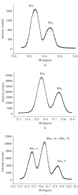
Figure 1a shows the diffraction pattern for the 440 re-flection at room temperature. The first peak corresponds to
the scattering of the plane (440) for Kα1 radiation and the
second one is the scattering of the same plane for Kα2
radia-tion. Figure 1b shows rocking curves for the 440 reflection at 125 K (near ferroelectric phase transition). The changes observed in Fig. 1b regarding to Fig. 1a are easily explained. The displacement of the peak position to higher angles oc-curs due to the reduction of the lattice parameters caused by thermal contraction. Its scattering intensity is related to the Debye-Waller factor.
Figure 1c shows the diffraction pattern below the trans-formation temperature. It shows three peaks where the first
is the scattering of the Kα1 radiation by a domain we call a.
The third peak is related to the diffraction of the Kα2
radia-tion by another domain that we call b. Finally the second
peak is the overlap of Kα2 diffracted by the domain a and
the Kα1 diffracted by the domain b6-9.
Before discussing X-ray results we need to show that under DC electric field, the KDP crystal undergoes a
C2v19→ D
2vj
≠19 phase transition. The idea of the experiment
Figure 1. X-ray diffraction pattern for: a) (440) plane at room tem-perature; b) (440) plane near transition at 124 K; c) (800) plane for ferroelectric phase at 119 K.
Vol. 6, No. 4, 2003 X-Ray Scattering Studies of the Metastable Ferroelectric Phase in KDP Induced by Static Electric Field 495
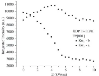
Figure 2 shows the diffraction patterns corresponding
to the a and b domains in the orthorhombic structure as a
function of the DC electric field up to 5 kV/cm at 119 K. The peaks emerge from the overlapping of the bands
corre-sponding to the Kα1 and Kα2 lines of Mo radiation. By
per-forming a spectral decomposition into Gaussian compo-nents, we can draw the variation of the integrated intensity of both reflections as a function of DC electric field, which is displayed in Fig. 3. It should be observed that the
inte-grated intensity corresponding to the a domain decreases
Figure 2. X-ray diffraction pattern for KDP obtained in the ferroelectric phase under electric field from 0-6 kV/cm.
with increasing the DC electric field up to 5 kV/cm, while
the b domain increases. In addition, for the electric field of
6 kV/cm, the peak corresponding to the a domain
disap-pears. These changes in the integrated intensity for the a
domain until 5 kV/cm and the total disappearance of this peak at 6 kV/cm indicate essentially that oxygen atoms on these planes moved out. This can be attributed to the result of rotation of the phosphate tetrahedron around the [010]
crystallographic direction relative to the C2v19 orthorhombic
structure.
4. Conclusion
We report here the mechanism which led to a confor-mational phase transition of KDP when an increase of DC electric field is applied along the orthorhombic c-axis. The huge variation in the integrated intensity of those peaks,
corresponding to the a and b domains, indicated the
occur-rence of changes in the atom positions on (800) planes. These changes were attributed to the rotation of the phosphate tet-rahedron around the b-axis, which also changes the local
site symmetries of phosphate ions from C2 in the C2v19 phase
to Cs in the C2v≠19 phase.
Acknowledgements
Financial support from Brazilian funding agencies CAPES, CNPq, and FUNCAP is gratefully acknowledged.
References
1. Busch, G. Helv. Phys. Acta, v. 11, p. 269, 1938. 2. Moreira, S.G.C.; Melo, F.E.A.; Mendes-Filho, J.; Moreira,
J.E. Ferroelectrics, v. 160, p. 47, 1994.
3. Melo, F.E.A.; Moreira, S.G.C.; Mendes-Filho, J.; Moreira,
J.E. Phys. Status Solidi B, v. 180, p. 371,1993.
4. Melo, F.E.A.; Moreira, S.G.C.; Chaves, A.S.; Guedes, I.;
Freire, P.T.C.; Mendes-Filho, J. Phys. Rev. B, v. 59,
p. 3276, 1999.
5. Varela, A.T.; Melo, F.E.A.; Barbosa Neto, N.M.; Guedes,
I.; Freire, P.T.C.; Mendes-Filho, J.; Sasaki, J.M. J.
Ra-man Spectrosc., v. 31, p. 915-919, 2000.
6. Bacon, G.E.; Pease, R.S. Proc. of Roy. Soc. Ser-A, v. 230:
p. 359-381, 1956.
7. Ubbelohde, A.R.; Woodward, I. Proc. Roy. Soc. A, v.12,
p. 188, 1946.
8. Bornarel, J. Ferroelectrics, v. 71, p. 255-268, 1987.
9. Kitaeva, G.Kh.; Kulik, S.P.; Penin, A.N.; Belinsky, A.V.
Phys. Rev. B, v. 51, p. 3362, 1995.