Immunotherapy, immunochemotherapy and chemotherapy
for American cutaneous leishmaniasis treatment
Imunoterapia, imunoquimioterapia e quimioterapia no
tratamento da leishmaniose tegumentar americana
Wilson Mayrink
1, Ana Cristina de Carvalho Botelho
1, 3, Paulo Araújo Magalhães
6†,
Sebastião Mariano Batista
1†, Antonio de Oliveira Lima
7†, Odair Genaro
1†, Carlos Alberto da Costa
4,
Maria Norma de Melo
1, Marilene Susan Marques Michalick
1, Paul Williams
1,
Magno Dias
2, Waleska Teixeira Caiaffa
8, Evaldo do Nascimento
1and George Luiz Lins Machado-Coelho
2ABSTRACT
The first choice of treatment for American cutaneous leishmaniasis is the pentavalent antimonial drug. Although it has been shown
that this treatment is mostly effective and indicated, some disadvantages should be taken into account such as side effects, long
term treatment inconveniences and counter-indication for patients suffering from cardiopathy, nephropathy; yet, aging, pregnancy
and other conditions. With the advent of the vaccine anti-American cutaneous leishmaniasis as a prophylactic measure, studies on
therapy using the vaccine associated or not with other drugs have been performed by many investigators and it is currently among
the alternative treatments and prevention measures for American cutaneous leishmaniasis. In conclusion, the association between
antimony and vaccine (immunochemotherapy) showed the same cure rate when compared with the standard treatment (100%)
and it was also able to reduce the salt volume in 17.9% and treatment length from 87 to 62 days, decreasing side effects.
Key-words:
American cutaneous leishmaniasis. Treatment. Immunotherapy.
RESUMO
O tratamento de primeira escolha para leishmaniose tegumentar americana é o antimonial pentavalente. Embora este tratamento
seja na maioria das vezes efetivo e indicado, devem ser consideradas as desvantagens tais como efeitos colaterais, longa
duração do tratamento e contra-indicação para cardiopatas, nefropatas, idosos, grávidas e outras condições. Com o advento
da vacina antileishmaniose tegumentar americana para fins profiláticos e terapêuticos, associando-a ou não a outros fármacos,
muitas pesquisas têm sido desenvolvidas, sendo a vacina a principal entre os atuais recursos no tratamento e prevenção da
leishmaniose tegumentar americana. Em conclusão, a associação do antimônio com a vacina (imunoquimioterapia) apresentou
o mesmo índice de cura em relação ao tratamento padrão (100%), e ainda reduziu o volume do sal em 17,9% e o tempo de
cura significativamente, de 87 para 62 dias; conseqüentemente, reduzindo os efeitos colaterais.
Palavras-chaves:
Leishmaniose tegumentar americana. Tratamento. Imunoterapia.
1. Departamento de Parasitologia do Instituto de Ciências Biológicas da Universidade Federal de Minas Gerais, Belo Horizonte, MG. 2. Departamento Farmácia da Escola de Farmácia da Universidade Federal de Ouro Preto, Ouro Preto, MG. 3. Centro Pesquisas René Rachou da Fundação Oswaldo Cruz, Belo Horizonte, MG. 4. Departamento de Parasitologia da Faculdade de Farmácia da Universidade Federal de Minas Gerais, Belo Horizonte, MG. 5. Departamento de Ciências Biológicas da Universidade Federal de Ouro Preto, Ouro Preto, MG. 6. Superintendência de Campanha Sanitária do Ministério da Saúde. 7. Fundação Ataulfo Paiva, Belo Horizonte, MG. 8. Departamento de Medicina Preventiva e Social da Faculdade de Medicina da Universidade Federal de Minas Gerais, Belo Horizonte, MG.
† In memoriam.
Supported by FAPEMIG, SMSC, CNPq, UFMG, CISMRC
Address to: Dr. Wilson Mayrink. Rua Equador 140/201, Bairro São Pedro, 30330-390 Belo Horizonte,MG, Brasil Tel: 55 31 3287-3038
Leishmaniasis is a disease caused by several digenetic
protozoan species of the genus
Leishmania
, which affect
humans and different domestic and sylvatic animal species.
The disease may arise in different forms and the most
important are: American visceral leishmaniasis (AVL) or
American kala-azar and American cutaneous leishmaniasis
(ACL). The latter, is primarily a disease that affects sylvatic
animals. Leishmaniasis is transmitted to humans through the
female bite of female haematophagous insect vectors of the
genus
Lutzomyia
, which are known, in Brazil, as ‘birigüi’,
‘mosquito-palha’ or ‘cangalhinha’.
ACL is a polymorphic disease of skin and mucous membranes
with single or multiple ulcerative lesions (simple cutaneous
form), nodular lesions (diffuse form) or mucocutaneous lesions
(mucocutaneous form), affecting nasopharynx regions
simultaneously with or just after an initial cutaneous infection.
Such variation is closely related to the patient’s immunological
status and the
Leishmania
species involved. ACL constitutes an
important health problem mainly because of its difficult
prevention. The World Health Organization (WHO) estimates
around 1-2 million annual new cases annually
78.
Up to 1912, leishmaniasis treatment had been
unsatisfactory. Row
62reported results on the curative properties
of promastigote forms, from
Leishmania
culture, used as a
vaccine in the treatment for Oriental sore; this therapy was
then abandoned due to a new systemic treatment with
antimonial drugs introduced that year by the Brazilian physician
Gaspar Vianna
76. This investigator used emetic tartar at 1%,
which proved to be more efficient than the vaccine.
Emetic tartar has been the only therapeutic alternative for
leishmaniasis for many years and because of its toxicity,
administration difficulties and unsatisfactory efficacy (frequent
recurrences), it was, at last, replaced with pentavalent
antimonial drugs
21 25. Despite its counter-indications for elderly
patients and other conditions, such as cardiopathy, nephropathy
and pregnancy, the pentavalent antimonial drug represents the
standard therapy for leishmaniasis nowadays.
Since the introduction of antimonial drugs for
leishmaniasis therapy, which have also been shown to produce
divergent results
2 3 8 17 18 20 24 26 29 33 44 49 51 52 60 63 65 66 70 74,
several
studies have been carried out in order to establish the best
therapeutic plan.
With the advent of the ACL vaccine for prophylactic aims,
idealized by Gomes
23, Pessoa and Pestana
55, modified by
Mayrink et al
35 36, and proved by Antunes et al
4and Armijos et al
5,
its therapeutic use has been under study, whether alone or
associated with other drugs; and it has also been regarded as
one of the current treatments, as well as one of the available
prophylactic measures for ACL
14 22 28 35 39 43.
Reports on the use of chemotherapy associated with
immunotherapy have shown considerable efficacy in some
cases of diffuse leishmaniasis
37 71 72.
Immunotherapy combined with the use of BCG, first
performed by Convit
14, showed cure rates and treatment length
comparable with those obtained through standard treatments
with Glucantime
.
The present work was aimed at: 1) using immunotherapy in
ACL patients for whom antimonial drugs are restricted and;
2) using immunotherapy combined with chemotherapy
(immunochemotherapy), since we observed that an
immunological stimulus associated with a chemotherapy
treatment leads to a reduction in antimony volume and treatment
period. This complex provides substantial benefits to the patient,
reducing their visits to the outpatient facility, thus, diminishing
ambulatory expenses, among many other advantages, when
compared to the standard therapy. This process is described in
detail in the present work.
The current work is aimed at evaluating the clinical effects
among different therapeutic plans and not at assessing the
immunological aspects involved in such responses, which has
been widely studied by other investigators
7 10 12 15 16 19 42 47 58 59 75.
PATIENTS AND METHODS
Population studied
. A sample of 542 patients diagnosed
with ACL, including males and females aged over five years
old (Table 1), was randomly selected in a open assay from a
group of subjects attending the Outpatient Facility Paulo
Araújo de Magalhães in Caratinga, Minas Gerais. The present
investigation and the protocols comply with the Research
Ethics Committee of Instituto de Ciências Biológicas of
Universidade Federal de Minas Gerais-ICB/UFMG.
Diagnosis.
Individuals with typical cutaneous lesions
were clinically diagnosed as being carriers of ACL. Clinical
diagnosis was confirmed through positive parasitological
examination and/or Montenegro skin test (MT).
Parasitological examination.
Parasitological
examination was confirmed by the presence of amastigote
forms of the parasite in skin biopsies from the lesions. The
examination was carried out by compressing the tissue from
biopsy between two histological slides, after removing blood
excess blood with filter paper. The material collected was
fixed in methanol, Giemsa stained and, then, examined under
light optical microscopy (magnification 1,000X).
Montenegro test.
Montenegro antigen was produced under
sterile conditions from cloned promastigote cultures of I-III-D
Leishmania (Leishmania) amazonensis
, which were killed,
sonicated and kept in merthiolate solution at 1:10,000
40. A volume
of 0.1mL of the antigen with 40
µ
g/mL of nitrogen
3841was injected
into intradermal layer in the inner side of the arm. Positivity was
indicated after nodule formation, 48 hours post test, without
considering erythematous areas.
Vaccine.
The vaccine was prepared by the Laboratory of
Leishmaniasis of the Department of Parasitology, ICB – UFMG,
using dead promastigote forms of
Leishmania (Leishmania)
amazonensis,
strain IFLA/BR/67/PH8
,
in accordance with the
technique proposed by Mayrink et al
36under quality control
patterns of the Pharmacy School of UFMG.
BCG.
BCG, used as coadjutant (BCG, Calmete Bacillus
-Guerin da Fundação Ataulfo Paiva, Rio de Janeiro, RJ), was
reconstituted by dilution in 1mL solution (100
µ
g/mL).
Antimoniate N-methyglucamine.
The pentavalent
antimonial (Sb
v) used here was N-methyglucamine
(Glucantime
; Rhodia, São Paulo, Brazil), comprising 85mg
pentavalent antimonial per mL.
Therapy.
Group 1 (Standard treatment) Chemotherapy I
(QT 1.0). This group consisted of 245 patients treated with
Glucantime
. It was slowly administered through
intramuscular route, at a daily dose of 1mL/5kg body weight,
without exceeding 10mL/day, for 10 days. After a 10-day
interval, this treatment was repeated. This period of 10 days
of treatment plus 10 days of interval was named
one
series
of
treatment
and it is carried out, and repeated until complete
clinical cure of the patients is achieved.
Group 2. Chemotherapy II (QT 0,5). This group
comprised 29 patients under treatment with Glucantime
,
at a daily dose of 0.5mL/5kg body weight. The administration
was performed as described above and the maximum daily
dose did not exceed 5mL/day.
Group 3. Serial immunotherapy (IT serial): in this group,
53 patients, underwent subcutaneous administration of the
vaccine (nitrogen total concentration: 360
µ
g/mL)
administered to the top outer forearm, according to the
method proposed by Mayrink
35as follows: first day- 100mL;
second day- 200
µ
L; third day- 300
µ
L; fourth day- 400
µ
L and
from the fifth to the tenth day- 500
µ
L.
After a 10-day interval, daily doses of 500mL were
administered.
Group 4. Immunochemotherapy I (IQT 1,0). In this group,
38 patients were administered anti-ACL vaccine through
subcutaneous route, as described for Group 3, simultaneously
with Glucantime
through intramuscular route at a dose of
1mL/kg body weight, as used for Group 1.
Group 5. Immunochemotherapy II (IQT 0.5). A group of 47
patients were given subcutaneous anti-ACL vaccine, as described
for Group 3, simultaneously with Glucantime
through
intramuscular route at a dose of 0,5mL/5kg body weight, as used
for Group 2.
Group 6. Colliquation (BCG + Glucantime
). A group of
47 patients were administered 100
µ
g intradermal BCG after
15 to 20 days of stimulation with the onset of colliquation,
Glucantime
was used at a dose of 1mL/5kg body weight, as
described for Group 1.
Group 7. Monthly immunotherapy with BCG (100
µ
g) (IT
and BCG monthly). Fifty-eight patients were submitted to
monthly intradermal doses of an association of 0.6mL of
vaccine with 100
µ
g BCG until clinical cure was achieved,
without exceeding a period of five months under treatment.
Group 8. Monthly BCG. Thirteen patients were treated with
monthly intradermal doses of 100
µ
g of BCG, administered
into the deltoid muscle.
Group 9. Monthly immunotherapy (IT monthly). In this group,
12 patients were treated with monthly doses of 0.6mL vaccine.
Therapeutic failure.
It should be noted that each
therapeutic plan was maintained until complete clinical cure
was achieved; if cure was not effected after two series, the
plan was, then, substituted for the standard therapy (serial
chemotherapy with Glucantime
).
Cure criterion.
The cure criterion was clinical, i.e., total
healing of cutaneous lesions and epithelization of the skin.
Statistical analysis.
Variance analysis for treatment
period analysis, adjusted by age, disease period, and MT area
was used
73. A significance level of 95% was taken into
account.
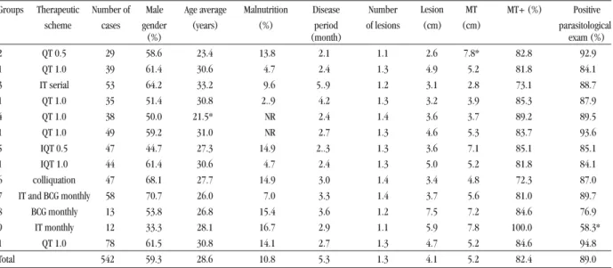
Table 1 - Demographic and clinical characteristics of the treated American cutaneous leishmaniasis patients.
Groups Therapeutic Number of Male Age average Malnutrition Disease Number MT+ (%) Positive scheme cases gender (years) (%) period of lesions parasitological
(%) (month) exam (%)
2 QT 0.5 29 58.6 23.4 13.8 2.1 1.1 2.6 7.8* 82.8 92.9
1 QT 1.0 39 61.4 30.6 4.7 2.4 1.3 4.9 5.2 81.8 84.1
3 IT serial 53 64.2 33.2 9.6 5..9 1.2 3.1 2.8 73.1 88.7
1 QT 1.0 35 51.4 30.8 2..9 4.2 1.3 3.2 3.9 85.3 87.9
4 QT 1.0 38 50.0 21.5* NR 2.4 1.4 3.6 3.7 89.2 89.5
1 QT 1.0 49 59.2 31.0 NR 2.7 1.3 4.6 5.3 83.7 93.6
5 IQT 0.5 47 44.7 27.3 14.9 2..3 1.3 3.6 7.1 85.1 85.1
1 IQT 1.0 44 61.4 30.6 4.7 2.4 1.3 5.0 5.2 81.8 84.1
6 colliquation 47 68.1 27.7 14.9 3.0 1.4 3.4 4.8 72.3 87.0 7 IT and BCG monthly 58 70.7 26.0 7.0 3.3 1.4 3.7 5.6 81.0 89.7
8 BCG monthly 13 53.8 26.8 15.4 3.6 1.2 7.5 7.2 84.6 76.9
9 IT monthly 12 33.3 28.1 16.7 2.9 1.1 5.9 7.8 100.0 58.3*
1 QT 1.0 78 61.5 30.8 14.1 2.7 1.3 4.7 5.2 84.6 94.8
Total 542 59.3 28.6 10.8 5.3 1.3 4.1 5.2 82.4 89.0
QT 1.0: Standard Group , * Significant difference when compared with standard group. MT: Montenegro Test.
RESULTS
A total of 542 patients with positive ACL diagnosis were
divided into 9 treatment groups. The demographic and clinical
characteristics of the groups studied are shown in Table 1. The
frequency of male gender was 59.3% and the mean age was 28.6
years old (ranging from 5 to 86 years old). No significant
relationship regarding the gender and age of the patients was
found among the groups tested, including the standard group.
Malnutrition, defined by body mass index (BMI = weight/
height
2), was observed in 10.8% of the patients, which was shown
to be similar for all the groups under study (Table 1).
The period of ACL disease, defined as the time taken
between the initial observation of the lesion, reported by the
patient, and clinical diagnosis, ranged from 10 days to 43.4
months with an average of 5.3 months. The number of lesions
varied from 1 to 3, and their mean diameter was 4.1cm. The
mean MT area, which was showen to be positive in 82.4% of
patients, was 5.2cm. The parasitological exam was positive in 89%
of patients. No relationship between the number of lesions, disease
period, and area of ulcers was observed among patients (Table 1).
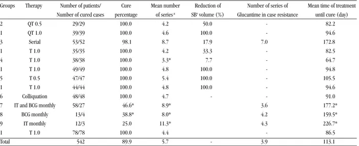
The treatment period of the groups under study and the volume
of antimony necessary to achieve cure are shown in Table 2.
The average length of treatment in days for clinical ACL cure
was: 94.6 for Group 1; 81.0 for Group 2; 172.8 for Group 3;
64.7 for Group 4; 105.5 for Group 5; 91 for Group 6; 177.2 for
Group 7; and 159.5 and 226.7 days for Groups 8 and 9,
respectively.
The therapy plans used in Groups 1, 4, 5, and 6 achieved
clinical cure in 100% of the patients with varying periods
(days) of 94, 64, 105 and 91 days, respectively.
Group 3, under immunotherapy, showed 98.1% of cured
patients, despite its increased treatment length when
compared with the standard group.
The average number of series, comprising all therapies,
ranged from 3.3 to 11.3.
Table 2 - Therapy plans under study for the treatment of American cutaneous leishmaniasis patients.
Groups Therapy Number of patients/ Cure Mean number Reduction of Number of series of Mean time of treatment Number of cured cases percentage of series a SBv volume (%) Glucantime in case resistance until cure (day)
2 QT 0.5 29/29 100.0 4.2 50.0 - 82.2
1 QT 1.0 39/39 100.0 4.6 100.0 - 94.6
3 Serial 53/52 98.1 8.7 17.9 7.0 172.8
1 T 1.0 35/35 100.0 4.2 33.3 - 82.5
4 T 1.0 38/38 100.0 3.3* 7.7 - 64.7
1 T 1.0 49/49 100.0 4.8 100.0 - 94.8
5 T 0.5 47/47 100.0 5.4 100.0 - 105.5
1 T 1.0 44/44 100.0 4.8 100.0 - 94.6
6 Colliquation 48/48 100.0 4.7 - - 91.0
7 IT and BCG monthly 58/27 46.6* 8.9* 3.6 177.2*
8 BCG monthly 13/4 38.8* 8.0* 4.2 159.5*
9 IT monthly 12/3 25.0 11.3* 4.3 226.7*
1 T 1.0 78/78 100.0 4.4 - 86.5
Total 542 89.9 5.7 - 3.9 113.1
1 QT 1.0: Standard Group, * Significant difference when compared with standard group.a Adjusted by disease time, lesion area and area of Montenegro test.1/ QT 1.0: Glucantime 1,0mL/5kg body weight Standard Therapy, 2/ QT 0.5: Glucantime 0,5mL/5kg body weight, 3/ IT serial: Vaccine serial, 4/ IQT 1.0: Vaccine + Glucantime 1mL, 5/ IQT 0.5: Vaccine + Glucantime 0,5mL, 6/ Colliquation: BCG + Glucantime, 7/ BCG IT montly: BCG+ vaccine monthly, 8/ Monthly BCG, 9/ Vaccine Monthly immunotherapy
DISCUSSION
No individual aspect of the patients or possible
correlations with age, gender, nutritional status and number
of lesions was shown to have a significant difference through
variance analysis among the groups under study.
Based on these results, immunotherapy (Group 3) may
be an alternative treatment for those patients for whom
Glucantime
is not indicated.
By associating the vaccine with the standard application
of Glucantime
(Group 4), a reduction in the volume of
antimony (17.9%) and treatment length for clinical cure
(from 94.6 to 64.7 days) was observed, when compared with
the standard treatment (Group 1).
In the case of immune-stimulation with vaccine, an
enhancement in interferon
γ
and interleukin 2 (IL-2)
production was observed potentializing the response type Th1
by activating macrophages through INF
γ
production, in
agreement with that shown by other investigators
45. This leads
to the destruction of parasites by increasing oxygen radical
levels, mainly nitric oxide. Furthermore, interferon ã induces
macrophages to produce IL 12, which acts on lymphocytes
and “Natural Killer” (NK) cells, potentializing the immune
response. These are ongoing findings in our works (data not
shown).
When meglumine antimoniate volume was diminished by
50% to treat Group 2 patients, treatment length was shown to
be as short (81 days) as that for Group 1, and cure rates
were the same in both groups.
Immunotherapy associated with half the dose of
meglumine antimoniate (Group 5) increased treatment
length, in contrast, antimony volume was decreased by 33.3%,
consequently, minimizing the side effects caused by
Glucantime
.
Group 6, receiving BCG for 15 to 20 days before
chemotherapy, showed a reduction of 7.7% in antimony
volume, with no statistical significance, and a slight increase
in treatment length for clinical cure in relation to Group 1.
The therapy comprising BCG and monthly immunotherapy
(Groups 8 and 9) did not contribute to a reduction in the
treatment period when compared with the standard treatment.
Considering the fact that other investigators only achieved 50.8%
of clinical cure at a dose of 20mg/kg/day, either through
intramuscular or intravenous route, for 20 days
60,
it
should be noted
that our service achieved a cure rate of 100% with Glucantime
therapy (Group 1). Literature data has shown that treatment with
meglumine antimoniate for 10 or 20 days, at a dose of 20mg/kg/day,
achieves clinical cures of 61% and 67%, respectively
53.
Similar results to those obtained here with the standard
treatment here (Group 1) were also achieved by our research
team in 2002
31while treating 29 patients from the municipality
of Araçuaí, State of Minas Gerais. These patients had no indication
for treatment with the standard antimonial therapy, mainly due
to the presence of electrocardiographic alterations (17),
antimony allergy (4), pregnancy (6) and nephropathy (2).
Among these patients, 10 underwent immunochemotherapy and
another 19 immunotherapy. The cure rate proved to be 100%
and the mean number of series up to clinical cure was 2.3
(ranging from 1 to 4 series), while for immunochemotherapy,
this was 2.47 series, with similar variation for both therapies.
Considering that antimony may produce side effects, a
concentration reduction in ACL treatments is quite important.
However, appropriate therapy should be carried out in order
to diminish likely recurrences of the mucous form
61as well
as other kinds of recurrences that have been frequently
reported
1 32 46 48 49 54 57 67 77.
Considering the broad range of therapies for ACL, there is no
consensus among those aimed at seeking a reduction in the side
effects of pentavalent antimonial drugs, there is no consensus.
Azeredo-Coutinho and Mendonça
6compared two therapies: a
continuous plan, as recommended by WHO
78, and a serial one.
These investigators concluded that the cure rate obtained with the
intermittent treatment (serial) proved to be significantly higher
than that with the continuous plan. The cure criterion was defined
by complete skin epithelization observed up to three months after
the end of treatment. Surprisingly, the follow-up of the patients
was greatly superior when they were submitted to the serial
treatment. Most of the patients under continuous treatment did
not attend the Outpatient Facility throughout the complete period
of treatment. Such data corroborate our findings
34concerning
serial treatment, although there are divergences regarding clinical
cure criteria. In the present study serial treatment was
maintained until complete healing of the cutaneous lesions
and the use of only three series was not followed.
Despite Machado-Coelho’s
31reports on resistance, have been
quite rare cases of resistance to Glucantime in chemotherapy
treatment have been quite rare in the municipality of Caratinga.
Patients from other regions usually seek this Out Patient Facility
due to previous failed treatments, performed in other localities.
In the case of ACL recurrence, patients attended at our
service are recommended to return to the Outpatient Facility
of Caratinga, as well as those from other regions, who are
addressed by different health units due to the popular
knowledge on treatments of cutaneous lesions provided by
this ambulatory in Caratinga. Some patients from different
regions, who underwent the treatment, indicated by the WHO
78(15mgSb/kg/day for 20 days and maximum daily dose of 15mL/
day), sought our services due to disease recurrence or
intolerance to the salt. Based on the WHO therapy, cure criterion
is the complete healing of cutaneous lesions observed up to 3
months after the treatment end, which lasts 20 days with or
without complete epithelization. Eight, from among these
patients, sought our ambulatory and underwent further
treatment with the antimony in a serial plan (a 10-day treatment
+ a 10-day rest) associated with the vaccine (0,5mL/day),
simultaneously administered with Glucantime
. The
Glucantime
daily dose was 1mL/5kg body weight without
exceeding 10mL/day. This treatment varied from 2 to 4 series,
which was only interrupted with clinical cure.
Several investigators have reported the side effects of
Glucantime
used in different therapy plans
27 26 32 45 51 64 66.
Considering the therapy plans of the current work, side effects
such as nausea, asthenia, myalgia, arthralgia and abdominal
discomfort were not observed throughout the follow-up every
20 days along with treatment until clinical cure. This might be
due to the serial plan with 10-day intervals between
administrations of N-methylglucamine antimoniate.
Due to the complexity involved in vector-host-parasite
interactions, investigators have been faced with different therapeutic
choices. No clear consensus exists among the various therapies
available in literature
9 11 13 29 79and those presented here. One of
the most serious problems is the lack of information regarding
daily doses of antimony
30 45 56 69. Our therapy plans are in accordance
with maximum daily dose of antimony (10mL - 850mg/Sb/day);
thus, an intravenous treatment using 20mg/kg/day would take
1,400mg/Sb/day, or 16.47 mL, for a patient weighing 70kg, though
most investigators do not mention such information.
The results from the patients under study and those from
a casuistic of 6,900 cases, since 1965, provide strong evidence
regarding the efficacy of the therapy used here. We have only observed
5 recurrence cases, which is frequently reported when the classic
therapy recommended by the WHO
1 45 48 50 54 67 68 77is used.
ACKNOWLEDGEMENTS
We are grateful to the Secretaria Municipal de Saúde of
Caratinga and the Consórcio Intermunicipal de Saúde of the
Micro-região of Caratinga, Minas Gerais State, Brazil, and Mr. Jair Cecílio
de Paula for his invaluable support during the field work.
REFERENCES
1. Aebscher T. Recurrent cutaneous Leishmaniasis: A Role for Persistent Parasites? Parasitology Today 10: 25-28, 1994.
2. Albanese G, Giorgetti P, Santagostinho L, Crippa D, Sala G. Cutaneous leishmaniasis. Treatment with itraconazole. Archives of Dermatology 125: 1540-1542, 1989.
3. Alving CR, Steck EA, Chapman WL JR, Waits VB, Hendricks LD, Swartz GM JR, Hanson WL. Therapy of leishmaniasis: superior efficacies of liposome-encapsulased drugs. Proceedings of the National Academy of Sciences of USA 75: 2959-2963, 1978.
4. Antunes CMF, Mayrink W, Magalhães PA, Costa CA, Melo MN, Dias M, Michalick MS, Williams P, Lima AO, Vieira JB. Controlled field of a vaccine against new world cutaneous leishmaniasis. International Journal Epidemiology 15: 572-580, 1986.
5. Armijos RX, Weiget MM, Aviles H, Maldonado R, Racines J. Field trial a vaccine against new world cutaneous leishmaniasis in at-risk child population: safety, immunogenicity, and efficacy during the first 12 months of follow-up. The Journal of Infectious diseases 177: 1352-1357, 1998. 6. Azeredo-Coutinho RBG, Mendonça SCF. Na intermittent schedule is better
than continuous regimen of antimonial therapy for cutaneous leishmaniasis in the municipality of Rio de Janeiro, Brazil. Revista da Sociedade Brasileira Medicina Tropical 35:477-481, 2002.
7. Barral-Netto M, Brodskyn C, Carvalho EM, Barral A. Human leishmaniasis @cytokines.Bahia.br. Brazilian Journal of Medical and Biological Research 31: 149-155, 1998.
8. Bassiouny A, EL-Meshad M, Talaat M, Kutty K, Metawaa B. Cryosurgery in cutaneous leishmaniasis. Brazilian Journal of Dermatology 107: 467-474, 1982.
9. Belazzoug S, Neal RA. Failure of meglumine antimoniate to cure cutaneous lesions due to Leishmania major in Algeria. Transactions of the Royal Society of Tropical Medicine and Hygiene 80: 670-671, 1986.
10. Bottrel RLA, Dutra WO, Martins FA, Gontijo B, Carvalho E, Barral-Netto M, Barral A, Almeida RP, Mayrink W, Locksley R, Gollob KJ. Flow Cytometric Determination of Cellular Sources and Frequencies of Key Cytokine-Producing Lymphocytes Directed Against Recombinant LACK and Soluble Leishmania Antigen in Human Cutaneous Leishmaniasis. Infection and Immunity 3232-3239, 2001.
11. Bourée P, Anciaux ML, Taugourdeau. Antimoniate de methyl-glucamine et stibo-gluconate de sodium dans le traitement des leishmanioses. Pathologie Biologue 33: 607-610, 1985.
12. Castes M, Tapia FJ. Immunopathology of American tegumentary leishmaniasis. Acta Científica Venezoelana 49: 42-56, 1998.
13. Chulay JD, Anzeze EM, Koech DK, Bryceson AD. High-dose sodium stibogluconate treatment of cutaneous leishmaniasis in Kenya. Transactions of the Royal Society of Tropical Medicine and Hygiene 77: 717-21, 1983. 14. Convit J, Rondon Ulrich M, Bloom B, Castellanos PL, Pinardi ME, Castes M, Garcia L. Immunotherapy versus Chemotherapy in localized cutaneous leishmaniasis. Lancet 21: 401-404, 1987.
15. Coutinho SG, Da-Cruz AM, Bertho AL, Santiago MA, De-Luca P. Immunologic patterns associated with cure in human American Cutaneous Leishmaniasis. Brazilian Journal of Medical and Biological Research 31: 139-142, 1998.
16. Da-Cruz AM, Conceição-Silva F, Bertho AL, Coutinho SG. Leishmania-reactive CD4+ and CD8+ T cells associated with cure of human cutaneous leishmaniasis. Infection and Immunity 62:2614-2618, 1994.
17. Dan M, Verrer E, el On J, Zuckerman F, Micheli. Failure of oral ketoconazole to cure cutaneous ulcers caused by Leishmania braziliensis. Cutis 38:198-199, 1986.
18. El Darouti MA, alRubaie SM. Cutaneous leishmaniasis. Treatment with combined cryotherapy and intralesional Stibogluconate injection. International Journal of Dermatology 29:56-59, 1990.
19. Elloso MM, Scott P. Expression and Contribution of 1 (CD80) and B7-2 (CD86) in the Early Immune Response to Leishmania major Infection. Journal of Immunology 162:6708-6715, 1999.
20. Furtado T. Terapêutica de Leishmaniose Americana - Medicamentos de Escolha. Revista Associação Médica de Minas Gerais 25:108-113, 1974. 21. Furtado TA. An appraisal of Chemotherapy of American Leishmaniasis.
Dermatology Ibero Latino Americano III: 2557-2558, 1968.
22. Genaro O, Toledo VP, Costa CA, Hermeto MV, Afonso LC, Mayrink W. Vaccine for prophylaxis and immunotherapy, Brasil. Clinical Dermatology 14: 503-512, 1996.
23. Gomes LS. A intradermorreação de Montenegro na Leishmaniose e outras pesquisas afins. Brasil-Médico 53: 1079-1087, 1939.
24. Guderian RH, Chico ME, Rogers MD, Pattishall KM, Grogl M, Berman JD. Placebo controlled treatment of Ecuadorian cutâneos leishmaniasis American Journal of Tropical Medicine and Hygiene 45: 92-97, 1991. 25. Gundz K, Afsar S, Ayhan S, Kandiloglu AR, Turel A, Filliz EE, Ok UZ.
Recidivans cutaneous leishmaniasis unresponsive to lipoma amphotericin B (AmBisone). Journal European Academic Dermatology Venereology 14: 11-13, 2000.
26. Hepburn NC. Cutaneous leishmaniasis. Clinical and Experimental Dermatology 25: 363-370, 2000.
27. Hepburn NC, Siddique I, Howie AF, Beckett GJ, Hayes PC. Hepatoxicity of sodium stibogluconate therapy for American cutaneous leishmaniasis. Transactions of the Royal Society of Tropical Medicine and Hygiene 88: 453-455, 1994.
28. Hermeto MV, Genaro O, Costa CA, Toledo VPCP, Michalick MSM, Melo MN, Magalhães PA, Resende FS, Dias M, Willians P, Mayrink W. Immunotherapy for American Cutaneous Leishmaniasis in the State of Minas Gerais, Brazil, using Leishvacin.Workshop National sobre Leishmanioses, Recife, Memórias do Instituto Oswaldo Cruz, 1993.
29. Herwaldt BL, Berman JD. Recommendations for treating Leishmaniasis with sodium stibogluconate (Pentostan) and review of pertinent clinical studies. American Journal of Tropical Medicine and Hygiene 46: 296-306, 1992. 30. Koff AB, Rosen T. Treatment of cutaneous leishmaniasis. Journal of
American Academic Dermatology 31: 693-708, 1994.
31. Machado-Coelho GLL, Assunção R, Mayrink W, Caiaffa WT. American cutaneous leishmaniasis (LTA) in Southeast Brazil: space-time clustering. International Journal of Epidemiology, Oxford 28: 982-989, 1999. 32. Marovisch MA, Lira R, Shepard M, Fuchs GH, Kruetzer R, Nutman TB,
Neva FA. Leishmaniasis recidivans recurrence after 43 years: a clinical and immunologic report after successful treatment. Clinical Infectious Diseases 33: 1076-1079, 2001.
33. Martinez S, Marr JJ. Allopurinol in the treatment of American Cutaneous Leishmaniasis. New England journal of Medicine 326: 741-747, 1992. 34. Mayrink W, Botelho ACC, Michalick MSM, Melo MN, Machado-Coelho GLL.
Imunoterapia, imunoquioterapia e Quimioterapia no Tratamento da Leishmaniose Tegumentar Americana. VII Reunião Anual de Pesquisa Aplicada em Leishmanioses. Uberaba, MG. Revista da Sociedade Brasileira de Medicina Tropical 35: 122-123, 2002.
35. Mayrink W, Costa CA, Magalhães PA, Melo MN, Dias M, Lima AO. Responses to Montenegro Antigen After Immunization With Killed Leishmania Promastigotes. Transactions of the Royal Society of Tropical Medicine and Hygiene 72: 676-676, 1978.
36. Mayrink W, Costa CA, Magalhães PA, Melo MN, Dias M, Oliveira-Lima A, Michalick MSM, Willians P. A field trial of a vaccine against American Dermal Leishmaniasis. Transactions of the Royal Society of Tropical Medicine and Hygiene 73: 385-387, 1979.
Immunoterapy as treatment of American cutaneous leishmaniasis: preliminary studies in Brazil. Parasitologia 34: 159-165, 1992. 38. Mayrink W, Melo MN, Costa CA, Coutinho SG, Hermeto MV, Genaro O, Toledo
VPCP, Guerra H. Multinational development of standart skin test antigen in American: preliminary results in the Minas Gerais state, Brazil. 2A. Reunião Anual sobre Pesquisa Básica em Doença de Chagas. Caxambu – MG. Memórias Instituto Oswaldo Cruz 88: 226 1993.
39. Mayrink W, Michalick MSM, Melo MN, Nascimento E, Magalhães PA, Costa CA, Oliveira-Lima A, Dias M. Tratamento da Leishmaniose Tegumentar utilizando vacina. Annals Brazilian Dermatology 66: 55-59, 1991. 40. Mayrink W, Resende FS, Costa CA, Hermeto MV, Genaro O, Toledo VPCR,
Michalick MSM, Melo MN, Nascimento E, Willians P, Oliveira-Lima A. Relatório de Atividades no Ambulatório de Leishmaninoses, Caratinga, Minas Gerais, Período1989-1992. Setor de Leishmanioses - Departamento de Parasitologia - Instituto de Ciências Biológicas, Universidade Federal de Minas Gerais, 1993.
41. Melo MN, Mayrink W, Costa CA, Magalhães PA, Dias M, Williams P, Araujo FG, Coelho MV, Batista SM. Padronização do antígeno de Montenegro. Revista Instituto Medicina Tropical de São Paulo 19: 161-164, 1977. 42. Mendonça SC, De Luca PM, Mayrink W, Restom TG, Conceição-Silva F,
Da-Cruz AM, Bertho AL, Da Costa CA, Genaro O, Toledo VP. Characterization of Human T Lymphocyte-mediated Immune Responses Induced by a Vaccine Against American Tegumentary Leishmaniasis. American Journal Tropical Medicine Hygiene 53: 195-201, 1995.
43. Michalick MSM, Mayrink W, Magalhães PA, Costa CA, Melo MN, Dias M, Willians P, Santos PH. Imunoterapia na leishmaniose tegumentar americana (LTA). In: Resumos do X Congresso da Sociedade Brasileira de Parasitologia, Salvador, BA, p. 119, 1987.
44. Momeni AZ, Aminjaveheri M. Treatment of recurrent cutaneous Leishmaniasis. International Journal of Dermatology 34: 129-133, 1995. 45. Mosman TR, Coffman RL. Th1 and Th2 cells: different functional properties.
Annals of Veterinary and Immunology 7:145, 1989.
46. Nadin A. Immunity to Cutaneous Leishmaniasis. Transactions of the Royal Society of Tropical Medicine and Hygiene 78:848, 1984.
47. Nascimento E, Mayrink W, Torres FV, Liu TY, Martins MT, Gazzinelli R, Fernandes AP, Genaro O, Costa CA, Toledo VPCP, Mc Master RW. Enhance Protection In Mice By Vaccination With Combined Parasite Recombinant Antigens (Rgp63 And Rp33) Against Leishmania (Leishmania) amazonensis. XXIII Congresso de Caxambu, MG. Memórias do Instituto Oswaldo Cruz 91: 163-163, 1996.
48. Netto EM, Marsden PD, Llanos-Cuentas EA, Costa JM, Cuba CC, Barreto AC, Badaro R, Jonhson WD, Jones TC. Long-term follow-up of patients with Leishmania (Viannia) braziliensis infection and treated with Glucantime. Transactions of the Royal Society of Tropical Medicine and Hygiene 84:367-370, 1990.
49. Neva FA, Wyler D, Nash T. Cutaneous Leishmaniasis-a case with persistent organisms after treatment in presence of normal immune response. The American Journal of Tropical Medicine and Hygiene 28: 467-471, 1979. 50. Oliveira-Neto MP, Mattos M, Souza CS, Fernandes O, Pirmez C. Leishmaniasis
recidiva cutis in New World cutaneous leishmaniasis. International Journal of Dermatology 37: 846-849, 1998.
51. Oliveira-Neto MP, Schubach A, Mattos M, Gonçalves-Costa SC, Pirmez C. Treatment of American Cutaneous Leishmaniasis: a comparation between low dosage (5mg/kg/peso and high dosage (20mg/kg/day) antimony regimens. Pathologie Biologue 45: 496-499, 1997.
52. Oster CN, Chulay JD, Hendricks LD, Pamplin CL 3rd, Ballou WR, Berman JD, Takafuji ET, Tramont EC, Canfield CJ. American cutaneous leishmaniasis: a comparison of three sodium stibogluconate treatment schedules. The American Journal of Tropical Medicine and Hygiene 34: 856-860, 1985. 53. Palacios R, Osorio LE, Gralalew LF, Ochoa MT, Treatment failure in children
in a randomized clinical trial with 10 and 20 days of meglumine antimoniate for cutaneous leishmanisasis due to Leishmania viannia species. The American Journal of Tropical Medicine and Hygiene 64: 187-193, 2001. 54. Passos VMA, Barreto SM, Romanha AJ, Krettli AU, Volpini AC, Costa MFFL.
American cutaneous leishmaniasis: use of a skin test as a predictor of relapse after treatment. World Health Organization 78: 968-974, 2000.
55. Pessoa SB, Pestana BR. A intradermo-reação de Montenegro nas campanhas sanitárias contra a Leishmaniose. Arquivos de Higiene e Saúde Pública 6:124-137 1941.
56. Pirmez C, Fernandes O, Oliveira-Neto MP. A reação em cadeia da polimerase (PCR) antes e após tratamento na leishmaniose tegumentar. Revista da Sociedade Brasileira Medicina Tropical 33:58-59, 2000.
57. Ramírez JL, Guevara P. Persistent Infections by Leishmania (Viannia) braziliensis. Memórias Instituto Oswaldo Cruz 92: 333-338, 1997. 58. Reed SG, Scott P. T-cell and cytokine responses in leishmaniasis. Current
Opinion in Immunology 5:524-531, 1993.
59. Ribeiro-de-Jesus A, Almeida RP, Lessa H, Bacellar O, Carvalho EM. Cytokine profile and pathology in human leishmaniasis. Brazilian Journal of Medical and Biological Research 31:143-148, 1998.
60. Rodriguez ME, In Guanzo P, Ramos A, Perez J. Treatment of cutaneous leishmaniasis with Co2 lazer radiation. Revista Cubana Medicina Tropical 42:197-202, 1990.
61. Romero GA, Guerra MV, Paes MG, Macedo VO. Comparation of cutaneous leishmaniasis due to Leishmania (Viannia) braziliensis and L. (V.) guyanensis in Brazil: therapeutic response to meglumine antimoniate. The American Journal of Tropical Medicine and Hygiene 65: 456-465, 2001. 62. Row R. Curative value of Leishmania culture vaccine in Oriental sore.
Brazilian Medical Journal 9:540-541, 1912.
63. Rzany B, Krutmann J, Goerttler E, Schopf E. Cutaneous leishmaniasis: treatment with cryotherapy and intramuscular injections of Meglumine Antimoniate (Glucantime). Hautarzt 41:98-101, 1990.
64. Saenz RE, Narváez E. Evaluation of the effectiveness and toxicity of Pentostan and Glucantime in the treatment of cutaneous leishmaniasis. Revista Medica do Panamá 12:148-157, 1987.
65. Saenz RE, Paz H, Berman JD. Efficacy of Ketoconazole against Leishmania braziliensis panamensis cutaneous leishmaniasis. American Journal of Medicine 89: 147-155, 1990.
66. Saldanha ACR, Romero GAS, Hamann EM, Magalhães AV, Macedo VO. Estudo comparativo entre estibogluconato de sódio BP 88R e antimoniato de meglumina no tratamento da leishmaniose cutânea: I. Eficácia e segurança. Revista da Sociedade Brasileira Medicina Tropical 32: 383-387, 1999. 67. Saravia NG, Weigle K, Segura I, Giannini SH, Pacheco R, Labrada LA,
Goncalves A. Recurrent lesions in human Leishmania braziliensis infection – reinfection? Lancet. 18: 398-402, 1990.
68. Schewach-Millet M, Fisher BK, Semanh D. Leishmaniasis recidivans treated with sodium stibogluconate. Cutis 28: 67-68, 1984.
69. Schubach A, Marzochi MC, Cuzzi-Maya T, Oliveira AV, Araujo ML, Oliveira AL, Pacheco RS, Momen H, Conceicao-Silva F, Coutinho SG, Marzochi KB. Cutaneous Scars in American Tegumentary Leishmaniasis patients: a site of Leishmania (Viannia) braziliensis persistence and viability eleven years after antimonial Therapy and clinical cure. American Journal of Tropical Medicine and Hygiene 58: 824-827, l998.
70. Sharquie KE, Al-Talib KK, Chu AC. Intralesional therapy of cutaneous leishmaniasis with sodium stibogluconate antimony. Brazilian Journal Dermatology 119: 53-57, 1988.
71. Silveira FT, Mayrink W. Leishmaniose Cutânea difusa no Estado do Pará, Brasil: Registro do primeiro caso de cura da doença após associação de quimioterapia com imunoterapia. XXIX Congresso da Sociedade Brasileira de Medicina Tropical. Revista da Sociedade Brasileira de Medicina Tropical 26 (supl):229, 1993.
72. Silveira FT, Mayrink W. Leishmaniose Cutâneo-Difusa No Estado do Pará, Brasil: Relato da Cura de Um Caso Depois de 24 Anos de Doença, Após Tratamento Combinado de Quimioterapia com Imunoterapia. In: Anais do XXXIII Congresso da Sociedade Brasileira de Medicina Tropical, p.129, 1997.
73. Snedecor GW, Cochran WG. Statistical Methods, 8th edition. Iowa State University Press, Ames, Owa, 1989.
75. Tapia FJ, Dittamar GS, Sánchez MA. Inadequate epidermal homing leads to tissue damage in human cutaneous leishmaniasis. Immunology Today 5:160-165, 1994.
76. Vianna G. Tratamento da leishmaniose tegumentar por injeções intravenosas de tártaro emético.In: Anais do 7O Congresso Brasileiro de Medicina e Cirurgia 4: 426-428, 1912.
77. Weigle KA, Valderrama L, Santrich C, Saraiva NG. Recurrences of Tegumentary Leishmaniasis. Lancet 2:557-558, 1985.