Rev Odontol UNESP. 2014 Nov.-Dec.; 43(6): 390-395 © 2014 - ISSN 1807-2577 Doi: http://dx.doi.org/10.1590/1807-2577.1041
Signs of Combination Syndrome and removable
partial denture wearing
Sinais da Síndrome da Combinação e o uso de prótese parcial removível
Camila Maria Bastos Machado de RESENDE
a*, Jaiane Augusta Medeiros RIBEIRO
a,
Kássia de Carvalho DIAS
b, Adriana da Fonte Porto CARREIRO
a, Michel Platini Pereira do REGO
a,
José Werbeson Nogueira de QUEIROZ
a, Gustavo Augusto Seabra BARBOSA
a,
Ângelo Giuseppe Roncalli da Costa OLIVEIRA
aaUFRN – Universidade Federal do Rio Grande do Norte, Natal, RN, Brasil bFaculdade de Odontologia, UNESP – Univ Estadual Paulista, Araraquara, SP, Brasil
Resumo
Introdução: A Síndrome da Combinação (SC) é uma condição patológica associada aos pacientes usuários de
prótese total maxilar e prótese parcial removível (PPR) mandibular. Objetivo: Observar e mensurar a prevalência dos sinais da Síndrome da Combinação encontrados em pacientes usuários de prótese total maxilar na presença ou ausência de PPR mandibular (Classe I de Kennedy); e averiguar uma possível associação entre a utilização de PPR e a prevalência dos sinais clínicos da síndrome. Material e método: A amostra foi composta por 62 pacientes atendidos no Departamento de Odontologia da Universidade Federal do Rio Grande do Norte (UFRN). O exame clínico intrabucal foi realizado para a verificação da presença ou ausência dos sinais clínicos específicos da Síndrome da Combinação, descrita por Kelly (1972): reabsorção óssea na região anterior da maxila, aumento das tuberosidades, hiperplasia papilar palatina, extrusão dos dentes naturais inferiores anteriores e reabsorção óssea posterior mandibular (variáveis dependentes). Para determinação da associação entre as variáveis dependentes e independentes (uso de PPR inferior e tempo de edentulismo superior), foi utilizado o teste qui-quadrado com significância de 95%. Resultado: A característica mais frequente foi a presença de reabsorção mandibular (93,5%). Quanto à associação entre o uso de PPR inferior e as características da Síndrome da Combinação, só houve diferença estatisticamente significativa entre portadores e não portadores de PPR com relação à extrusão dos dentes inferiores anteriores (p = 0,045). Conclusão: Dentro das limitações deste estudo, verificou-se que os sinais clínicos da Síndrome da Combinação foram bastante prevalentes, e não foi observada associação entre o uso de PPR e as características da Síndrome.
Descritores: Prótese parcial removível; prótese total; reabsorção óssea.
Abstract
Introduction: Combination Syndrome (CS) is a pathological condition observed in maxillary complete denture
(CD) and mandibular removable partial denture (RPD) wearers. Purpose: The aim of this study was to observe and measure the prevalence of CS signs in treatment-seeking wearers of maxillary CD associated or not with RPD (mandibular Kennedy Class I). The association between RPD wearing and the number of CS clinical signs was also evaluated. Material and method: The sample included 62 patients seen at the Department of Dentistry, Federal University of Rio Grande do Norte (UFRN). A clinical oral examination was conducted to assess the presence of specific clinical signs of CS as described by Kelly (1972): bone resorption in the maxillary anterior region, tuberosity overgrowth, palatal papillary hyperplasia, extrusion of mandibular anterior teeth and bone resorption in the mandibular posterior region. The chi-square test at the 95% level of significance was used to test the association between dependent and independent variables. Result: Mandibular resorption was the most frequent complication (93.5%). There was a statistically significant difference between RPD wearers and non-wearers with regard to extrusion of mandibular anterior teeth (p = 0.045). Conclusion: Within the limitations of the present study, a high prevalence of CS clinical signs was observed, but no association between RPD wearing and syndrome characteristics was found.
INTRODUCTION
h e Glossary of Prosthodontic Terms1 dei nes Combination
Syndrome as a set of characteristics that occur when an edentulous maxilla is opposed by mandibular anterior teeth. Kelly,2 in 1972,
introduced the term Combination Syndrome when analyzing six patients wearing a maxillary complete denture occluding with a distal-extension removable partial denture (Kennedy Class I RPD). h e characteristic features of this syndrome include: loss of bone from the anterior portion of the maxillary ridge, overgrowth of the tuberosities, papillary hyperplasia of the mucosa of the hard palate, extrusion of mandibular anterior teeth, and loss of alveolar bone and ridge height beneath the removable partial denture bases. Some years later, new features were attributed to Combination Syndrome: loss of vertical dimension of occlusion, occlusal plane discrepancies, spatial repositioning of the mandible in the anterior region, poor denture i t and periodontal alterations3.
Prevention of degenerative changes caused by complete dentures occluding with bilateral distal-extension removable partial dentures is possible through an appropriate treatment plan and with periodic review of the RPDs and remaining teeth. Treatment alternatives such as preservation of posterior teeth to support mandibular RPDs and use of overlay-type complete dentures provide more adequate occlusal stability and should be considered as treatment options2.
Although Kelly2 mentioned the use of an adequate RPD
as a way to prevent development of signs of the syndrome, the scientii c evidence on this ef ect is still limited. Salvador et al.4
studied the presence of Combination Syndrome signs in patients rehabilitated with mandibular RPDs. h e authors concluded that patients with Kennedy Class II mandibular RPDs did not show Combination Syndrome, whose prevalence was 25% among the patients attended. h e most prevalent CS characteristic was maxillary anterior bone resorption, which was present in 81% of the Kennedy Class I patients and in 75% of the Kennedy Class II patients. Kelsey5 stated that intolerable forces produced by
poorly i tted dentures can cause pressure and inl ammation of the supporting tissues, which can make bone loss on the alveolar ridge inevitable. Tolstunov6 stated that use of poorly i tted dentures
for a prolonged period may contribute to mandibular posterior bone resorption. It is essential to identify the initial symptoms of CS and take steps for its immediate correction6. Shen, Gonglof 7
found changes in 24% of their patients with natural mandibular anterior teeth and maxillary complete dentures, and found that this rate was not signii cantly dif erent for patients that did or did not use mandibular removable partial dentures. Faced with these conl icting results, studies are needed that relate the Combination Syndrome signs to RPD wearing, in order to evaluate whether wearing a mandibular denture could minimize the development of syndrome signs. h us, diagnosis of this condition can facilitate implementation of appropriate clinical conduct and check the destructive process.
h e present study’s objective was to observe and measure the prevalence of Combination Syndrome or Kelly signs found
in patients wearing maxillary complete dentures (CD) with or without mandibular Kennedy Class I removable partial dentures (RPDs) and i nd out whether there is an association between RPD wearing and the prevalence of clinical signs of the syndrome.
MATERIAL AND METHOD
A cross-sectional study was conducted in the Department of Dentistry, Federal University of Rio Grande do Norte (UFRN), with the approval of the Research Ethics Committee of the institution, under Protocol 409896. h e sample was selected from patients with a completely edentulous maxilla wearing maxillary complete dentures (CD) and with a bilateral partially edentulous mandible (Kennedy Class I), wearing or not wearing a mandibular removable partial denture (RPD), who were seeking treatment at the UFRN Prosthodontics Clinic between April and June 2011. Patients wearing mandibular RPDs that were not made with a metal structure and/or patients who showed signs of poor general health were excluded.
Based on the sample selected to evaluate the association between Combination Syndrome signs and mandibular RPD wearing, considering a 95% coni dence interval, a power of 80% and an estimated 50% rate of individuals who did not wear dentures (control), the size of the sample is sui cient to indicate a signii cant dif erence when the dif erence is greater or equal to 30%, hence adequate to generate hypotheses.
At er signing a free and informed consent form, the individuals participated in an interview on their denture history and time of edentulism. h en they were submitted to a clinical oral examination conducted by two trained researchers who made a joint analysis to determine the presence or absence of the clinical signs specii c for Combination Syndrome (CS), as described by Kelly2: bone resorption in the maxillary anterior
region, tuberosity overgrowth, palatal papillary hyperplasia, extrusion of natural mandibular anterior teeth and mandibular posterior bone resorption (Chart 1). If there was any inconformity with regard to the presence of any of the established criteria, a third examiner, expert in the matter, would resolve it.
h e variables were presented in a descriptive manner by means of absolute numbers and proportions. To determine the association between the dependent (clinical signs of CS) and independent (mandibular RPD wearing and time of maxillary edentulism) variables, the chi-square test was used with a signii cance level of 5%.
RESULT
h e sample was composed of 62 patients with a mean age of 58.9 years, a minimum of 39 and a maximum of 78 years, where 21% (n = 13) were men and 79% (n = 49), women. h ese individuals had maxillary edentulism for a mean period of 28.4 years, with a minimum of 2 years, a maximum of 53 years and a standard deviation of 10.1.
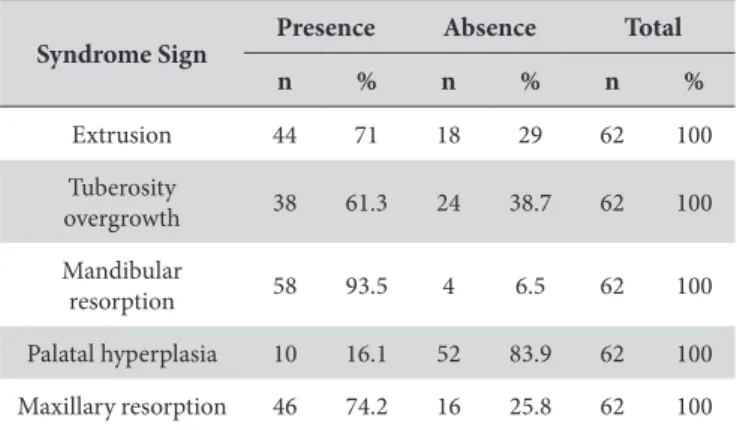
the sample, except for palatal papillary hyperplasia, which was present in only 16.1% (n = 10; Table 1).
he patients were not evaluated with regard to diagnosing CS by counting speciic signs to determine whether they had the syndrome or not, but rather with regard to the presence or absence of each sign and the number of signs per individual.
Of the characteristics, the most frequent was the presence of mandibular resorption (93.5%; Table 1) and the most frequent occurrences were the presence of 3 signs (45%) and 4 signs (32%) per individual, representing a total of 77% of the sample, a value that demonstrates the high prevalence of CS clinical features in patients presenting the proile studied (Table 2).
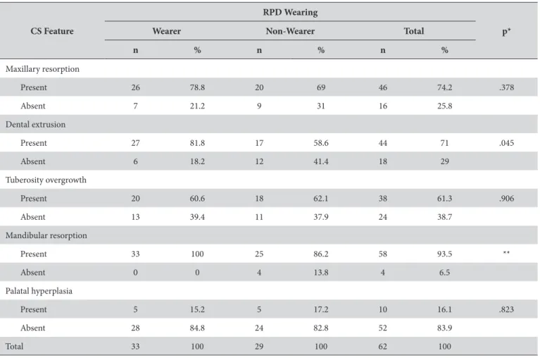
With regard to the association between mandibular RPD wearing and CS features, a statistically signiicant diference between RPD wearers and non-wearers was observed only with regard to extrusion of mandibular anterior teeth (p = 0.045; Table 3). he mandibular resorption data could not be analyzed by the statistical test (Table 3), since 100% of the patients wearing RPDs showed this characteristic, making it invariable, thus having a cell with zero value, and making it impossible to use the test.
DISCUSSION
he term syndrome signiies a set of signs and symptoms that are found associated to a known or unknown entity. By this deinition, CS would only be diagnosed when all its signs are present. Nevertheless, there is still no clear indication in the dental literature that all its signs must be present for CS to be diagnosed4. Consequently, in the present study, the prevalence of
CS signs was assessed rather than the diagnosis of CS.
In the present study, 93.5% of the sample showed mandibular posterior bone resorption, the CS clinical sign most prevalent among the dependent variables studied (Table 1). his clinical feature is common among these patients owing to the natural mandibular bone resorption process that occurs primarily in height. Another factor that inluences the presence of this clinical feature is the quality of denture it5. he greater the denture
misit, the greater will be the induction of forces on the residual ridge and its resorption. In the sample, 100% of the individuals wearing RPDs showed this characteristic. Tolstunov6 stated that
bone is deposited and resorbed according to the tensions placed upon it. he use of poorly itted dentures for a prolonged period
may contribute to this type of resorption. In the sample studied, the technical quality of the patients’ dentures was not assessed, and the fact that the sample was drawn among patients seeking new dentures may have contributed to the high resorption value found for the patients wearing dentures. his result was not very diferent from that found for the patients who did not wear dentures (86.2%), showing that lack of a denture is also detrimental. he statistical signiicance with regard to denture wearing and this clinical feature was not evaluated owing to a zero cell in the data distribution (Table 3). Shen, Gonglof7 observed
no diference with regard to manifestation of this characteristic between wearers (56%) and non-wearers (46%) of a bilateral distal-extension mandibular RPD.
Chart 1. Description of the Clinical Examination
Syndrome Characteristics Clinical Evaluation
Bone resorption in the maxillary anterior region Observation of laccid tissue in the anterior region of the residual ridge
suscep-tible to displacement
Tuberosity overgrowth Vertical and/or horizontal growth of ibrous or bone tissue in the right and/or let tuberosity region
Palatal papillary hyperplasia Observation of erythematous mucosa with a papillary surface in the hard palate
Extrusion of the remaining natural mandibular teeth Observation of dental wear at the enamel or dentin level
Mandibular posterior bone resorption Observation of accentuated bone resorption in the posterior edentulous region
Table 1. Prevalence of signs of Combination Syndrome. Absolute and relative values
Syndrome Sign
Presence Absence Total
n % n % n %
Extrusion 44 71 18 29 62 100
Tuberosity
overgrowth 38 61.3 24 38.7 62 100
Mandibular
resorption 58 93.5 4 6.5 62 100
Palatal hyperplasia 10 16.1 52 83.9 62 100
Maxillary resorption 46 74.2 16 25.8 62 100
Table 2. Frequency of clinical signs of Combination Syndrome per individual. Absolute and relative values
Combination Syndrome
No. of signs n %
0 1 2
1 3 5
2 7 11
3 28 45
4 20 32
5 3 5
Reduction of mandibular posterior support results in a gradual reduction of occlusal load in this region and a consequent increase in load in the anterior region, which can result in excessive pressure on the anterior portion of the maxilla, accelerating the process of maxillary anterior bone resorption and promoting the appearance of loose hyperplastic tissue3. his
provides an explanation for the research indings that show a high prevalence of 74.2% (n = 46) of maxillary anterior resorption (Table 1). his high prevalence is in accordance with the indings of Kelly,2 but disagrees with this author when he identiies this
characteristic as being the most prevalent3. No statistically
signiicant diference between wearers and non-wearers was observed with regard to this syndrome manifestation. his may occur owing to the absence of posterior support among the patients who had no mandibular arch rehabilitation or had unsatisfactory rehabilitation, generating similar problems in the anterior region.
An overgrowth of tuberosities was present in 61.3% of the sample, representing 60.6% of the patients wearing an RPD and 60.6% of those who did not (Table 1 and 3). here was no association between the presence of this characteristic and mandibular RPD wearing, as can be seen from the quite similar values. his similarity may have occurred because poorly itted mandibular dentures may have a biomechanical behavior compatible with denture absence. Shen, Gonglof7 found a similar
prevalence (56%) when they evaluated patients with bilateral distally edentulous mandibular arches wearing a maxillary CD and a mandibular RPD, but found a lower value (22%) for patients who did not use an RPD. he diference between the results of the present study and those found by Shen, Gonglof7 may be justiied
by the relatively small sample of mandibular RPD wearers and non-wearers of the latter study (n = 25).
he sign least frequently found in the present study was palatal papillary hyperplasia (16.1%), an inlammatory change caused by wearing poorly itted dentures, oten combined with poor hygiene and some other predisposing factors. In epidemiological studies conducted on mucosa changes, primarily among denture users, the prevalence of hard palate papillary hyperplasia is low8,9. Xie et al.9
found values that ranged from 5% to 10%. MacEntee et al.8 found
hyperplasia in 8% of 155 denture wearers. Coelho et al.10 observed
inlammatory ibrous hyperplasia in 16.7% of the sample in a prevalence study with 334 individuals. Another study, conducted in Turkey in 2009, consisting of 170 complete denture wearers, showed that the incidence of papillary hyperplasia increased with time of denture wearing: 13.3 % from 0 to 10 years of denture wearing and 86.7% for more than 10 years11.
he literature points to speciic clinical data that deine CS, but there is no evidence that an individual must show the ive clinical signs simultaneously to be considered as having the Combination Syndrome. In this study, it was found that 77%
Table 3. Prevalence of CS signs and their association with mandibular RPD wearing. Absolute and relative values and statistical signiicance. Natal, RN, 2011
CS Feature
RPD Wearing
p*
Wearer Non-Wearer Total
n % n % n %
Maxillary resorption
Present 26 78.8 20 69 46 74.2 .378
Absent 7 21.2 9 31 16 25.8
Dental extrusion
Present 27 81.8 17 58.6 44 71 .045
Absent 6 18.2 12 41.4 18 29
Tuberosity overgrowth
Present 20 60.6 18 62.1 38 61.3 .906
Absent 13 39.4 11 37.9 24 38.7
Mandibular resorption
Present 33 100 25 86.2 58 93.5 **
Absent 0 0 4 13.8 4 6.5
Palatal hyperplasia
Present 5 15.2 5 17.2 10 16.1 .823
Absent 28 84.8 24 82.8 52 83.9
Total 33 100 29 100 62 100
(n = 48) of the patients showed 3 to 4 clinical signs characteristic of CS. Shen, Gonglof7 evaluated the presence of some clinical CS
signs deined by Kelly2 in 1972 and others added by Saunders.3
Considering only Kelly’s CS signs, a greater prevalence of 3 signs was found in patients with a maxillary CD and the presence or absence of a bilateral distal-extension RPD. he high number of signs per individual in our study also may be justiied by the long period of edentulism of the sample and the consequent greater establishment of degenerative processes resulting from complete maxillary and partial mandibular edentulism without adequate treatment.
In relation to the use of a mandibular removable partial denture, the literature indicates that it prevents the appearance of some signs3. he present study found no evidence that RPD
wearing provided beneits in preventing CS. he only clinical sign that showed an association with RPD wearing was tooth extrusion. However, the association was related to RPD wearing; in other words, RPD wearers had more extrusion than non-wearers. his result indicates that the dentures probably were unsatisfactory or that the dental arches were not simultaneously rehabilitated, or even that the individuals already showed CS characteristics when irst rehabilitated. In a comparative study, Jozefowicz12 investigated the inluence of denture wearing on
residual ridge resorption and found that individuals who did not wear dentures had signiicantly higher residual ridges than those who wore dentures, except for the female group from 60 to 79 years of age. Previously, Cambell13 found a similar result, but the
diference between the wearer and non-wearer groups was not statistically signiicant, perhaps owing to the small sample used. Alveolar bone resorption is an inevitable process ater tooth loss, but it can be minimized with the construction of well-itted dentures, scheduled follow-up sessions and guidance on denture wearing and care5. In 1972, Kelly2 observed extrusion in 100%
(n = 6) of individuals evaluated radiographically ater a period of 3 years, which is in accordance with our indings.
As Tolstunov14 stated, prevention of posterior occlusion loss
and anterior hyperfunction are considered the main treatment approaches for CS. he RPD is one of the treatment modalities to correct and treat CS among those available, as long as it is planned to preserve stability, including a maxillary complete denture as antagonist, with a balanced distribution of occlusal tensions and careful maintenance in order to preserve posterior occlusion6. For patients who already show CS features, these
can be minimized with surgical procedures, special anatomical impression-taking techniques for laccid tissue, and correct surveying and planning of the rests and clamps of the mandibular RPD infrastructure. Periodic return visits to the dental oice to check the need for relining, the integrity of the occlusal contacts, and denture hygiene and it are also important for these patients.
here is also the possibility of treating these patients by placing implants in the mandibular posterior region, eliminating the need for a distal extension, and impeding the vertical and lateral movements responsible for accelerated bone resorption below the RPD resin base15. his alternative changes the Kennedy Class I
coniguration to a Class III one, with a biomechanical advantage, improving masticatory eiciency as well as denture stability and esthetics, depending on the positioning of the implant16.
Palmqvist et al.17 observed that, in patients who received
mandibular implant-supported ixed dentures, bone resorption in the posterior part of the mandible practically ceased. Consequently, treatment with implants on both arches is a factor to be considered to control bone resorption associated with Combination Syndrome18.
One of the limitations of this study is the fact that the sample was not probabilistic and representative of the general population. In addition, the fact that the patients were seeking treatment may have made it more likely that they would have CS signs and have been using inadequate dentures. Also, because it was a cross-sectional study, all the measurements were made at the same time, and, therefore, there was no follow-up period for the individuals.
In spite of the limitations, the large number of signs characteristic of the syndrome present in this research is a fact that calls attention to the range of damage already caused, resulting from the lack of follow-up of the dentures installed, insuicient or inadequate treatment, or even the lack of an early diagnosis. his situation will change when the clinician dealing with a susceptible patient is aware of the importance of preventing, diagnosing and intervening early so that damage does not occur, or is minimized.
To evaluate the beneit of RPD wearing in preventing CS signs, controlled and randomized clinical trials evaluating the presence of clinical signs immediately ater installation and ater prolonged denture-wearing periods are warranted, together with monitoring of the quality of these dentures. hus, further research should be conducted to assess the association of syndrome features and RPD wearing in longitudinal studies monitoring patients right ater they lose their teeth, where the technical quality of the dentures constructed should also be evaluated.
CONCLUSION
REFERENCES
1. The glossary of prosthodontic terms. J Prosthet Dent. 2005 July; 94(1): 10-92. http://dx.doi.org/10.1016/j.prosdent.2005.03.013. PMid:16080238
2. Kelly E. Changes caused by a mandibular removable partial denture opposing a maxillary complete denture. 1972. J Prosthet Dent. 2003 Sept; 90(3): 213-9. http://dx.doi.org/10.1016/S0022-3913(03)00240-3. PMid:14503534
3. Saunders TR, Gillis RE Jr, Desjardins RP. The maxillary complete denture opposing the mandibular bilateral distal-extension partial denture: treatment considerations. J Prosthet Dent. 1979 Feb; 41(2): 124-8. http://dx.doi.org/10.1016/0022-3913(79)90292-0. PMid:366110 4. Salvador MCG, do Valle AL, Ribeiro MCM, Pereira JR. Assessment of the prevalence index on signs of combination syndrome in patients
treated at Bauru School of Dentistry, University of Sao Paulo. J Appl Oral Sci. 2007 Feb; 15(1): 9-13. http://dx.doi.org/10.1590/S1678-77572007000100003. PMid:19089092
5. Kelsey CC. Alveolar bone resorption under complete dentures. J Prosthet Dent. 1971 Feb; 25(2): 152-61. http://dx.doi.org/10.1016/0022-3913(71)90101-6. PMid:4924826
6. Tolstunov L. Combination syndrome: classification and case report. J Oral Implantol. 2007; 33(3): 139-51. http://dx.doi.org/10.1563/1548-1336(2007)33[139:CSCACR]2.0.CO;2. PMid:17674680
7. Shen K, Gongloff RK. Prevalence of the ‘combination syndrome’ among denture patients. J Prosthet Dent. 1989 Dec; 62(6): 642-4. http:// dx.doi.org/10.1016/0022-3913(89)90582-9. PMid:2585321
8. MacEntee MI, Glick N, Stolar E. Age, gender, dentures and oral mucosal disorders. Oral Dis. 1998 Mar; 4(1): 32-6. http://dx.doi. org/10.1111/j.1601-0825.1998.tb00252.x. PMid:9655042
9. Xie Q, Ainamo A, Tilvis R. Association of residual ridge resorption with systemic factors in home-living elderly subjects. Acta Odontol Scand. 1997 Oct; 55(5): 299-305. http://dx.doi.org/10.3109/00016359709114968. PMid:9370028
10. Coelho CM, Sousa YT, Daré AM. Denture-related oral mucosal lesions in a Brazilian school of dentistry. J Oral Rehabil. 2004 Feb; 31(2): 135-9. http://dx.doi.org/10.1111/j.1365-2842.2004.01115.x. PMid:15009597
11. Canger EM, Celenk P, Kayipmaz S. Denture-related hyperplasia: a clinical study of a Turkish population group. Braz Dent J. 2009; 20(3): 243-8. http://dx.doi.org/10.1590/S0103-64402009000300013. PMid:19784472
12. Jo’ zefowicz W. The influence of wearing dentures on residual ridges: a comparative study. J Prosthet Dent. 1970 Aug; 24(2): 137-44. http:// dx.doi.org/10.1016/0022-3913(70)90136-8. PMid:5271099
13. Campbell RL. A comparative study of the resorption of the alveolar ridges in denture-wearers and non-denture-wearers. J Am Dent Assoc. 1960 Feb; 60: 143-53. PMid:13807301.
14. Tolstunov L. Management of biomechanical complication of implant-supported restoration of a patient with combination syndrome: a case report. J Oral Maxillofac Surg. 2009 Jan; 67(1): 178-88. http://dx.doi.org/10.1016/j.joms.2008.09.013. PMid:19070766
15- Cunha LDAP, Rocha EP, Pellizzer EP. Prevalência da Síndrome de Kelly em usuários de prótese parcial removível. RGO – Rev Gaúcha Odontol. 2007 Out-Dez; 55(4):325-8.
16. Cunha LD, Pellizzer EP, Verri FR, Falcón-Antenucci RM, Goiato MC. Influence of ridge inclination and implant localization on the association of mandibular Kennedy class I removable partial denture. J Craniofac Surg. 2011 May; 22(3): 871-5. http://dx.doi.org/10.1097/ SCS.0b013e31820f7d6a. PMid:21558932
17. Palmqvist S, Carlsson GE, wall B. The combination syndrome: a literature review. J Prosthet Dent. 2003 Sept; 90(3): 270-5. http://dx.doi. org/10.1016/S0022-3913(03)00471-2. PMid:12942061
18. Tolstunov L. Combination syndrome symptomatology and treatment. Compend Contin Educ Dent. 2011 Apr; 32(3): 62-6. PMid:21560744.
CONFLICT OF INTEREST
he authors declare no conlicts of interest.
*CORRESPONDING AUTHOR
Camila Maria Bastos Machado de Resende, Rua Ilíria Taveres Galvão, 52/701, Bloco B, Tirol, 59022-460 Natal - RN, Brasil, e-mail: [email protected]