INTRODUCTION
Free-end removable partial dentures (RPDs), especially mandibular ones, continue to challenge practitioners in the field of dental prosthetics not only on account of their biomechanical complex-ity, for they are supported by two distinct types of tissue (dental and fibromucosal), but also in view of the high failure rate observed with this type of denture in rehabilitative oral treatment.
The free-end fibromucosal support system is of major importance not only because of the in-creased exposure to forces as teeth are gradually lost but also as a result of the difference in me-chanical operation between the edentulous area
and the support teeth. It is acknowledged, none-theless, that the alveolar bone is the true support for such forces2.
The morphology of the alveolar bone in the residual ridge is directly affected by anatomical and systemic factors, masticatory habits and the wearing of dentures3. Pietrokovski23 (1975) gauged the density of the residual ridge bone and observed that it is intermediate between that of the trabecu-lar bone inside the mandible and that of the corti-cal bone of the outer casing. The bone available in the posterior edentulous area of the mandible, according to the Misch20 (1999) classification,
con-Analysis of the prevalence of different topographical
characteristics of the residual ridge in mandibular free-end arches
Análise da prevalência de diferentes características topográficas
do rebordo residual em arcos mandibulares com extremidades
livres
Carlos Gramani Guedes* Artêmio Luiz Zanetti** Pedro Paulo Feltrin***
ABSTRACT: This study observed the prevalence of different types of residual ridge inclination in free-ends of man-dibles and reported possible correlative factors that may affect resorption. For this purpose, periapical radiographs and individual data collected from a sample of 64 hemiarches were used. Two radiographs were taken of each free-end, and tracing was employed to determine the angles formed by the resorption configuration in the area of the 1st mandibular molar. The following conclusions were drawn: 1) the great majority of alveolar ridges were distally
descending; 2) the average angle was wider for users of mandibular removable partial dentures; 3) the results obtained suggest that the type of opposing maxillary arch affects the inclination of mandibular ridges; 4) greater inclination was observed when the 2nd bicuspids of the mandible were the abutment teeth; 5) no significant
cor-relation was established between age, sex and residual ridge resorption.
DESCRIPTORS: Denture, partial, removable; Radiography.
RESUMO: Esta pesquisa constatou a prevalência dos tipos de rebordos residuais no sentido ântero-posterior em extremidades livres inferiores, além de correlacionar alguns fatores que possam influenciar as suas reabsorções. Para isso, utilizamos radiografias periapicais e dados individuais colhidos na amostra de 64 hemiarcos; foram ob-tidas 2 radiografias de cada área de extremidade livre e, a partir de traçados, determinamos os ângulos formados pela reabsorção na altura do 1º molar inferior. Podemos constatar que: 1º) a grande maioria de inclinação encon-trada foi de rebordos descendentes para distal; 2º) a média de angulação foi maior para usuários de prótese parcial removível inferior; 3º) os resultados sugeriram existir influência do tipo de arco antagônico superior na inclinação dos rebordos inferiores; 4º) observou-se aumento de inclinação quando os segundos pré-molares inferiores eram os dentes adjacentes ao espaço protético; e 5º) não foram encontradas correlações significantes entre idade, sexo e reabsorção do rebordo residual.
DESCRITORES: Prótese parcial removível; Radiografia.
sists of thick, dense or porous cortical bone at the crest of the ridge and fine trabecular bone inside, in addition to another type of bone of lower density displaying thin, porous cortical bone at the ridge and encasing a fine trabecular bone.
Nevertheless, biomechanical studies show that mandibular removable partial dentures (RPDs) in action form a system of levers on the fibromuco-sal tissue in which the abutment tooth acts as a rotational fulcrum, while the angle of inclination of the ridge forms a slanted plane system, which increases proportionally to the inclination of the residual ridge and may cause deflections at the point where bite force is applied19,26.
Although the form of residual ridge is affect-ed by the type of fibromucosal support in each individual, this is the real support for the RPD. Studying the angle is therefore relevant to aid prosthodontists in designing prosthetic devices and also to supply valuable data for research on the biomechanical behaviour of dentures subjected to bite force.
METHODOLOGY
Selection of sample
Patients were selected from clinics run by the Discipline of Dentistry, University of Brasília, based on the presence of uni- or bilateral free-ends when the abutment teeth were mandibular cuspids or 2nd bicuspids, whether or not the patients wore mandibular RPDs. All the individuals examined reported good health and had no systemic distur-bances when the selection took place. A total of 64 hemiarches were classified by the consecutive method, divided into two groups of 32 hemiarches for each situation, of both genders and from dif-ferent age groups. Cases in which the 1st bicuspid was the abutment tooth were excluded from the sample, since the intention was to investigate the relation between the sagittal length of the eden-tulous segment and the degree of inclination. All the individuals selected were informed about the research and signed a declaration of consent, duly approved by the Ethics & Research Committee. Casts and tooth arrangement
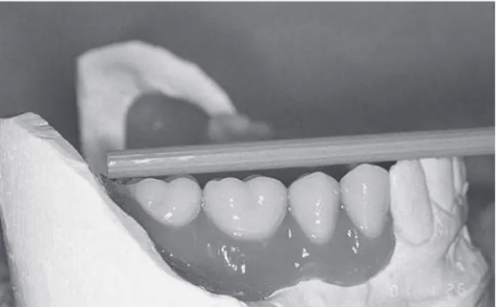
Plaster casts were obtained for all the patients, on which an acrylic resin denture base was made and artificial teeth were arranged. The occlusal plane was used as a reference in all the casts, standardised by tracing an imaginary line through the incisal angle of the cuspid to the end of the 2nd third of the retromolar pad26,28, as shown in
Figure 1. Base points were created by setting 3 mm ball-bearings at the end of the 2nd third of the ret-romolar pad, fixed in the internal part of the resin base. The central cavity of the 1st artificial molar was used as the base reference point for measuring this angle; another ball-bearing was set under this area. This region was chosen because it receives a greater magnitude of bite force16, and is more likely to have greater resorption since the 1st per-manent molars are the first to be extracted from the mandible13.
Radiographic shots
The radiographic shots were taken on ultra-rapid periapical film (Kodak Ektaspeed Plus, EP-21P, type 2, Rochester, New York, USA), with a 0.7 second exposure time. The area radiographed ex-tended from the cuspid to the retromolar pad, and the artificial tooth-base appliance was positioned in the mouth. Since this is a considerable area to be radiographed, occlusal film (Kodak Ektaspeed occlusal radiographic film, Rochester, New York, USA) was used, cut lengthwise in a darkroom and spliced using dark isolating tape.
The radiographic technique employed was the parallel technique with the aid of a film-holder/ positioner (Jon brand radiographic positioner, São Paulo, Brazil) and a long-cone locator. For each hemiarch 2 radiographic shots were taken, as rec-ommended by El Basty, Eid10 (1985). Since full occlusal or incisal surfaces were required, cases in which the remaining teeth did not possess ad-equate clinical crowns or had crowns diverging from the occlusal plane were eliminated from the sample. Tooth extrusions in the maxillary arch
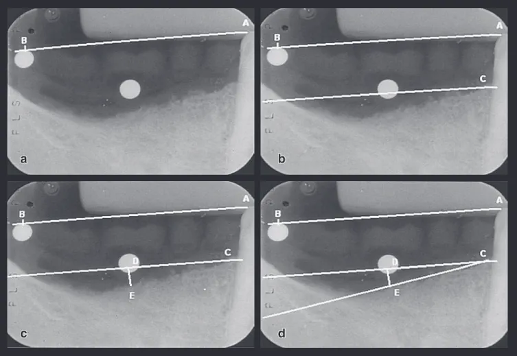
ence plane, joining the lowest point of the sphere placed at the 1st molar level to the highest point of the edentulous segment (line DE) (Figure 2c).
A line was then traced from point C to point E to form the angle DCE, thus measuring the ridge inclination angle (Figure 2d).
RESULTS AND DISCUSSION
From the results obtained, it can be observed that among the 64 hemiarches examined, there were only 3 cases of distally ascending ridges and 1 case of a horizontal (i.e. 0° inclination) ridge, with respective prevalences of 4.68% and 1.56%. Dis-tally descending ridges presented a general aver-age inclination of 17.95° (19.8° when 2nd bicuspids were the abutment teeth and 16.10° when cuspids were the abutment teeth).
These data help elucidate certain aspects were likewise corrected by abrasion or prosthetic
means to prevent them from interfering in the po-sitioning of the artificial denture base for the ra-diographic shots in occlusion.
The radiographs were developed by the visual method and assessed by a single observer in a dark environment. They were subsequently fixed on tracing paper to obtain the tracings.
Tracings
A straight line AB was traced corresponding to the occlusal reference plane (Figure 2a).
Using a ruler and a square, a line was traced parallel to the AB segment, using the distal alveo-lar crest of the abutment tooth as a reference to determine the individual’s current bone loss (point C) (Figure 2b).
A line was traced perpendicular to the
refer-FIGURE 2 - a) radiographic image with tracing representing the occlusal plane (line segment AB); b) line segment parallel to the occlusal plane taking the distal alveolar crest of the abutment tooth (point C) as the point of reference;
c) line traced perpendicular to the occlusal plane parallel (line segment DE); d) tracing of line CE to form angle DCE corresponding to the ridge inclination angle.
b
a
the tangent for the rectangular triangle – the wider the angle obtained. Therefore, if free-ends where cuspids are the abutment teeth are compared with free-ends where 2nd bicuspids serve as the abut-ment teeth, it is geometrically logical to expect the angle to be wider in the second case.
Table 3 shows a clearly significant statistical difference in resorption between users and non-users of mandibular RPDs when the cuspid is the abutment tooth. A variety of explanations for this phenomenon are found in literature. These include: the effect of dentures on residual ridge morphol-ogy23; poor proprioceptive reaction of the fibro-mucosal membrane, which fails to signal occlusal described in the literature. Elbrecht11 (1943) and
Zanetti, Laganá29 (1988) observe that the inclina-tion of the residual ridge directly affects the force exerted on it, producing a distal effect on distal-ly descending ridges when a mandibular RPD is subjected to bite force transmitted in the form of compression. Cecconi et al.7 (1971), Christidou et
al.8 (1973), Plotinick et al.24 (1975) and Feingold
et al.12 (1988) use experimental data to emphasise
that ridge inclination affects the movement of the abutment tooth of a mandibular RPD.
The sagittal inclination measurements ob-tained for each hemiarch and the individual data recorded for patients (type of opposing arch, use of mandibular RPD, abutment tooth, age and sex) were tabulated and processed statistically by vari-ance analysis.
Table 1 shows that, in the sample examined, there was greater resorption (and consequently a wider angle) among patients using mandibular RPDs (19.3° on average) than among non-users of mandibular dentures (16.47°). This can be ex-plained by the lever systems formed by free-end dentures in action19, as well as by the direct, pro-portional relation observed between the length of use of the RPD and bone resorption3,5,6,23. It should be stressed that the average resorption angle was not negligible (approximately 16.5°) among non-wearers of mandibular removable partial dentures. This finding may corroborate the theory of atrophy due to disuse1, reduced mechanical stimulus fail-ing to provide the necessary conditions for normal remodelling.
The relation between the type of opposing arch and denture use and angle of resorption is shown in Tables 1 and 2, which show a patently greater level of resorption for natural teeth and maxil-lary removable partial dentures in comparison to complete dentures in the superior arch. From a biomechanical perspective, this finding suggests that the magnitude of pressure the opposing arch exerts is directly related to resorption of the re-sidual ridge in the lower arch. This is in line with the findings Plotinick, Beresin24 (1975) recorded. The results likewise confirmed a lower level of re-sorption with complete dentures in the opposing arch because the bite forces among patients are considerably smaller14.
The recording of wider angles when the 2nd bicuspid is the abutment tooth (Table 3) can be ex-plained by the shorter distance between the meas-urement points. Figure 3 shows that the shorter the distance between the points – by the rules of
TABLE 1 - Average measurements (in degrees) of angles under diverse conditions and critical figure (thresh-old) for contrast.
Opposing arch
Tukey 5% Natural
teeth Complete denture RPD
17.65 16.05 19.95 2.63
Denture use
2.37
Yes No
19.30 16.47
Abutment tooth
Cuspid Bicuspid
16.10 19.67
RPD: removable partial denture.
TABLE 2 - Average measurements (in degrees) for main factors (opposing arch and denture use) and critical figure (threshold) for contrast.
Denture use
Opposing arch
Tukey 5% Natural
teeth Complete denture RPD
Yes 20.00 15.80 22.10
4.53
No 15.30 16.30 17.80
RPD: removable partial denture.
TABLE 3 - Average measurements (in degrees) for main factors (abutment tooth and denture use) and critical figure (threshold) for contrast.
Abutment tooth Denture use Tukey 5%
Yes No
Cuspid 18.53 13.67
3.32
overload of the supporting tissues9 and provokes excessive pressure on the bone and mucosa, which in turn impairs circulation of the blood; the osteo-cytes become unable to supply the tissues with sufficient oxygen4, or even prostaglandin synthesis by the cells of the fibromucosal membrane is jeop-ardized21. The most obvious explanation, however, is the lever effect a mandibular RPD produces, the force moment exerted increasing proportionally to the distance between the point where the pressure is applied and the rotational fulcrum25,29.
Tables 4 and 5 show that among women the angle of resorption is discreetly increased. Al-though Helkimo et al.15 (1977) detected less bite force among women, non-mechanical factors may be associated with this phenomenon. These include calcium mobilisation due to hormonal causes, especially estrogen deficiency during and after menopause17,27.
Furthermore, regarding the interpretation of data in Table 4, a slight difference was observed in patients over 60 years old in comparison to young-er patients. Nonetheless, data in Table 5 – which compares the use of dentures and the age of indi-viduals – failed to show greater resorption among patients over 60 years of age, from which, in fact, a less pronounced angle was recorded. These find-ings may be explained by what Owall et al.22 (1997) consider to be a diminished masticatory capacity among older patients leading to less compression of the bone tissue as a result of diminished mus-cular activity. Klemetti18 (1994) has observed that reduced bone density is to be expected in women
after the menopause but is not regularly encoun-tered in older men, amongst whom there are even cases of increased bone density. Therefore, in light of the results obtained and the findings recorded in literature, it cannot be claimed that there is a cause-effect relation between resorption of the residual ridge and age.
CONCLUSIONS
Given the methodology employed, the meas-urements recorded and the statistical treatment of the data produced, it is reasonable to conclude that:
1. There is a prevalence of distally descending free-end mandibular residual ridges.
2. There is a statistically significant wider angle of distally descending ridges among users of mandibular RPDs than among non-users, and this difference is more evident when cuspids are the abutment teeth.
3. The results suggest that the magnitude of ridge inclination among users of mandibular RPDs is affected by the type of opposing maxil-lary arch.
4. Ridge inclinations were, on average, more pro-nounced when 2nd bicuspids, as opposed to cuspids, were the abutment teeth, regardless of the use of mandibular RPDs.
5. No significant data were found to correlate age and sex, in isolation from other factors, to residual ridge resorption.
TABLE 4 - Measurements (in degrees) of angles accord-ing to sex, age and supportaccord-ing tooth.
Sex Abutment tooth Age
Cuspid Bicuspid years< 60 years> 60
Female 18.00 19.50 19.19 22.00
Male 12.30 18.75 18.75 20.50
TABLE 5 - Measurements (in degrees) of angles accord-ing to denture use, sex and age.
Denture
use FemaleSexMale < 60 years > 60 yearsAge
Yes 18.50 16.00 19.62 19.50
No 17.23 14.12 17.00 15.88
BC CD tg��
A
B C D
� � r
BC AC tg��
REFERENCES
1. Applegate OC. An evaluation of the support for the remov-able partial denture. J Prosthet Dent 1960;10:112-23. 2. Applegate OC. Evaluating oral structures for removable
partial dentures. J Prosthet Dent 1961;11:882-5. 3. Atwood DA, Coy WA. Clinical, cephalometric, and
densi-tometric study of reduction of residual ridges. J Prosthet Dent 1971;26:280-95.
4. Beerstecher E Jr, Bell RW. Some aspects of the biochemi-cal dynamics in the periodontal ligament and alveolar bone resulting from traumatic occlusion. J Prosthet Dent 1974;32:646-50.
5. Carlsson GE, Hedegärd B, Koivumma KK. Studies in den-tal prosthesis. IV. Final results of a 4-year longitudinal investigation of dentogingivally supported partial den-tures. Acta Odontol Scand 1965;23:443-72.
6. Carlsson GE, Ragnarson N, Astränd P. Changes in height of the alveolar process in edentulous segments. Svenk Tand Tid 1969;62:125-36.
7. Cecconi BT, Asgar K, Dootz E. Removable partial den-ture abutment tooth movement as affected by inclination of residual ridges and type of loading. J Prosthet Dent 1971;25:375-81.
8. Christidou L, Osborne J, Chamberlain JB. The effects of partial denture design on the mobility of abutment teeth. Br Dent J 1973;135:9-18.
9. Crum RJ, Rooney GE. Alveolar bone loss in overdentures: a 5-year study. J Prosthet Dent 1978;40:610-3.
10. El Basty S, Eid HI. The shape of the rest seat prepara-tion and the percentage of alveolar bone loss of cuspid abutments receiving indirect retainers in lower bilateral free-end saddle cases. Egypt Dent J 1985;31:63-81. 11. Elbrecht A. De la próthesis parcial y de su construcción.
Trib Odontol 1943;31:15-32.
12. Feingold GM, Grant A, Johnson W. The effect of variation of residual ridge angle on partial denture abutment tooth movement. J Oral Rehabil 1988;15:379-84.
13. Glisic B. Analysis of factors influencing the amount and localisation of residual ridge reduction of mandible. Sto-matol Glas Srb 1989;36:419-26.
14. Haraldson T, Karlsson V, Carlsson GE. Bite force and oral function in complete denture wearers. J Oral Rehabil
1979;6:41-8.
15. Helkimo E, Carlsson GE, Helkimo M. Bite force and state of dentition. Acta Odontol Scand 1977;35:297-303. 16. Howell AM, Brüdevold F. Vertical forces used during
chew-ing of food. J Dent Res 1950;29:133.
17. Jahangiri L, Devlin H, Ting K, Nishimura I. Current per-spectives in residual ridge remodeling and its clinical im-plications: a review. J Prosthet Dent 1998;80:224-37. 18. Klemetti E, Vainio P, Lassilla V. Mineral density in the
mandibles of partially and totally edentate postmeno-pausal women. Scand J Dent Res 1994;102:64-7. 19. Kratochvil FJ. Influence of occlusal rest position and clasp
design on movement of abutment teeth. J Prosthet Dent 1963;13:114-24.
20. Misch CE. Implant Dentistry. 2nd ed. St. Louis: Mosby;
1999.
21. Nishimura I, Szabo G, Flynn E, Atwood DA. A local patho-physiologic mechanism of the resorption of residual ridg-es: prostaglandin as a mediator of bone resorption. J Prosthet Dent 1988;60:381-8.
22. Owall B, Käyser AF, Carlsson GE. Prótese dentária: prin-cípios e condutas estratégicas. São Paulo: Artes Médicas; 1997.
23. Pietrokovski J. The bony residual ridge in man. J Prosthet Dent 1975;34:456-62.
24. Plotinick IJ, Beresin VE. The effects of variations in the opposing dentition on changes in the partially edentulous mandible. Part 1. Bone changes observed in serial radio-graphs. J Prosthet Dent 1975;33:278-84.
25. Shaw FG. Simple levers and partial dentures. Part 1. Dent Tech 1968;24:24-7.
26. Stewart TK, Rudd KD, Kubker WA. Clinical removable partial prosthodontics. St. Louis: Mosby; 1983.
27. Xie Q, Ainamo A, Tilvis R. Association of residual ridge resorption with systemic factors in home-living elderly subjects. Acta Odontol Scand 1997;55:299-305. 28. Weinberg LA. Atlas of removable partial denture
prostho-dontics. St. Louis: Mosby; 1969.
29. Zanetti AL, Laganá DC. Planejamento: prótese parcial removível. São Paulo: Sarvier; 1988.