116
www.scielo.br/rsbmt INTRODUCTION
Address to: Dra. Melissa Orzechowski Xavier. Lab. Micologia/FM/FURG. Campus Saúde, Visconde de Paranaguá 102, 96201-900 Rio Grande, RS, Brasil.
Phone: 55 53 3233-8871
e-mail: [email protected]
Received in 08/02/2012
Accepted in 21/05/2012
Atypical presentation of sporotrichosis:
report of three cases
Melissa Orzechowski Xavier
[1],[2], Laura Riffel Bittencourt
[2],[3],
Cláudio Moss da Silva
[1],[4], Roseli Stone Vieira
[4]and Hugo Cataud Pacheco Pereira
[1][1]. Área Interdisciplinar de Ciências Biomédicas, Área de Clínica Médica, Faculdade de Medicina, Universidade Federal do Rio Grande, Rio Grande, RS. [2]. Laboratório de Micologia, Faculdade de Medicina, Universidade Federal do Rio Grande, Rio Grande, RS. [3]. Curso de Medicina, Universidade Federal do Rio Grande, Rio Grande, RS. [4]. Serviço de Infectologia , Hospital Universitário Dr. Miguel Riet Corrêa Jr, Universidade Federal do Rio Grande, Rio Grande, RS.
ABSTRACT
Sporotrichosis occurs after fungal implantation of Sporothrix spp. in the skin, and is the main subcutaneous mycosis in Latin America. Here we describe three atypical cases of the disease. The irst case report an extra-cutaneous occurrence of the disease with joint infection; the second one describes a patient with bilateral lymphocutaneous form of sporotrichosis; and the third shows a zoonotic cutaneous case with the development of an erythema nodosum as a hypersensitivity reaction. These cases show the disease importance on the region and the necessity of fungal culture to the diagnosis conirmation.
Keywords: Sporothrix sp. Septic arthritis. Erythema nodosum. Itraconazole.
Sporotrichosis, acquired by inoculation of the dimorphic fungus Sporothrix spp., is the main subcutaneous mycosis in Brazil. Clinically the typical presentation of sporotrichosis is a reddish lesion that appear near to the inoculation site and which tends to progress to erithematous nodules and ulcers with new similar lesions ascending in lymphatic vessels1,2.
Although ascending nodular lymphangitis is the commonest clinical presentation, the disease can manifest with distinct characteristics, which turns the diagnosis more dificult. Here we describe three atypical cases of sporotrichosis diagnosed in 2010 and 2011 at the University Hospital of Universidade Federal do Rio Grande (HU-FURG), Rio Grande, State of Rio Grande do Sul, Brazil. This study was approved by the Ethic Committee of the Institution (CEPAS-FURG 175/2011).
Case RepORT
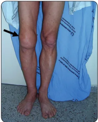
A male patient, 51-years-old, isherman, was admitted with a complaint of pain and swelling on his right knee that had started one year before, since the occurrence of a local bruising trauma in a wood boat, but without skin penetration. Serology for human immunodeiciency virus (HIV) and hepatitis C virus (HCV) were negative, but the patient suffered of diabetes, and was drinker and smoker in the past (two years and seven years before, respectively). Previous ultrasonography suggested septic arthritis, and patient was treated with surgical procedure and cefalotin without clinical resolution. In that occasion, laboratorial
exams of the sinovial luid showed no microorganism growth in bacterial and fungal culture. Clinical examination revealed pain with high intensity, dificulty walking, and logistic signs on the affected joint (Figure 1). Ultrasound imaging showed an extensive suffusion with a viscous luid and suprapatellar bursa distension. Suspecting of a septic arthritis reactivation an arthrocentesis was done. Cytology of synovial luid revealed a count of 800 erythrocytes/mm3 and of 3,200 leukocytes/mm3 with 53% of lymphocytes and 47% of segmented neutrophil. Biochemical examination was not done due to the synovial
FIGURE 1 - Black row showing swelling in the right knee of the patient with monoarthritis by Sporothrix sp. (Case 1).
Revista da Sociedade Brasileira de Medicina Tropical 46(1):116-118, Jan-Feb, 2013
http://dx.doi.org/10.1590/0037-868215282013
117
www.scielo.br/rsbmt
Xavier MO et al - Atypical sporotrichosis
DIsCUssION
thickening. At the microbiology lab, none bacterial growth was detected, but a great number of fungal colonies grew in Sabouraud agar. The characteristics of the colonies in Sabouraud agar at 25ºC and in Brain-Heart Infusion (BHI) agar at 37°C associated with its micromorphology allowed us to conirm the diagnosis of a Sporotrhix sp. articular infection. The patient was scheduled to treat his arthritis with itraconazole (200mg twice a day - BID) for one year. After six months of treatment, it was observed a signiicant improvement of his articular symptoms.
Case 2
Male patient, 43 years-old, soldier, was admitted in the HU-FURG ambulatory showing ulcerated, papular and nodular lesions, with violaceous borders in both arms and distributed by the lymphatic pathway. The patient reported the practice of exercises crawling and rolling on the ground with vegetation. This professional practice occurred one week before the appearance of the irst lesion on his left arm, which progressed following the lymphatic vessels and also appeared in the right arm. About ten lesions (1-3cm) in each arm were observed in the clinical examination, some with intact skin and others ulcerated with exudates (Figure 2). Regional lymphadenomegaly was also detected. According to the patient, the lesions had an evolution about three or four months without clinical resolution with previous antibacterial treatment. Clinical suspicion of bilateral lymphocutaneous sporotrichosis was done and patient was submitted to a skin biopsy. Histopathological evaluation showed mixed multifocal inlammatory process without any identiiable microorganism or malignity evidence. At mycology lab a great number of fungal colonies grew in Agar Sabouraud and were phenotipically identiied as Sporothrix sp., conirming the initial clinical suspicion. Itraconazole was prescribed at 200mg/day for three months. Patient did not return to the evaluation of his clinical evolution.
FIGURE 2 - Ulcerated, erythematous nodular lesions in right (A) and left (B) arm of the patient with bilateral lymphocutaneous sporotrichosis (Case 2).
A
B
Case 3
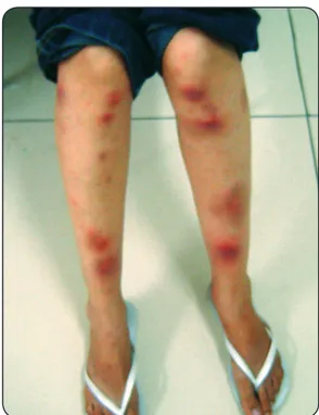
Female patient, 28 years-old, student, referred the appearance of a skin lesion about 15 days after a scratch of a cat with sporotrichosis. Physical examination revealed a 3cm pustular lesion with violaceous borders in the trauma region in her forearm, associated with a more recent 2cm lesion, nodular, erythematous and irm, following the lymphatic vessel. After eight days of the initial skin lesions at forearm, multiple painfull reddish nodules appeared in her both legs (Figure 3), featuring a typical case
of erythema nodosum. Histopathology of the biopsy of the ulcerated lesion in forearm showed an inlammatory process with great number of multinucleated foreign body giant cells, and it cultive resulted in Sporothrix sp. growth, conirming the diagnosis of sporotrichosis. The immunological condition of the patient was investigated due to the probability of a fungal dissemination, but immunossupression was ruled out. Nonsteroidal antiinlammatory drugs resolved the eryhtema nodosum in a few weeks, and treatment with itraconazole 200mg/day for six months resulted in a clinical cure.
FIGURE 3 - Skin rash due to a hypersensitivity reaction in a zoonotic case of sporotrichosis (Case 3).
The clinical presentation of sporotrichosis described here, as septic arthritis, bilateral lymphatic lesions and hypersensitivity reaction to Sporothrix sp. infection, are rare with scarce reports in the literature.
118
www.scielo.br/rsbmt Rev Soc Bras Med Trop 46(1):116-118, Jan-Feb, 2013
ReFeReNCes
monoarthritis case as the only evidence of sporotrichosis without the presence of a skin lesion has been previously reported in the literature4, and authors also did not found a speciic source of infection.
Arthritis due to Sporothrix sp. is often associated with immunosuppressed patients, especially those with acquired immunodeiciency syndrome (AIDS). In our case 1, patient despite being negative for HIV, had a history of chronic alcoholism and diabetes, factors also cited as a predisposition to the onset of extra-cutaneous sporotrichosis1,5. Differential diagnosis of monoarthritis include other infectious causes, and noninfectious, as hypothyroidism, amyloidosis, multiple myeloma, paraneoplastic syndrome, among others4.
According to the majority of reported cases of bilateral sporotrichosis, this presentation may occur by self-inoculation, multiple inoculations or re-infection7. The crawling exercise in soil with decaying vegetation, performed by our patient 2 prior to the onset of the bilateral lymphocutaneous lesions, suggests that there has been simultaneous multiple inoculations of the fungus into the dermis through microtrauma by plant material, since this is the natural habitat of Sporothrix sp., similar to that reported by other authors7,8. The bilateral presentation of sporotrichosis is described as atypical and rare, although it have already been reported in children by zoonotic transmission9 or by trauma with plants in adults7,8. Differential diagnosis of this clinical presentation of sporotrichosis include cutaneous tuberculosis, atypical mycobacterial infection, prurigo, nocardiosis, and cutaneous leishmaniasis in itssporothrichosis-like clinical form 1,7.
Hypersensitivity reactions to infection by Sporothrix sp.
as occurred in our case 3 may present themselves as different clinical manifestations. Case of a reactional episode of polyarthritis was described in an immunocompetent patient with lymphocutaneous sporotrichosis that showed the involvement of ankles and elbows10. Three cases of erythema multiforme (EM) associated with Sporothrix sp. infection had been described in adult patients that developed a skin rash after 7-30 of the primary infectious cutaneous11. And, similar to our case 3, hypersensitivity fungal triggering erythema nodosum (EN) associated with sporotrichosis was irst described in 2002, in three patients in Rio de Janeiro12. According to our case reported, the lesions of EN described in the literature may arise after 7 to 40 days of the initial injury12.
Although few cases of hypersensitivity to Sporothrix sp. infection are reported in the literature, as in our case 3, all of them10-12 occurred through zoonotic transmission from sick cats. A hypothesis that could explain this association is the constant and intense stimulation of the immune system of patients when they are in touch with cats with sporotrichosis11,12. This could occur due to the fact that this species tends to have a great amount of fungal propagules in their lesions, triggering an intense exposure of humans to fungus antigens and even culminating with subclinical infection and reinfection, causing hypersensitivity2,11,12.
We must remark that the gold standard for diagnosis of sporotrichosis in any clinical presentation is mycological culture for fungi isolation and identiication1, as occurred in the three
cases reported here. The fungal culture is required because the sporotrichosis lesions in humans tend to have few yeast cells which provide a dificulty in observe them either in cytological or histopathological examination1,4.
However, the dificulty of diagnosis of atypical sporotrichosis described in this study are also frequently reported in the literature, either in bilateral case of sporotrichosis7,9 or in the osteoarticular involvement3,4, especially given the numerous other causes more commonly associated with, which are raised as the main diagnostic hypotheses2,4. In this scenario, it is common the occurrence of a late diagnosis, and, especially in relation to osteoarticular involvement, the delay in identifying the etiology of the injury can lead to unfavorable prognosis and may result in loss of function of the affected area4,5, similar to that observed in our patient that showed signs of muscle atrophy by disuse as a consequence of chronic dificulty in walking.
In conclusion, the three reported cases of atypical sporotrichosis described here emphasize the disease importance in the region, and highlight the need to include it as differential diagnosis even in cases of non-classic lesions of the disease. Thus, fungal culture will be conducted leading in the diagnosis conirmation and favoring the prognosis through an early and correct treatment.
1. Kauffman CA, Bustamante B, Chapman SW, Pappas PG. Clinical Practice Guidelines for the Management of Sporotrichosis: 2007 Update by the Infectious Diseases Society of America. Clin Infect Dis 2007; 45:1255-1265.
2. Lopes-Bezerra LM, Schubach A, Costa RO. Sporothrix schenckii and Sporotrichosis. Anais Acad Bras Ciências 2006; 78:293-308.
3. Appenzeller S, Amaral TN, Amstalden EMI, Bertolo MB, Marques Neto JF, Samara AM, et al. Sporothrixschenckii infection presented as monoarthritis: report of two cases and review of the literature. Clin Rheumatol 2006; 25:926-928. 4. Costa RO, Mesquita KC, Damasco PS, Bernardes-Engemann AR, Dias CMP,
Silva IC, et al. Infectious arthritis as the single manifestation of sporotrichosis: Serology from serum and synovial luid samples as an aid to diagnosis. Rev Iberoam Micol 2008; 25:54-56.
5. Howell SJ, Toohey JS. Sporotrichal arthritis in south central Kansas. Clin Orthop Relat Res 1998; 346:207-214.
6. Gamo R, Aguilar A, Cuétara M, Gonzalez-Valle O, Houmani M, Martín L, et al. Sporotrichosis Following Mesotherapy for Arthrosis. Acta Derm Venereol 2007; 87:430-431.
7. Haruna K, Shiraki Y, Hiruma M, Ikeda S, Kawasaki M. A case of lymphangitic sporotrichosis occurring on both forearms with a published work review of cases of bilateral sporotrichosis in Japan. J Dermatol 2006; 33:364-367.
8. Alves SH, Aurélio PL, Tecchio MZ, Zuchetto A, Schirmer R, Santurio JM. Subcutaneous bilateral sporotrichosis: A rare presentation. Mycopathologia 2004; 158:285-287.
9. Borrego JAB, Mayorga J, Tarango-Martínez VM. Esporotricosis linfangítica bilateral y simultánea. Rev Iberoam Micol 2009; 26:247-249.
10. Oroino-Costa R, Bóia MN, Magalhães GAP, Damasco PS, Bernardes-Engemann AR, Benvenuto F, et al. Arthritis as a hypersensitivity reaction in a case of sporotrichosis transmitted by a sick cat: clinical and serological follow up of 13 months. Mycoses 2009; 53:81-83.
11. Galhardo MCG, Barros MBL, Schubach AO, Cuzzi T, Schubach TMP, Lazéra MS, et al. Erythema multiforme associated with sporotrichosis. Eur Acad Dermatol Venereol JEADV2005; 19:507-509.