Arq Bras Cardiol 2004; 82: 291-4.
Fontes et al Vascular prosthesis infection in thoracic aorta surgery
2 9 1
Instituto do Coração do Hospital das Clínicas - FMUSP
Mailing address: Noedir Antônio Groppo Stolf - InCor - Av. Dr. Eneas C. Aguiar, 44 Cep 05403-000 - São Paulo, SP, Brazil - E-mail: [email protected]
Received: 11/25/2002 Accepted: 3/10/2003
Arq Bras Cardiol, volume 82 (nº 3), 291-4, 2004
Ronaldo Ducceschi Fontes, Noedir Antônio Groppo Stolf, Júlio Cesar Marino, David Pamplona, Luis Francisco Ávila, Sérgio Almeida Oliveira
São Paulo, SP - Brazil
Vascular Prosthesis Infection in Thoracic Aorta Surgery.
Review of the Experience and a Case Report Illustrating
treatment with an Unconventional Technique
Case Report
We report the case of a 37-year-old-female patient who had undergone a Bentall procedure at our service and re-turned with intense chest pain and acute aortic dissection type III, which was diagnosed and clinically treated. One year after this episode, this dissection expanded, and the pa-tient underwent surgery with interposition of a Dacron graft in the descending aorta. In the immediate postoperati-ve period, the patient experienced left bronchopneumonia and was discharged afebrile and in good condition. One month after discharge, she returned with fever and toxemia. Pleural empyema was diagnosed, and she underwent an ex-ploratory thoracotomy that did not confirm this diagnosis, but revealed intense effusion thickening. Four months after the exploratory thoracotomy, Klebsiella pneumoniae and Enterobacter sp were isolated in a blood culture. Magnetic resonance imaging revealed shapes compatible with peri-graft infection. With this clinical and laboratory picture, graft removal was indicated as was axillo-bifemoral graf-ting. Surgery was successfully performed, the patient was discharged in good condition, and remains well after a 57-month follow-up without complications. The methods used for diagnosis and treatment of prosthesis infection in thora-cic aorta surgery are discussed.
In recent years, significant progress has been achie-ved in the treatment of aneurysms and aortic dissections 1,2. The advances are related to surgical techniques, vascular prostheses in the postoperative period, antibiotic therapy, as well as methods of diagnostic investigation using com-puted tomography and magnetic resonance 3.
Despite these advances, severe 4 postoperative
com-plications sometimes still occur with surgical procedures in the aorta. Among them, graft infection is one of the most se-vere, resulting in great morbidity and mortality 3.
Some methods may be used to treat this complication with favorable results 3,5. The authors report their service’s experience with vascular prosthesis infection and point out the case of a patient who evolved with prosthesis infection in the postoperative period of distal dissection surgery, with treatment performed with an alternative surgical technique.
Case Report
A 37-year-old, white female patient with Marfan’s syn-drome underwent surgery in 1985 to treat annuloaortic ecta-sia with the Bentall procedure. The patient was discharged in good condition and was being followed-up when she re-turned in 1990 complaining of intense thoracic pain. A De Bakey type III aortic dissection was diagnosed.
Since no complications were present, clinical treat-ment was maintained until May 1991, when she returned with pain and underwent a chest X-ray, digital angiography, and CT scan demonstrating an 80-mm dilation of the des-cending thoracic aorta.
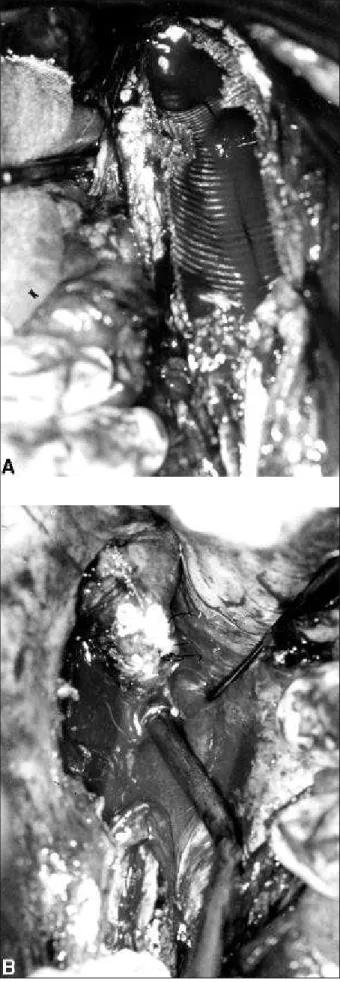
The patient was referred for a surgical procedure and underwent correction of the dissection through interposi-tion of a Dacron graft, replacing the descending aorta (fig. 1A and B). The surgery was performed using simple aortic clamping between the left carotid and the left subcla-vian artery. Poor tissue was verified in the proximal stump, and because the distal stump was near the diaphragm, access was hindered. During this period, cerebrospinal pressure was monitored and maintained below 10 mmHg through drainage of 50 mL of liquid.
The patient evolved with bronchopneumonia, recei-ved cefoxitin for 15 days, and was discharged in excellent clinical condition.
hospi-2 9 hospi-2
Fontes et al
Vascular prosthesis infection in thoracic aorta surgery
Arq Bras Cardiol 2004; 82: 291-4.
tal after 1 month with fever suggesting empyema effusion. She underwent exploratory thoracotomy that did not con-firm the diagnosis but demonstrated only intense pleural thickening. During this procedure, puncture of the perigraft region was performed without loss of fluid.
She remained in the hospital to recover from the thora-cotomy and experienced fever and bacteremia crises. Three months later, Klebsiella pneumoniae and Enterobacter sp
were isolated in a serial blood culture, and treatment with an-tibiotics was initiated; however, the clinical picture did not improve. Investigation continued with the use of CT scan-ning that revealed a large number of thrombi in the perigraft region and the presence of a small amount of air (fig. 2). This picture led to the diagnosis of prosthesis infection, and its removal was indicated so that an axillo-bifemoral graft could be performed due to the difficulties expected. The surgery was successfully performed in 1991 (fig. 3A and B). The Dacron graft was withdrawn, the proximal and distal aortic stumps were closed, and the axillo-bifemoral graft was pre-formed. The patient remained in the hospital for 45 days with antibiotics, the fever ceased, and the patient progressi-vely improved until she was discharged.
She is still being followed-up, and she has been asymptomatic for 7 years. A late angiographic study de-monstrated that the graft was pervious and the thoracic
Fig. 1 - A) Aspect of the dissected aorta, the great amount of thrombi in the false lumen; B) final apsect of the operation to correct descending aorta dissection, demonstrating extensive Dacron graft.
Arq Bras Cardiol 2004; 82: 291-4.
Fontes et al Vascular prosthesis infection in thoracic aorta surgery
2 9 3
Fig. 3 -A) The graft is partially withdrawn. Detail of the reoperation to treat infection; B) the interruption of the descending aorta right after emergency of the left subclavian artery is noticed.
artery was excluded (fig. 4). She is currently taking oral anti-coagulant medication and beta-blockers.
Discussion
Postoperative infection of a graft in thoracic artery surgery is a rare complication. It has occurred in between 0.5 and 5% of the patients undergoing this type of surgery 6,7.
Large surgeries used to treat thoraco-abdominal or even abdominal aneurysms, requiring anastomoses with fe-moral and thoracic arteries, are usually more susceptible to infections, especially when infected cutaneous lesions are present in the abdominal region, together with the inadequa-te use of central venous catheinadequa-ters 1,3.
The infectious process may start in the suture site lea-ding to dehiscence and false aneurysm formation. These fal-se aneurysms may tear in cavities or adjacent organs, lea-ding to hemoptysis, hematemesis, or melena accorlea-ding to their location 8.
2 9 4
Fontes et al
Vascular prosthesis infection in thoracic aorta surgery
Arq Bras Cardiol 2004; 82: 291-4.
Infection of aortic prostheses is evident by the presen-ce of fever and thoracic pain. The infectious propresen-cess must therefore be carefully investigated. This investigation must entail complementary examinations that will indicate the ap-propriate conduct. The first important measure is to try to isolate the responsible germ by using serial blood cultures. The most frequently found germs are Staficoco Aureus, S.
Epidermidis, Streptococo, Enterobacter, E. Coli, Proteus,
and Pseudomonas. Clinical treatment includes specific anti-biotics for a minimum 30-day period 9.
Complementary examinations through images must be used, and the most indicated are CT scan, and magnetic re-sonance, where the presence of air and liquid in the perigraft region can be observed. Other methods may contribute to the diagnosis, such as radioisotope study, aortography in the cases where false aneurysm or aortic obstruction is sus-pected; digestive endoscopy when stomach, esophagus, or duodenum erosion is suspected 3.
In almost all cases, the treatment for this complication is surgical, performed with graft replacement as well as graft withdrawal followed by extraanatomical derivation, and, oc-casionally, the prosthesis is covered with vascular grafts followed by extraanatomical derivation 3,4,10.
In our case, the clinical picture that suggested perigraft infection was hindered by the radiologic image, which sug-gested the presence of empyema in the hemi-thorax apex. The patient previously underwent exploratory thoracotomy.
Surgery was indicated after the germs had been isola-ted in the blood culture, in association with the clinical pic-ture and the examination using images. However, the risk of a third thoracotomy; the clinical picture of malnutrition; dif-ficulty reconstructing the vascular graft from the diseased descending aorta; the ascending aorta with graft, paraple-gia, and also the intolerance to synthetic material 11; before the positive result of blood cultures made us wait too long before withdrawing the graft.
We believe that the increased time of clamping and bleeding during the first intervention were the factors that contributed to the development of infection and that other investigation methods, such as radiology with labelled leu-kocytes, could help early treatment 12.
We have concluded that the incidence of perigrafts in surgeries to correct thoracic aorta aneurysms is low and, in the present case, axillo-bifemoral was the adequate option with excellent late and immediate results.
1. Fontes RD, Stolf NAG, Lourenço Filho DD, Tranchesi R, Mady C, Pereira Barreto AC, Pileggi FJC, Jatene AD. Dez anos de cirurgia dos aneurismas da aorta ascendente no Instituto do Coração-FMUSP. Rev Bras Cir Cardiovasc 1991; 6: 24-9. 2. Pêgo-Fernandes PM, Stolf NAG, Fontes RD, Verginelli G, Jatene AD. Cirugia
das dissecções crônicas da aorta ascendente com insuficiência valvar. Rev Bras Cir Cardiov 1990; 5: 149-13.
3. Constantino MJ. Recurrent aortic graft infection following descending thoracic aorta to femoral artery bypass a case report and review: J Cardiovasc Surg 1991; 32: 477-81.
4. Svensson LG, Crawford ES, Hess KR, Coseli JS, Safi HJ. Dissection of the aorta and dissecting aortic aneurysm. Circulation 1990; 82(suppl IV) 5: IV24-IV46. 5. Matley PJ, Beningfield SJ, Lourens S, Immelman EJ. Successful treatment of infec-ted thoracoabdominal aortic graft by percutaneous catheter drainage. Jvasc Surg 1991; 13: 513-5.
6. Reilly LM, Altman H, Lusby RJ, Kers RA. Late results following surgical mana-gemento of vascular graft infections; J Vasc Surg 1984; 1: 36-44.
References
7. Ilgenfritz FM, Jordan FT. Microbiological monitoring of aortic aneurysm wall and contents during aneurismectomy. Arch Surg 1988; 123: 3506-8.
8. Tollefson DF, Bank DF, Kaebnick HW, Seabrook GR, Towne JB. Surface biofilm disruption: enhanced recovery of miocroorganismos from vascular prostheses. Arch Surg1987; 122: 38-43.
9. Olah A, Vogt M, Laske A, Carrell T, Bauer E, Turina M. Axillo-femoral bypass and simultaneous removal of the aorto-femoral vascular infection site: is the procedu-re safe? Eur J Vasc Surg 1992: 252-4.
10. O´Hara PJ, Hertez NR, Beven EG, Krajewaski LP. Surgical management of infec-ted abdominal aortic grafts: review of a 25-year of experience. J Vasc Surg 1986; 725-31.
11. Vollmar PE, Mohr W, Haman H, Brecht-Kraus D. Perigraft reation: incompatibi-lity of synthetic grafts? New aspects on clinical manifestations, pathogenesis and therapy. World J Surg 1982; 12: 750-5.