Cop
yright
© ABE&M t
odos os dir
eit
os r
eser
vados
.
Can SPECT change the surgical
strategy in patients with primary
hyperparathyroidism?
O SPECT pode mudar a estratégia cirúrgica nos pacientes com hiperparatireoidismo primário?
Letícia Iervolino1, Nilza Maria Scalisse1, Sergio Setsuo Maeda1
SUMÁRIO
O hiperparatireoidismo primário (HPTP) é a causa mais comum de hipercalcemia diagnosticada ambulatorialmente. É mais frequente no sexo feminino e na pós-menopausa e a prevalência é de 1 a 4:1000 na população geral. O adenoma solitário esporádico secretor de PTH corresponde a 90% dos casos de HPTP, enquanto a doença multiglandular é mais comum nas síndromes de hiperparatireoidismo familiar (5%) e o carcinoma de paratireoide representa menos de 1% dos casos. Somente após a certeza da autonomia funcional de uma ou mais glândulas paratireoides é que devem ser realizados exames de imagem localizatórios, com a inalidade de planejar o procedimento cirúrgico. Além disso, esses exames apresentam limitações e podem resultar em falsos-positivos e negativos. Há casos em que a localização da glândula paratireoide é de ex-trema diiculdade, sendo necessária a associação de métodos de imagem para localização pré--operatória como o uso do 99mTc-pertecnetato, SPECT, SPECT/CT e a ultrassonograia. Descreve-mos um caso de paciente feminina, 50 anos, com diagnóstico de hiperparatireoidismo primário, submetida a um procedimento cirúrgico sem sucesso, com manutenção da hipercalcemia e do hiperparatireoidismo, em que, somente após a realização da cintilograia associada ao SPECT/ CT, foi possível localizar a glândula paratireoide hiperfuncionante em região retrotraqueal. Arq Bras Endocrinol Metab. 2012;56(4):265-9
SUMMARY
Primary hyperparathyroidism (PHPT) is the most common cause of hypercalcemia in outpa-tients. It is more common in females, after menopause, and the prevalence is 1 to 4:1000 in the general population. Patients with PHPT have abnormal regulation of PTH secretion, resulting in elevated serum calcium and inappropriately high or normal PTH in relation to the calcium value. Sporadic PTH-secreting adenoma alone accounts for 90% of cases of PHPT, while mul-tiglandular hyperplasia is more common in familial hyperparathyroidism syndromes (5%) and parathyroid carcinomas represent less than 1% of cases. Only after making sure there is func-tional autonomy of one or more parathyroid glands, localization imaging tests should be per-formed to guide a possible surgical procedure. It is important to highlight that these tests have limitations and can yield false-positive and false-negative results. There are cases in which the parathyroid gland is dificult to be located, requiring a combination of imaging methods for pre-operative localization, such as 99mTc-pertechnetate, SPECT, SPECT/CT, and US. We describe the case of a 50-year-old female patient diagnosed with PHPT, who underwent a surgical procedure without success, with maintenance of hypercalcemia and hyperparathyroidism. In this case, the hyperfunctioning parathyroid was located in the retrotracheal region only after scintigraphy combined with SPECT/CT were used. Arq Bras Endocrinol Metab. 2012;56(4):265-9
1 Discipline of Endocrinology,
Department of Medicine, Faculdade de Medicina da Santa Casa de São Paulo (FMSCSP), São Paulo, SP, Brazil
Correspondence to:
Letícia Iervolino Av. Angélica, 696, ap. 61. 01228-000 – São Paulo, SP, Brazil [email protected]
Cop
yright
© ABE&M t
odos os dir
eit os r eser vados .
INTRODUCTION
P
rimary hyperparathyroidism (PHPT) is the most common cause of hypercalcemia in outpatients (1,2). It is more common in females, especially post menopausal ones (3), and the prevalence is 1 to 4:1000 in the general population (1).PHPT is characterized by abnormal regulation of PTH secretion, leading to elevated serum calcium and normal or inappropriately high levels of PTH in rela tion to the corresponding calcium values. Sporadic PTHsecreting adenoma accounts for 90% of the ca ses, whereas multiglandular disease is more common in familial syndromes (5%), and parathyroid carcinomas represent less than 1% of the cases (3).
Only after making sure that one or more para thyroid glands have functional autonomy, imaging stu dies should be performed to locate it and facilitate the surgical approach. It is important to highlight that the se tests have limitations and can result in falsepositives and falsenegatives (2).
Scintigraphy with 99mTcSestamibi is the gold stan
dard method for localization, but most of the services perform only planar images (two dimensions). The association with SPECT (Single Photon Emission Computed Tomography) or SPECT/CT allows more accurate threedimensional anatomical localization of abnormal areas in scintigraphy. The advantage of this association is that it combines assessment of glandular function with anatomic location, signiicantly contribu ting to surgical planning, especially when cervical ana tomy is distorted, in cases of prior cervical surgery and ectopic adenomas (4).
The presence of an ectopic hyperfunctioning para thyroid gland or multiglandular hyperplasia is the main cause of persistent and recurrent hyperparathyroidism. The inferior parathyroid glands are derived from the epithelium of the dorsal bulbar part of the third pha ryngeal pouches, and the superior parathyroid glands, from the dorsal part of the fourth pouches. Para thyroid glands are usually located posteriorly to the thyroid, but their location may vary. The position of the superior glands is more constant than that of the inferior ones. They can be found anywhere near or wi thin the thyroid or thymus. Occasionally, the inferior parathyroids may be close to the bifurcation of the common carotid artery, and in other cases, they may follow the thymus into the chest, considering to be ectopic parathyroids (5).
CASE REPORT
This case report was previously approved by the Ethics Committee of Faculdade de Medicina da Santa Casa de São Paulo, and the patient signed an informed consent form.Afemale patient, MLPG, 50 years old, who was born and raised in São Paulo, and presented mental re tardation of unknown etiology, underwent total thyroi dectomy for multinodular goiter in the past.
In 2008, she was diagnosed with osteoporosis in a bone densitometry scan. During the investigation, hypercalcemia and high PTH were found (Table 1). Pa rathyroid scintigraphy (99mTcSestamibi) showed a high
uptake area near the sternal notch to the right, which may have corresponded to the lower right hyperfunc tioning parathyroid (Figure 1). The patient underwent parathyroidectomy (pathology study: thymus fragments with diffuse lymphoid hyperplasia and areas of distorted architecture). However, after the irst procedure, the la boratory proile was not resolved (Table 1).
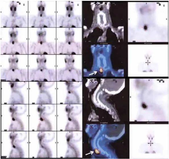
In March 2010, continuing the investigation, cer vical MRI detected a 0.7 cm nodular image in the left paratracheal region with mild contrast uptake. It was not possible to differentiate between a lymphonode and an increased parathyroid. Due to the unsuccessful previous surgical treatment, a 99mTcsestamibi scintigra
phy complemented with SPECT/CT was performed, and a right retrotracheal parathyroid adenoma was de tected (Figure 2). The patient underwent another sur gery (pathology study: parathyroid adenoma) in July 2010. The parathyroid adenoma was localized with a signiicant drop in intraoperative PTH (IOPTH time 0 = 299 pg/mL, IOPTH 15 minutes after parathyroi dectomy = 21 pg/mL).
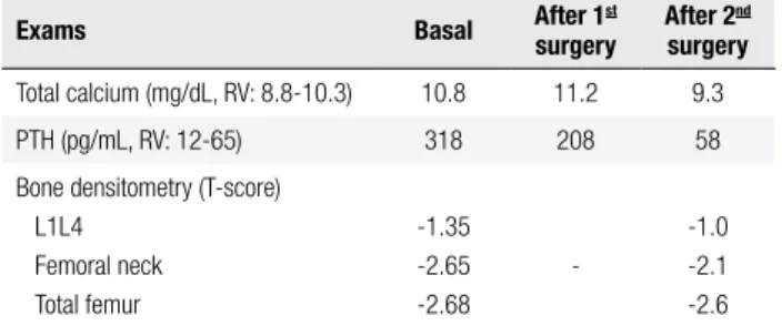
Currently, the patient evolved to normocalcemia and, six months after surgery, bone densitometry sho wed improvement in bone mass with a 12% increase in bone mineral density in the femoral neck, a 36% increa se in total femur, and a 4% increase in the lumbar spine after 6 months of the second surgery (Table 1).
Table 1. Pre- and post-operative laboratory exams
Exams Basal After 1surgeryst After 2surgerynd
Total calcium (mg/dL, RV: 8.8-10.3) 10.8 11.2 9.3 PTH (pg/mL, RV: 12-65) 318 208 58 Bone densitometry (T-score)
Cop
yright
© ABE&M t
odos os dir
eit
os r
eser
vados
.
Figure 2.99mTc-Sestamibi scintigraphy complemented with SPECT/CT.
Cop
yright
© ABE&M t
odos os dir
eit
os r
eser
vados
.
DISCUSSION
We described the case of a patient who, after under going a surgical procedure without success, progressed with persistent hypercalcemia and elevated PTH. The second preoperative localization of hyperfunctioning parathyroid adenoma was possible with scintigraphy with SPECT/CT, which featured an ectopic gland in the retrotracheal region. After new surgical procedure, with excision of the adenoma, the clinical picture was improved.
Multiple methods for localizing parathyroid glands can be used, such as scintigraphy, ultrasonography, com puted tomography, and magnetic resonance imaging. Scintigraphy in two phases (15 and 120 minutes) is per formed with 99mTcSestamibi. The high uptake in hyper
functioning parathyroid is explained by that in this cells there is a large number of abnormal mitochondria for which Sestamibi has high afinity, since the washout of the thyroid is faster (6,7). In most services, only two dimensional planar images are made, which caused the irst scintigraphy in this case to be misinterpreted.
Although planar images of the parathyroid have a relatively high accuracy, intrathyroid adenomas with low 99mTcsestamibi uptake and ectopic adenomas may
be neglected. The use of double tracer (99mTcSestami
bi and 99Tcpertechnetate) associated with SPECT and
computed tomography allow the localization of the pa rathyroid adenoma in three dimensions, the differentia tion from thyroid lesions (only the thyroid can uptake the 99mTcpertechnetate), and increases sensitivity for
the detection of the affected gland in 90%, aiding in surgical planning (8).
However, the routine use of SPECT before surgery is still controversial. According to Lorberboym and cols. (9), combined planar scintigraphy identiied 79% of the adenomas and the use of SPECT increased sen sitivity to 96%, with SPECT being superior to the pla nar image in 17% of the cases, mainly in patients with ectopic parathyroid or multiglandular goiter. Another study evaluated the detection of parathyroid adenomas smaller than 1 g in 92 patients with PHPT. The correct localization of the adenoma using planar images was 87% while with the association of SPECT, the sensi tivity increased to 95%, allowing us to conclude that SPECT can improve the investigation eficiency (10).
On the other hand, some authors believe that SPECT does not bring additional information, not improving the detection rate for adenomas. Chen and
cols. (11) concluded that the latter images, subtraction images and SPECT are not necessary, given they bring little information other than early images with 99Tc
pertechnetate. Staudenherz and cols. (12)evaluated patients with thyroid disease (50% of the cases) and pre vious cervical surgery (21% of the cases) submitted to planar images with 99mTcMIBI, SPECT, US, and sub
traction scintigraphy with 201Ta and 99Tcpertechnetate.
The images were then compared with the histopatho logy of the surgical specimen. The analysis showed that the use of SPECT provided additional information for tumor localization in 39% of the patients with thyroid disease or previous cervical surgery, but did not increa se overall detection rates, suggesting that this exam should not be used for all patients with PHPT.
Recent advances in the techniques of preoperative localization of parathyroid adenomas allow better plan ning and surgical success. The combination of ultra sonography and scintigraphy is the most appropriate approach, and the ideal situation is to ind a good cor relation between these methods. The cases of ectopic adenomas and adenomas located deep in the cervical region are rare, accounting for 3%6% of cases. Howe ver, they are a challenge for the surgeon and a frequent cause of failure if accurate localization tests are not performed before the procedure. In these patients, it is useful to perform SPECT or SPECT/CT after con ventional scintigraphy, because this approach provides useful information for the surgeon (13).
It is important to highlight that conventional pa rathyroid scintigraphy can result in falsepositive and falsenegative results. The most common cause of false positive results is the solitary and solid thyroid nodu le or the multiglandular goiter. Benign or malignant tumors can capture the radiotracer and may result in falsepositives, including thymoma, breast, lung, head and neck cancer, as well as their metastases to bone and lymphonodes, and bronchial carcinoid tumors (8).
Cop
yright
© ABE&M t
odos os dir
eit
os r
eser
vados
.
tein, and the multidrug resistance related to protein expression and celular cycle (8).
The adoption of techniques that combine images with
99mTcpertechnetate, SPECT scintigraphy and conven
tional computed tomography (99mTcSestamibi), increa
se sensitivity, provide useful topographical information for the differentiation between parathyroid and thyroid tissue, and may help to decrease the percentage of un successful surgeries due to ectopic adenomas not found intraoperatively (7). Finally, the use of IOPTH can help detecting the surgical cure or the need to keep surgical exploration when there is no decrease in the tracer.
Disclosure: no potential conlict of interest relevant to this article was reported.
REFERENCES
1. Taniegra ED. Hyperparathyroidism. Am Fam Physician. 2004;69(2):333-9.
2. Khan A, Bilezikian J. Primary hyperparathyroidism: pathophysio-logy and impact on bone. CMAJ. 2000;163(2):184-7.
3. Ferris RL, Simental AA Jr. Molecular biology of primary hyperpa-rathyroidism. Otolaryngol Clin North Am. 2004;37(4):819-31. 4. Radan L, Gorenberg M. The value of SPECT/CT 99mTc-sestamibi
scintigraphy in the diagnosis of ectopic parathyroid adenoma. Isr Med Assoc J. 2009;11(7):448.
5. Moore KL, Persaud TVN. Embriologia clínica. 7ª ed. Rio de Janei-ro: Elsevier; 2004.
6. Ng P, Lenzo NP, McCarthy MC, Thompson I, Leedman PJ. Ec-topic parathyroid adenoma localized with sestamibi SPECT
and image-fused computed tomography. Med J Aust. 2003;179(9):485-7.
7. Oliveira MA, Maeda SS, Dreyer P, Lobo A, Andrade VP, Hoff AO, et al. Importância da complementação com SPECT e 99m Tc na cin-tilograia das paratireóides e da correlação clínica, laboratorial, ultrassonográica e citológica na localização pré-operatória do adenoma de paratireoide – ensaio pictórico. Arq Bras Endocrinol Metabol. 2010;54(4):352-61.
8. Eslamy HK, Ziessman HA. Parathyroid scintigraphy in patients with primary hyperparathyroidism: 99mTc sestamibi SPECT and SPECT/CT. Radiographics. 2008;28(5):1461-76.
9. Lorberboym M, Minski I, Macadziob S, Nikolov G, Schachter P. Incremental diagnostic value of preoperative 99mTc-MIBI SPECT in patients with a parathyroid adenoma. J Nucl Med. 2003;44(6):904-8.
10. Moka D, Voth E, Dietlein M, Larena-Avellaneda A, Schicha H. Tech-netium 99m-MIBI-SPECT: a highly sensitive diagnostic tool for lo-calization of parathyroid adenomas. Surgery. 2000;128(1):29-35. 11. Chen CC, Holder LE, Scovill WA, Tehan AM, Gann DS. Comparison
of parathyroid imaging with technetium-99m-pertechnetate/ses-tamibi subtraction, double-phase technetium-99m-sestechnetium-99m-pertechnetate/ses-tamibi and technetium-99m-sestamibi SPECT. J Nucl Med. 1997;38(6):834-9. 12. Staudenherz A, Abela C, Niederle B, Steiner E, Helbich T, Puig S,
et al. Comparison and histopathological correlation of three para-thyroid imaging methods in a population with a high prevalence of concomitant thyroid diseases. Eur J Nucl Med. 1997;24(2):143-9. 13. Rubello D, Casara D, Pagetta C, Piotto A, Pelizzo MR, Shapiro B. Determinant role of Tc-99m MIBI SPECT in the localization of a retrotracheal parathyroid adenoma successfully treated by radio-guided surgery. Clin Nucl Med. 2002;27(10):711-5.