The merits of cytology in the workup for upper tract urothelial
carcinoma - a contemporary review of a perplexing issue
_______________________________________________
E.F. Sverrisson, T. Kim, P.N. Espiritu, W.J. Sexton, J.M. Pow-Sang, J. Dhillon, P.E. Spiess
Genitourinary Oncology Program, Moffitt Cancer Center, Tampa, FL, USA
ABSTRACT
ARTICLE
INFO
______________________________________________________________ ______________________ Introduction: The importance of upper tract cytology for evaluating tumors is
uncle-ar. We correlated upper tract cytology with histologic findings in patients who un-derwent nephroureterectomy for upper tract urothelial carcinoma (UTUC) at a single tertiary care referral center.
Materials and Methods: 137 patients underwent nephroureterectomy between 2004 and 2012. 18 patients were excluded (benign tumors, atrophic kidneys with the re-maining 119 patients serving as our study population). Upper tract cytology from the renal pelvis and/or ureter were retrospectively reviewed and analyzed with final pathology data in the remaining patients with UTUC.
Results: 57% (68/119) had preoperative upper tract cytology collected. 73% (50/68) patients had abnormal cytology (positive, suspicious) with a sensitivity of 74% (whi-ch increased to 90% if atypical included), specificity of 50% and a positive predictive value of 98%. High grade tumors were more common than expected (77% high grade vs. 20% low grade). Abnormal cytology did not predict T stage or tumor grade. In-terestingly, positive upper tract cytology was found in all of the UTUC CIS specimen.
Conclusions: Upper tract cytology has been utilized to support the diagnosis of upper tract urothelial carcinoma. Our data demonstrates that abnormal cytology correlates well with the presence of disease but does not predict staging or grading in these res-pective patients.
Key words:
cytology [Subheading]; Carcinoma, Transitional Cell; Urinary Tract
Int Braz J Urol. 2014; 40: 493-8
_____________________
Submitted for publication: September 30, 2013
_____________________
Accepted after revision: March 05, 2014
INTRODUCTION
Upper tract urothelial carcinoma (UTUC) is uncommon, representing less than 5% of uro-thelial tumors. The standard workup for UTUC in-cludes upper tract imaging (either cross-sectional or retrograde pyelogram), upper tract (selective) cytology (UTC) and possible ureteroscopy with tumor biopsy. Endoscopic management is an ac-ceptable option for low grade and smaller tumors, especially in patients where nephron sparing is an imperative indication (i.e. patients with renal insufficiency or multiple comorbidities). These patients can be treated either by a retrograde or
an antegrade approach, depending on tumor size, location and accessibility. To appropriately select these candidates, a thorough workup is required which yields reliable information regarding loca-tion, size, stage, and grade of the tumor.
outcomes in 2010, looking at both voided urinary cytology and selective UTC. The sensitivity of se-lective UTC was 71% and 78% for detecting high grade disease and muscle invasive disease, respec-tively. They concluded that urinary cytology in iso-lation lacked performance characteristics to accu-rately predict invasiveness and grade of UTUC (2).
The purpose of this study is to investigate the diagnostic performance of UTC in patients with UTUC.
MATERIALS AND METHODS
A retrospective chart review of all patients who underwent a radical nephroureterectomy (RNU) at a single institution between 2004 and 2012 was performed. One-hundred-thirty-seven patients were identified in our IRB approved ins-titutional database and 18 of them were excluded. The patients who were excluded had undergone surgery revealing benign pathology (benign tu-mors, chronic infection, atrophic kidneys). Upper tract cytology from the renal pelvis and/or the ureter were reviewed and correlated with final pa-thology data. The specimens were selectively col-lected with an open end catheter under fluorosco-pic guidance from the tumor side. A minimum of 3-5mL of sterile saline was used to wash (barbo-tage) 3-5 times on average, before the retrograde pyelogram was performed. The washing was done by 3 experienced urologic oncologists, supervi-sing fellows and residents in training. The spe-cimens were sent fresh to pathology, where they were centrifuged and the supernatant was pou-red off and vortexed to resuspend the cell pellet. CytoLyt solution was added and the specimen was centrifuged again. The supernatant was poured off and 1-2 drops of the pellet were added to the Pre-servCyt solution. After 15 minutes the specimen was run on a Thinprep processor and the slide was stained with modified Papanicolau staining me-thod and coverslipped to be screened.
The cytology results were reported as being positive, suspicious, atypical, reactive or benign and the histology was staged and graded accor-ding to the American Joint Committee of Cancer AJCC/TNM staging and 2004 World Health Orga-nization/International Society of Urologic
Patho-logy (WHO/ISUP) grading systems. No particular classification scheme for urine cytology has been followed by the cytopathologists. Positive and suspicious cytologies were defined as abnormal, but atypical and reactive were defined as benign.
The sensitivity, specificity and positive pre-dictive values were calculated using Microsoft® Ex-cel software (version 2007). We used the Student’s t-test to compare means of groups and the Fisher exact test was used to compare proportions.
RESULTS
A total of 119 patients were submitted to RNU for UTUC at a mean age at diagnosis of 70 years (range 42-90 years). Muscle invasive tumors were found in 52 (44%) of 119 patients and interestingly these tumors were more prominent in patients who did not have an upper tract cytology collected as a part of their workup when compared to the patients who did (51% (26/51) vs. 38% (26/68), P = 0.08), al-though this finding was not statistically significant. Age did not affect the tumor stage and muscle in-vasive disease was seen in 41% of patients less than 65 years compared to 44% of those greater than 65 years (P = 0.39). Sixty-eight patients (57%) had UTC collected as a part of their preoperative workup and 73% of the specimens were abnormal. No difference was noted between males and females except that the disease was more prevalent amongst males (70% versus 30%, p < 0.002) (Table-1). Table-2 demons-trates the correlation of UTC with final pathological tumor stage. Five patients had CIS and all of them had positive UTC.
Table 1 - Characteristics of the eligible patients.
Male (%) Female (%) P – value
Number 83 (70) 36 (30)
Age, years (mean) 69 72.6 0.1
Tumor stage
T0 1 (1.2) 1 (2.7) 0.52
Ta 27 (32.5) 10 (27) 0.67
Tis 2 (2.4) 3 (8.3) 0.16
T1 16 (19.3) 7 (19.4) 1.00
≥ T2 37 (44.6) 15 (41.7) 0.38
Tumor grade
HG 66 (79.5) 28 (77.8) 0.42
LG 17 (20.5) 8 (22.2) 0.42
Upper tract cytology available 47 (56.6) 21 (58.3) 0.43
Abnormal 36 (76.6) 14 (66.7) 0.19
Atypical 8 (17) 3 (14.3) 1.00
Benign 3 (6.4) 4 (19) 0.19
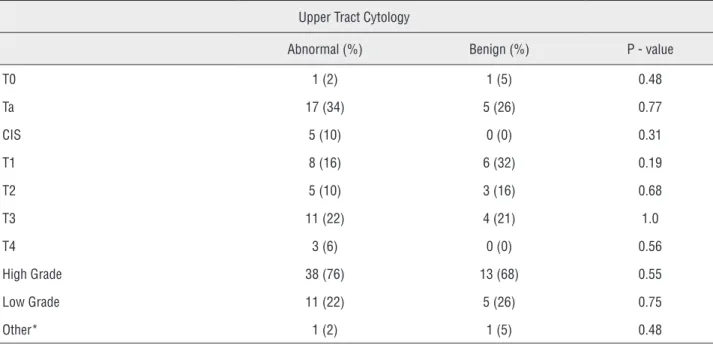
Table 2 - Correlation of UTC with tumor stage and grade.
Upper Tract Cytology
Abnormal (%) Benign (%) P - value
T0 1 (2) 1 (5) 0.48
Ta 17 (34) 5 (26) 0.77
CIS 5 (10) 0 (0) 0.31
T1 8 (16) 6 (32) 0.19
T2 5 (10) 3 (16) 0.68
T3 11 (22) 4 (21) 1.0
T4 3 (6) 0 (0) 0.56
High Grade 38 (76) 13 (68) 0.55
Low Grade 11 (22) 5 (26) 0.75
Other* 1 (2) 1 (5) 0.48
DISCUSSION
The diagnosis of UTUC can be difficult and upper tract selective cytology has been utilized to support the diagnosis of disease presence. With ad-vancement in technology and better understanding of the pathogenesis of UTUC, the role of endoscopic
management has become a more popular treatment option, especially in patients with solitary kidneys, impaired renal function and in patients with mul-tiple comorbidities who are at higher risk during prolonged and more extensive surgeries. Accuracy in diagnosis is therefore especially important and better diagnostic tools may be needed.
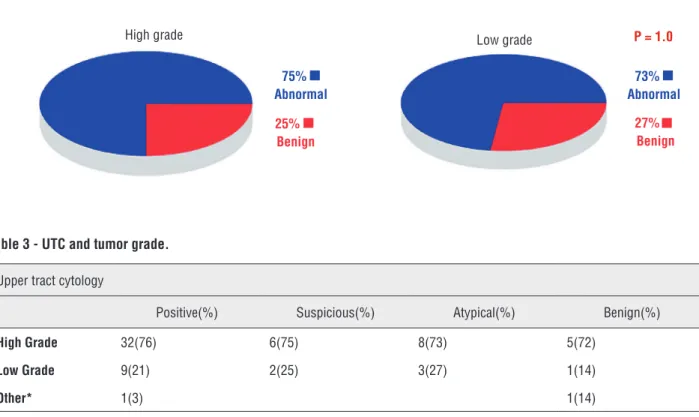
Figure 1 - UTC and tumor grade.
High grade
25% Benign
75% Abnormal
Low grade P = 1.0
27% Benign
73% Abnormal
Figure 2 - UTC and tumor stage.
Muscle invasive tumors Superficial tumors
P = 1.0
28% Benign 27%
Benign
72% Abnormal 73%
Abnormal Table 3 - UTC and tumor grade.
Upper tract cytology
Positive(%) Suspicious(%) Atypical(%) Benign(%)
High Grade 32(76) 6(75) 8(73) 5(72)
Low Grade 9(21) 2(25) 3(27) 1(14)
Other* 1(3) 1(14)
According to our study, 73%, of patients who underwent RNU for UTUC had abnormal UTC with a calculated sensitivity and specifici-ty of 74% and 50%, respectively. The low speci-ficity may be explained by various reasons, for example, lower stage and grade tumors, which may be less prone to shedding tumor cells into the collecting system, or sampling error. Skola-rikos et al. evaluated 62 patients who were tre-ated for UTUC and underwent an ureteroscopic biopsy and/or collection of UTC as a part of their preoperative workup. Only 40% (19/48) of their patients had positive/suspicious UTC but their data demonstrated that positive cytology pre-dicted high grade tumors in 14 (67%) of 21 ca-ses. They also demonstrated improved sensitivity and specificity of detecting high grade UTUC by combining biopsy results and upper tract cytolo-gy. The sensitivity and specificity increased from 43% to 55% and from 23% to 85%, respectively. Their biopsy grade correlated well with tumor in-vasiveness. None of the 6 specimens with biopsy grade 1 compared to 11 of 13 with grade 3 were found to be muscle invasive (5).
A more recent retrospective study by Williams et al. showed a higher prediction rate and correlation with tumor stage of positive upper tract cytology in patients who underwent a nephroureterectomy for UTUC. They correla-ted UTC with histologic findings and 21 (70%) of 30 specimens were positive. Positive UTC was associated with high grade tumors (82%) and predicted stage pT1 or greater in 15 (75%) of 20 cases (6).
Straub and associates investigated the ac-curacy of upper tract cytology and ureteroscopic biopsy in predicting the correct tumor grade in patients with UTUC. Their data showed the sen-sitivity of cytology and biopsy to be 64% and 74%, with a combined sensitivity of 84%. The accuracy of cytology in predicting high grade tumors was 53% which improved to 68% when combined with biopsy results and more impor-tantly 15% of high grade tumors were misinter-preted as being low grade (3). Our findings show that abnormal UTC correlates with the presence of UTUC and was found in 73% of the patients but it did not predict tumor stage or grade
whi-ch is contrary to prior studies. Interestingly, all of the CIS specimens had positive UTC, but the number (N = 5) was too low to reach statistical significance. Our study also demonstrates that superficial upper tract tumors were as likely to have abnormal cytology as muscle invasive tu-mors (72% of superficial and 76% muscle invasi-ve tumors) which emphasizes the importance of combining all preoperative data (cytology, cross--sectional imaging, ureteroscopic findings and biopsy results) when making a decision if endos-copic surgery is a suitable therapeutic option. Xu et al. demonstrated higher sensitivity with VUC in combination with fluorescence in situ hybri-dization analysis (FISH) for detecting upper tract tumors with an overall sensitivity 85.9% com-pared to 45.1% for VUC and 78.9% for FISH (4). Table-4 summarizes published studies focusing on diagnostic utilities of UTC.
The present study has some limitations including its retrospective design and the inhe-rent selection bias for patients to undergo a ne-phroureterectomy and therefore more likely to have higher grade and more advanced disease which may not accurately reflect all patients tre-ated at outside institutions. Moreover, the urine cytology was not routinely graded, and therefore we were unable to correlate the grade of the po-sitive cytology with final pathology.
CONCLUSIONS
The diagnosis of UTUC may be challen-ging and according to our data, selective cytolo-gy correlates with the presence of urothelial car-cinoma but does not accurately predict the stage or grade of these respective tumors. At present, UTC is one of the more important diagnostic to-ols to work up upper tract tumors and should be used in conjunction with other diagnostic moda-lities and clinical findings whenever technically possible. The continual goal for novel biomarkers of diagnostic, therapeutic, and prognostic utility remains a mainstay of current research efforts.
CONFLICT OF INTEREST
REFERENCES
1. Jovanovic M, Soldatovic I, Janjic A, Vuksanovic A, Dzamic Z, Acimovic M, et al.: Diagnostic value of the nuclear matrix protein 22 test and urine cytology in upper tract urothelial tumors. Urol Int. 2011; 87: 134-7.
2. Messer J, Shariat SF, Brien JC, Herman MP, Ng CK, Scherr DS, et al.: Urinary cytology has a poor performance for predicting invasive or high-grade upper-tract urothelial carcinoma. BJU Int. 2011; 108: 701-5.
3. Straub J, Strittmatter F, Karl A, Stief CG, Tritschler S: Ureterorenoscopic biopsy and urinary cytology according to the 2004 WHO classification underestimate tumor grading in upper urinary tract urothelial carcinoma. Urol Oncol. 2013; 31: 1166-70.
4. Xu C, Zeng Q, Hou J, Gao L, Zhang Z, Xu W, et al.: Utility of a modality combining FISH and cytology in upper tract urothelial carcinoma detection in voided urine samples of Chinese patients. Urology. 2011; 77: 636-41.
5. Skolarikos A, Griffiths TR, Powell PH, Thomas DJ, Neal DE, Kelly JD: Cytologic analysis of ureteral washings is informative in patients with grade 2 upper tract TCC considering endoscopic treatment. Urology. 2003; 61: 1146-50.
6. Williams SK, Denton KJ, Minervini A, Oxley J, Khastigir J, Timoney AG, et al.: Correlation of upper-tract cytology, retrograde pyelography, ureteroscopic appearance, and ureteroscopic biopsy with histologic examination of upper-tract transitional cell carcinoma. Endourol. 2008; 22: 71-6. 7. Raica M, Mederle O, Ioiart I: Upper urinary tract washing
in the diagnosis of transitional cell carcinoma of the renal pelvis and calices. A comparison with voided urine. Rom J Morphol Embryol. 1995; 41: 101-5.
8. Keeley FX Jr, Bibbo M, McCue PA, Bagley DH: Use of p53 in the diagnosis of upper-tract transitional cell carcinoma. Urology. 1997; 49: 181-6.
9. Highman WJ: Transitional carcinoma of the upper urinary tract: a histological and cytopathological study. J Clin Pathol. 1986; 39: 297-305.
_______________________ Correspondence address:
Philippe E. Spiess, MD Associate Member, Dept of GU Oncology Moffitt Cancer Center 12902 Magnolia Drive, office 12538 Tampa, FL 33612, USA Fax: +1 813 745-8494 E-mail address: [email protected] Table 4 - Summary of published studies.
Study Number Sensitivity (%) Specificity (%) PPV (%) Accuracy of bx predicting high grade
Raica (7) 34 97 - -
-Keeley (8)* 28 82 - -
-Messer (2)** 168 71 - 53
-Williams (6) 30 70 - - 75%
Skolarios (5) 48 40 - - High****
Highman (9) 24 58 - -
-Xu (4)*** 71 86 97.8 -
-Straub (3) 77 64 - - 58%
Our study 68 74 50 98
-* Additional washes obtained after a biopsy was performed ** Only high grade disease
***Voided cytology and FISH combined