Value of color doppler ultrasound, kub and urinalysis in
diagnosis of renal colic due to ureteral stones
_______________________________________________
Mahmoud Abdel-Gawad
1, Ravi Kadasne
1, Chandrashekar Anjikar
1, Emad Elsobky
21Department of Urology, Emirates International Hospital, Al Ainvand 2Department of Urology, Al-Noor Hospital, Abu Dhabi, United Arab Emirates
ABSTRACT
ARTICLE
INFO
______________________________________________________________ ______________________
Purpose: Despite the routine use of helical CT in diagnosis of renal colic, there are recent concerns regarding the radiation exposure, overuse and costs. We attempted in this retrospective study to evaluate the accuracy of ultrasound (gray-scale and color Doppler with twinkling), KUB and urinalysis in diagnosis of renal colic due to ureteral calculi presented in Emergency Room.
Materials and Methods: A total of 939 consecutive cases of renal colic presented to ER have been managed and evaluated by ureteral ultrasound, KUB and urinalysis for the presence of ureteral stones. Non-confirmatory cases were subjected to Helical CT examination.
Results: Renal and ureteral ultrasound (gray-scale) alone detected ureteral calculi in 615 cases (65.4%) and after utilizing Color Doppler Ultrasound with twinkling the diagnosis was made with confidence in 935 cases (99.6%) but 4 (0.4%). KUB showed radiopaque stones in 503 (53.6%) patients and no stones were detected in 436 (46.4%). Microhematuria presented in 835 (88.9%) cases while absent in 102 (10.9%). There were 190 (20.3%), 77 (8.2%) and 671 (71.5%) patients with upper, middle and lower ureteral stones respectively. The simultaneous positive findings in US and KUB with microhematuria were found only in 453 (48.2%) cases.
Conclusions: The use of Color Doppler ultrasound with twinkling increased the de-tection rate of ureteral stones in acute renal colic patients presented to ER with less radiation exposure. Ultrasound examination as a single modality is superior to KUB and urinalysis in initial diagnosis of renal colic.
Key words:
Renal Colic; Ultrasonography; Calculi; Ureter; Hematuria
Int Braz J Urol. 2014; 40: 513-9
_____________________
Submitted for publication: July 26, 2013
_____________________
Accepted after revision: December 10, 2013
INTRODUCTION
Acute flank pain is one of the most com-mon presentations in ER. CT is generally accepted as the imaging study of choice to evaluate pa-tients with flank pain in emergency department (1). In 1995, Smith et al. described the use of he-lical CT in patients suspected of having acute re-nal colic (2). Recently, there is growing awareness and concerns about the overuse of CT in
a triangular shape when highly evident (4). Many recent studies have reported and encouraged the use of twinkling sign to improve the accuracy of ultrasound to detect renal and ureteral stones (5). In this retrospective study, we evaluated the use of US (gray-scale and Color Doppler), KUB and urine analysis in diagnosis of renal colic due to ureteral calculi in patients presented to ER.
MATERIALS AND METHODS
Patients’ data
After approval of Institutional Review Bo-ard, we conducted this retrospective study by re-viewing the medical records of patients diagnosed with acute renal colic in the emergency room from January 2003 to December 2010. The patients were identified by using ICD-9 codes and were confir-med by charts review. The patients were evaluated for flank pain in the form of general and local examinations. After control of pain, the patient underwent immediate radiological examinations in the form of plain x-ray of the abdomen (KUB) and ultrasound scanning (gray-scale and color Doppler) of both flanks. Microscopic urine exa-mination was performed and more than 3 RBCs/ HPF was considered significant and counted. The findings of KUB were interpreted by two separate independent conventional radiologists who were blinded to the results of the ultrasound findings. US examinations (gray-scale and color Doppler with twinkling) were performed by another two senior ultrasonographists.
Ultrasound Technique
No bowel preparation or full bladder was mandatory as most of the patients arrived with acute renal colic. All US examinations were per-formed by using three models of Ultrasound ma-chines that were available during the study period (Toshiba Power Vision 6000, Model: 370A-SSA, Japan, Toshiba Aplio, Model USEL-790A, Japan and Philips iU22 - Bothell WA, USA). A multi--frequency convex abdominal transducer with frequency of 1 to 6 MHz was used. A diagnostic criterion was direct visualization of the calculus in the ureteric lumen with or without associated hydronephrosis. Symptomatic side was screened
to assess the degree of hydronephrosis and for possible stones in the kidney, pelvis or upper ure-ter. If no definite stone was visualized, the bladder and the vesicoureteral junction were screened for possible calculi. If no stone was identified at these sites, the effort was made to trace the ureter from the renal pelvis till the iliac crossing. Graded com-pression with insinuation helped in localization of the stone at this site by decreasing the distance be-tween the transducer and ureter. Ultrasound scan-ning of full bladder was not necessary in every case as filling of the bladder was not possible in all patients due to severe pain and possible lower ureteric calculus causing frequency of urination; in addition, an over distended bladder hindered detection and limited the amount of pressure that can be applied to the bladder. Once the stone was identified, the gray-scale ultrasound was used to measure the long axis of the stone and its leng-th was recorded. The location of leng-the stone was recorded as upper, middle and lower accordingly. For visualization of the posterior acoustic shado-wing of the stone, focal zones were always placed at the depth of or slightly deeper than the stone level, with careful control of the B-mode gain set-ting. Color Doppler was applied over the site of the stone and twinkling sign was observed, a red and blue color map was used. The color window size was adjusted to cover the concerned area and adjacent tissue. The color Doppler gain was set to the point just below the threshold for color noise. When the twinkling sign was clearly obtained, po-wer Doppler was also used to differentiate betwe-en the surrounding vessels and the ureter. In addi-tion, color Doppler also helped in differentiation between stasis and prominent vessels at the renal hilum in certain cases. If there was no evidence of stones by Doppler ultrasound, then the patient was submitted to helical CT for further evaluation. The patients were followed up for a period of 7-8 weeks. A definite diagnosis was made when the patient passed a stone either spontaneously or af-ter inaf-tervention.
Statistical analysis
retrieved from the medical charts. Data were analyzed by using SPSS version 15.0 (SPSS, Chicago Illinois, USA). The correlation between diagnostic tools (KUB, US), patients’ characteristics (age and sex) and stones characteristics (site, size and side) was performed by using chi-square test. A value of p ≤ 0.05 was consi-dered statistically significant.
RESULTS
Out of 939 patients, there were 825 males (87.9 %) and 114 females (12.1%). Male to female ratio was 7:1. Age range (mean ± SD) was 9-80 years (37.9 ± 11). Our study found that renal ultra-sound (gray-scale) when used alone detected calcu-li in 615 cases (65.4%) and after uticalcu-lizing CDU with twinkling sign, the diagnosis was made with con-fidence in 935 cases (99.6%) which is statistically significant (p < 0.05) (Table-1) and the twinkling was consistent in all confirmed cases (Figures 1A, 1B and 2). The overall sensitivity and specificity of CDU were 99.6% and 100% respectively. There were 4 (0.4%) patients in whom the stones could not be found neither by gray scale or color Dop-pler ultrasound and were not seen in KUB but the clinical scenario of pain and microhematuria war-ranted the employment of helical CT. In the latter group, the stones were confirmed by helical CT. The stones were located in the lower ureter in all 4 pa-tients and the size of stones ranged from 3-5mm. The KUB showed radiopaque stones in 503 (53.6%) cases and no stones were visualized in 436 patients (46.4%) (Table-2). Microhematuria was present in 835 cases (88.9%) with specificity of 89.1%, while in
Table 1 - Detection rate and distribution of ureteral stones in 935 cases (bilateral in 4 cases) after comparison between gray scale ultrasound and color Doppler ultrasound.
Gray-scale ultrasound Color Doppler ultrasound
Site Number % Number %
Upper 105 11.1 % 190 20.3 %
Middle ureter 30 43.1 % 77 8.2 %
Lower 480 51.2 % 672 71.5 %
Total 615 (65.4%) 939 (99.6%)
Figure 1A - Grey scale ultrasound of stone in the left middle ureter with posterior shadowing.
102 cases (10.9%) the urine was negative (Table-3) . There were 190 (20.3%), 77 (8.2%) and 671 (71.5%) upper, middle and lower ureteral stones respecti-vely (Table-1). Right side stones were seen in 438 (46.7%) patients while left side stones were seen in 499 (53.3%). Stones were detected bilaterally in 4 cases (0.4%). The range of stones size (mean ± SD) was 3-26mm (7.5 ± 2.7). Simultaneous positive findings in US and KUB combined with microhe-maturia were found only in 453 (48.2%) cases. US (gray and color Doppler) results were independent from any other factor while KUB results of stones were significantly related to site, side, size of sto-nes, age of patient and urine analysis (p = 0.000, 0.042, 0.005, 0.000, 0.016 respectively). Microhe-maturia was related to stone size (p = 0.008).
Table 2 - Site and size of ureteral stones by KUB in 939 cases.
Site Radiopaque % Radiolucent % Mean stone
Size (mm)
Upper 139 77.6 % 51 11.7 % 11.7 ± 1.8
Middle ureter 42 8.3 % 35 8.1 % 7.1±0.8
Lower 322 64.1 % 350 80.2 % 7.6 ± 0.29
503 (53.6%) 436 (46.4%)
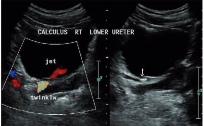
Figure 2 - Stone in the lower end of the ureter by gray scale ultrasound and the twinkling sign after the application of the color Doppler ultrasound.
DISCUSSION
The diagnostic approach of acute flank pain is controversial and can vary from center to center, city to city or country to country depen-ding on what is considered acceptable. Important factors include the local prevalence of stone dise-ase, medical resources available, relative costs in a particular system, and the merits and limitations of each diagnostic modality (6). Although CT is the gold standard for diagnosis of renal colic, it is not available outside hospital facilities and is costly (7). There are recent concerns regarding the radiation exposure during CT examinations and its indiscriminate use. Moreover, many patients
may receive an additional radiation dose during follow-up studies (if a calculus is not expelled) or with new episode of colic and the cumulative effective doses of radiation from imaging proce-dures increased with advancing age (8,9).
equipments and technologies (13).In our study, gray-scale ultrasound examination alone detected ureteral calculi in 65.4% of patients presented to ER with low specificity. The twinkling artifact was first described in 1996 by Rahmouni et al., as a sign generated by a highly reflective object that, despite being stationary, would generate a rapid alteration between red and blue when interroga-ted with color Doppler sonography (4). Although it was first described many years ago, the diagnos-tic value of twinkling in renal lithiasis is generally unacknowledged (14,15). In the present study, the gray-scale Ultrasound sensitivity and specificity increased significantly when CDU was used with twinkling, where 99.6% of stones could be located with great confidence which is similar to results of recent studies (5,9). Regarding the echographic identification of ureterolithiasis, there are nume-rous studies with quite contradictory results and with a very wide range of values, the sensitivity of the method ranging between 19% and 96% (16-18). Our study showed the echographic identifica-tions of ureteral calculi in 65.4% only. As there are different stone sizes and shapes, the twinkling sign can not differentiate between different types of stones albeit the results were consistent and in-dependent of stones sizes. In 4 cases of our stu-dy (false negative), localization of ureteral stones could not be defined neither after using gray scale nor application of CDU, afterward helical CT lo-calized the stones at lower ureter. These patients were obese and their BMI was over 35. Similarly, Lee et al. reported that 4 of 20 renal stones and 2 of 16 ureteral stones did not show any twinkling sign. The authors suggested that ureteral stones may be influenced more than renal stones by ul-trasonic attenuation of interposed tissues because the ureter is deeply seated below abundant
fat-ty tissue without a proper acoustic window (19). Others suggested reservation of CT for those pa-tients who first have negative or equivocal results for KUB and sonography and this notion was ap-plied in some of our cases (20). The size of stones in our study ranged from small sized to large ones (3-26mm) with twinkling artifact consistently ge-nerated in 99.6% of cases which was independent of stone size and site. In the same way, Park et al. have documented the usefulness of the twinkling artifact in confirming the presence of small stones in 86% of cases (9). The application of CDU with twinkling needed a systematic screening of the ureter from the renal pelvis till the vesicoureteral junction with graded compression on the ureter especially if the stone is suspected in the mid-ure-ter or at the crossing of iliac vessels to reduce the distance between transducer and the ureter. This maneuver is similar to Puylaert technique of gra-duated compression in diagnosis of acute appen-dicitis (21). In recent studies, one group waited for 24 hours then scanned the patient, another study prepared the patient before ultrasound examina-tion with intravenous drip infusion (5,9). Conver-sely, in our series the patients were scanned within 2 hours of presentation to ER without special pre-paration or specific protocols of hydration. The-refore, this uniformity of ultrasound examination minimized the possibility of changes in stone loca-tion or the degree of ureteral dilataloca-tion that could affect the twinkling artifact quality and detection. Moreover, we did not find any association between the location of the stone and twinkling genesis. The twinkling generation was not related to ureteral di-latation or hydronephrosis. Lee et al. have reported the same conclusion (19).
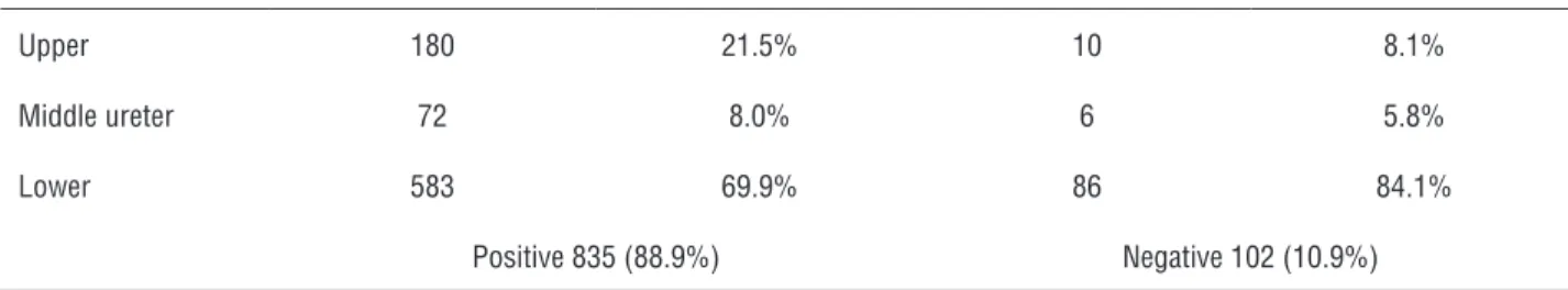
In our study we found that 20.3% of the stones were located in upper ureter and only 8.2% Table 3 - Relation between microhematuria and stone site in Color Doppler examined cases.
Upper 180 21.5% 10 8.1%
Middle ureter 72 8.0% 6 5.8%
Lower 583 69.9% 86 84.1%
in the middle ureter while most (71.5%) were situ-ated in the lower ureter. Saita et al. determined the success rates of US according to the localization of the stone and they reported success rates of 82.2% in the proximal and 68% in the distal ureter (20). Other authors reported that approximately 65% of acutely presenting calculi impact in the lower seg-ment of the ureter, therefore US has the potential to provide diagnostic follow up in a substantial number of individuals (22). Interestingly, we have used three ultrasound machines of different models and the results of twinkling artifact were reprodu-cible. Although Aytac et al. (15) reported that the twinkling sign depends on the color sensitivity and the acoustic output of the ultrasound unit,we did not notice such observation.
KUB, when used alone in renal colic, is of limited diagnostic value with a sensitivity of 53-62% and specificity of 67-69% for the detection of ureteral calculi; in our series KUB detected radio-paque stones in 53.6% of renal colic cases which is similar to others (23,24). KUB was sufficient to document the size and site of radiopaque calculi as upper ureteral stones were more common than lower ureteral stones in this study (p < 0.001). Ho-wever, 46.4% of stones were not visible in x-ray of our cases. KUB in young patients was signi-ficantly better in detection of stones than older age group (p = 0.005). This improvement may be either due to less soft tissue density or stone types but its significance should be weighed against ex-penses of radiation exposure.
Urinalysis has been widely accepted as a standard test for diagnosing acute renal colic, unli-kely the incidence of negative hematuria in patients with ureterolithiasis has been reported to be 9% to 33% (25,26). In our study, microhematuria was de-tected in 88.9% of patients and absent in 10.9%. We also have observed that microscopic hematuria was common with lower ureteral stones (69.6%) and was related to stone size and noteworthy, was com-mon with radiopaque than radiolucent stones (p = 0.016). Interestingly, in this retrospective study the urinalysis was requested at the initial presentation of renal colic to minimize possibility of false nega-tive results since other authors found the incidence of negative hematuria is highest on the days 3 and 4 after initial presentation of renal colic (27).
Finally, there are some limitations of our study; it is a retrospective with its inherited shor-tcomings, the ultrasound did not show the associa-tion between the twinkling signs and the type of the stones, lastly we did not report on the outcome of renal colic cases despite the sufficient period of follow-up. However, our study was designed to determine the value of CDU with twinkling in the initial diagnosis of acute renal colic and is the largest cohort of renal colic patients diagnosed by using this technique. We feel that these encoura-ging results are of potential usefulness in the cli-nical practice with less cost, easy availability and mobility of ultrasound.
CONCLUSIONS
The present study emphasized that utiliza-tion of color Doppler ultrasound with twinkling in trained hands can provide an excellent alternative modality with high sensitivity and specificity in diagnosis of acute renal colic and should be em-ployed at initial presentation in ER.
ABBREVIATIONS
KUB = Kidney, Ureter, Bladder
CDU = Color Doppler Ultrasound
CT =Computed Tomography
ER = Emergency Room
CONFLICT OF INTEREST
None declared.
REFERENCES
1. Ahmad NA, Ather MH, Rees J: Unenhanced helical computed tomography in the evaluation of acute flank pain. Int J Urol. 2003; 10: 287-92.
2. Smith RC, Levine J, Dalrymple NC, Barish M, Rosenfield AT: Acute flank pain: a modern approach to diagnosis and management. Semin Ultrasound CT MR. 1999; 20: 108-35. 3. Ferrandino MN, Bagrodia A, Pierre SA, Scales CD Jr,
4. Rahmouni A, Bargoin R, Herment A, Bargoin N, Vasile N: Color Doppler twinkling artifact in hyperechoic regions. Radiology. 1996; 199: 269-71.
5. Mitterberger M, Aigner F, Pallwein L, Pinggera GM, Neururer R, Rehder P, et al.: Sonographic detection of renal and ureteral stones. Value of the twinkling sign. Int Braz J Urol. 2009; 35: 532-9; discussion 540-1.
6. Shokeir AA, Abdulmaaboud M: Prospective comparison of nonenhanced helical computerized tomography and Doppler ultrasonography for the diagnosis of renal colic. J Urol. 2001; 165: 1082-4.
7. Grisi G, Stacul F, Cuttin R, Rimondini A, Meduri S, Dalla Palma L: Cost analysis of different protocols for imaging a patient with acute flank pain. Eur Radiol. 2000; 10: 1620-7. 8. Levine JA, Neitlich J, Verga M, Dalrymple N, Smith RC;
Radiology: Ureteral calculi in patients with flank pain: correlation of plain radiography with unenhanced helical CT. 1997; 204: 27-31.
9. Park SJ, Yi BH, Lee HK, Kim YH, Kim GJ, Kim HC: Evaluation of patients with suspected ureteral calculi using sonography as an initial diagnostic tool: how can we improve diagnostic accuracy? J Ultrasound Med. 2008; 27: 1441-50.
10. Aslaksen A, Göthlin JH: Ultrasonic diagnosis of ureteral calculi in patients with acute flank pain. Eur J Radiol. 1990; 11: 87-90.
11. Katz DS, Lane MJ, Sommer FG: Non-contrast spiral CT for patients with suspected renal colic. Eur Radiol. 1997; 7: 680-5. 12. Assi Z, Platt JF, Francis IR, Cohan RH, Korobkin M:
Sensitivity of CT scout radiography and abdominal radiography for revealing ureteral calculi on helical CT: implications for radiologic follow-up. AJR Am J Roentgenol. 2000; 175: 333-7.
13. Patlas M, Farkas A, Fisher D, Zaghal I, Hadas-Halpern I: Ultrasound vs CT for the detection of ureteric stones in patients with renal colic. Br J Radiol. 2001; 74: 901-4. 14. Kamaya A, Tuthill T, Rubin JM: Twinkling artifact on color
Doppler sonography: dependence on machine parameters and underlying cause. AJR Am J Roentgenol. 2003; 180: 215-22. Erratum in: AJR Am J Roentgenol. 2003; 180: 54. 15. Aytaç SK, Ozcan H: Effect of color Doppler system on the
twinkling sign associated with urinary tract calculi. J Clin Ultrasound. 1999; 27: 433-9.
16. Yilmaz S, Sindel T, Arslan G, Ozkaynak C, Karaali K, Kabaalioğlu A, et al.: Renal colic: comparison of spiral CT, US and IVU in the detection of ureteral calculi. Eur Radiol. 1998; 8: 212-7.
17. Ripollés T, Agramunt M, Errando J, Martínez MJ, Coronel B, Morales M: Suspected ureteral colic: plain film and sonography vs unenhanced helical CT. A prospective study in 66 patients. Eur Radiol. 2004; 14: 129-36.
18. Sheafor DH, Hertzberg BS, Freed KS, Carroll BA, Keogan MT, Paulson EK, et al.: Nonenhanced helical CT and US in the emergency evaluation of patients with renal colic: prospective comparison. Radiology. 2000; 217: 792-7. 19. Lee JY, Kim SH, Cho JY, Han D: Color and power Doppler
twinkling artifacts from urinary stones: clinical observations and phantom studies. AJR Am J Roentgenol. 2001; 176: 1441-5.
20. Saita H, Matsukawa M, Fukushima H, Ohyama C, Nagata Y: Ultrasound diagnosis of ureteral stones: its usefulness with subsequent excretory urography. J Urol. 1988; 140: 28-31. 21. Puylaert JB: Acute appendicitis: US evaluation using graded
compression. Radiology. 1986; 158: 355-60.
22. Eisner BH, Reese A, Sheth S, Stoller ML: Ureteral stone location at emergency room presentation with colic. J Urol. 2009; 182: 165-8.
23. Dalla Palma L, Stacul F, Bazzocchi M, Pagnan L, Festini G, Marega D: Ultrasonography and plain film versus intravenous urography in ureteric colic. Clin Radiol. 1993; 47: 333-6. 24. Svedström E, Alanen A, Nurmi M: Radiologic diagnosis of
renal colic: the role of plain films, excretory urography and sonography. Eur J Radiol. 1990; 11: 180-3.
25. Press SM, Smith AD: Incidence of negative hematuria in patients with acute urinary lithiasis presenting to the emergency room with flank pain. Urology. 1995; 45: 753-7. 26. Li J, Kennedy D, Levine M, Kumar A, Mullen J: Absent
hematuria and expensive computerized tomography: case characteristics of emergency urolithiasis. J Urol. 2001; 165: 782-4.
27. Kobayashi T, Nishizawa K, Mitsumori K, Ogura K: Impact of date of onset on the absence of hematuria in patients with acute renal colic. J Urol. 2003; 170(4 Pt 1): 1093-6.
_______________________ Correspondence address: