Key words:
Female; Ureteral Obstruction; Urodynamics; Urinary Incontinence
Int Braz J Urol. 2013; 39: 498-505
__________________
Submitted for publication: January 30, 2013
__________________
Accepted after revision: June 14, 2013
Objective: To analyze the benefit of voiding chain cystourethrography (VCC) [placing a radiographic opaque chain into the urethra and bladder and asking the patient to void un-der fluoroscopy] in the urodynamic evaluation of female bladun-der outlet obstruction (BOO). Materials and Methods: Females with post anti-incontinence operation voiding dys-function who underwent urodynamic evaluation augmented with VCC and later had urethrolysis were identified. Six diagnostic criteria for obstruction were applied to each patient: (1) VCC ( obstructed: chain was angulated and could not be voiding out) (2) Video urodynamic study (VUDS) (detrusor contraction combined with radiographic obstruction) (3) maximum flow (Qmax) ≤ 15 cc/sec, detrusor pressure (pDet)@ Qmax≥ 20 cm H20 (4) Qmax≤ 11 cc/sec, pDet@ Qmax≥ 25 cm H20 (5) Qmax≤ 12 cc/sec, pDet@ Qmax
≥ 25 cm H20 (6) Blaivas-Groutz (B-G) nomogram. Urethrolysis results were reviewed. Agreement in assessment of BOO criteria was assessed by estimating the proportion of pair-wise agreements along with an exact binomial 95% confidence interval (CI) and by estimating kappa along with a 95% CI.
Results: Twenty-one patients were identified. Twenty of the 22 urethrolyses (91%) were clinically successful. Diagnosis of BOO was most common for VCC (86%) and then B-G Nomogram (67%). Agreement with the VCC was relatively poor for each of the five other methods (14%-62%) with the video urodynamic study (VUDS) being the best. Three patients with successful urethrolysis were diagnosed only by the VCC. All of kappa values regarding agreement with the VCC were low; the highest value of 0.15 was observed for VUDS.
Conclusion: VCC may augment selection criteria for urethrolysis.
INTRODUCTION
Selection for urethrolysis in symptomatic females with a history of an anti-incontinence procedure may be challenging, especially in the absence of immediate temporal association and/ or with a history of previous failed urethrolysis. While recognizing the value of the various pressu-re flow criteria developed for female bladder
ou-tlet obstruction (BOO), our clinic relies principally on the video urodynamic appearance of the blad-der and urethra with voiding as described by Nitti et al. (1) and the Blaivas Groutz (B-G) nomogram (2). In some patients with a high index of clinical suspicion, the pressure flow criteria for obstruc-tion were not strictly met and/or it was difficult to identify the urethra during testing. One exam-ple would be patients that voided with minimal or
Voiding Chain Cystourethography: Assessing A Historical
Test’s Role in Selection for Urethrolysis
_______________________________________________
Steven P. Petrou, Karen Ryan, Dorothea Metz-Kudashick, Michael G. Heckman, David D. Thiel
Department of Urology (SPP, DDT); Department of Radiation and Oncology (KR), and Section of Biostatistics (MGH) Mayo Clinic, Jacksonville, Florida, USA; No affiliation (DMK)
ABSTRACT
ARTICLE
INFO
no detrusor contraction. To augment the evalua-tion and radiographic appearance of the urethral and bladder anatomy we incorporated the use of a historical test, the voiding chain cystourethro-gram (VCC). We report on our experience using this classic historical test (3) in selection for ure-throlysis.
MATERIALS AND METHODS
IRB approval was obtained for this re-trospective review of 21 patients who underwent urethrolysis performed by a single surgeon and had VCC as part of their video urodynamic study (VUDS) for voiding dysfunction following an anti--incontinence procedure. All patients underwent a complete preoperative clinical and videourodyna-mic evaluation including a catheter free uroflow and catheterized post-void residual videourody-namics with pressure flow analysis, and fluoros-copically visualized VCC.
Urodynamic Evaluation
Video urodynamic study (VUDS) was completed using a multi-channel 7F transurethral catheter and a rectal air charged catheter. The filling portion of the cystogram was performed at 50 mL per minute using contrast medium. Whe-re clinically needed (e.g. seveWhe-re sensory urgency/ detrusor overactivity/small capacity bladder), the fill rate was reduced to 12.5 - 25 mL per minu-te. Intermittent fluoroscopic imaging was utilized to quantify trabeculation, vesicoureteral reflux, and voiding anatomy. During the voiding portion of the study, the patient was asked to void to the best of her ability. If the patient was unable to generate an uroflow, the maximum detrusor pressure generated was quantified. Notation was made of the patient’s voiding characteristics such as voiding with a detrusor contraction, Valsalva maneuvers, or a combination of both. The pa-tient then underwent VCC using the below tech-nique.
Voiding chain cystography technique
The urodynamics (UDS) catheter was removed, and the bladder was filled with ap-proximately 250ml of diluted Omnipaque (G.E.
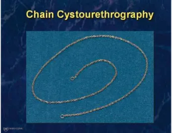
Healthcare, Inc., 25 ml contrast with 100 ml ste-rile water) using a steste-rile 14Fr catheter. Dilution optimized visualization of the chain through the contrast. After the instillation catheter was remo-ved, the cystourethrography chain was dipped in 2% lidocaine and inserted into the urethra using a rounded forceps. The chain used was a 14kt gold (rhodium-plated) link chain measuring 60 mm in length and 2.15 mm in width; it weighs approximately 7.40 grams and has one round link on each end; the end links have an outsi-de diameter of 5.00 mm and an insioutsi-de diameter of approximately 3.20 mm (Figure-1). The chain was secured with tape loosely to the inner leg.
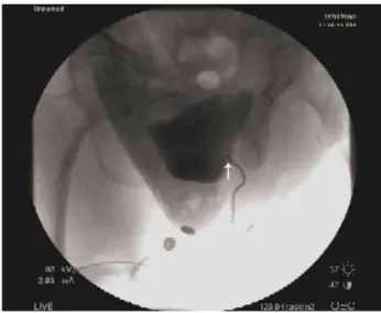
The UDS table was elevated to 90 degrees and the patient was turned to the side, using flu-oroscopy to check chain placement. The patient was radiographically examined while coughing, with Valsalva maneuvers, and with voiding. The VCC was used to identify the bladder, bladder neck, and urethral position (including the pre-sence or abpre-sence of urethral angulation) during voiding, and notation was made whether the pa-tient was able to void the chain out of the bla-dder with micturition (Figures 2-4). The VUDS data was retrospectively analyzed separately by multiple criteria for female BOO.
Criteria for obstruction:
1. UDS obstruction: radiographic evidence for obstruction between the bladder and distal urethra with a sustained detrusor contrac-tion (1,4);
2. Maximum flow (Qmax) ≤ 15 cc/sec, detrusor pressure (pDet) @ Qmax≥ 20 cm H20 (Pressu-re Flow Criterion 1) (5);
3. Qmax≤ 11 cc/sec, pDet @ Qmax≥ 25 cm H20 (Pressure Flow Criterion 2) (6);
4. Qmax≤ 12 cc/sec, pDet @ Qmax≥ 25 cm H20 (Pressure Flow Criterion 3) (7);
5. Blaivas-Groutz nomogram obstructed (2); 6. VCC obstructed: a combination of
radio-graphic chain angulation and inability to void the chain out on command (Figures 2a, 2b and 3).
Successful urethrolysis was determined by resolution of presenting urinary complaints and normalization of physical examination in-cluding the absence of urethral tethering.
Statistical analysis
Post-void residual values were compared before and after surgery using a Wilcoxon signed rank test.
Agreement in assessment of BOO between the VCC and each of the five other methods was assessed by estimating the proportion of pair-wi-se agreements along with an exact binomial 95% confidence interval (CI), and also by estimating kappa along with a 95% CI. All statistical analy-ses were performed using SAS (version 9.2; SAS Institute, Inc., Cary, North Carolina).
Figure 2a and 2b - shows the chain in position for the VCC and then the chain angulation with the attempted voiding during the VCC. Figure 2a – white arrow shows chain in urethra with the patient relaxed. Figure 2b - white arrow shows angulation of chain, consistent with obstruction during voiding attempt with VCC.
Figure 3 - Demonstrates chain angulation from a VCC test that is consistent with obstruction. White arrow shows chain angulation consistent with obstruction from patient with previous suburethral sling.
RESULTS
A total of 21 female patients who un-derwent a video urodynamic study for voiding dysfunction and went on to have urethrolysis were considered for inclusion in this study. Median age was 65 years (Range: 43 - 83 years). One patient in the series had an unsuccessful urethrolysis and underwent a second successful urethrolysis; only the set of measurements corresponding to the first urethrolysis were considered in these analyses in order to satisfy the statistical assumption of inde-pendent measurements. Twenty of the 22
opera-Figure 4a, 4b, and 4c - Reveals a VCC test that is consistent with no obstruction: the chain in place, voiding without angulation and then being voided from the bladder. Figure 4a reveals chain in urethra with the patient relaxed, with no angulation. Figure 4b shows patient beginning to void, white arrow shows no angulation. Figure 4c – white arrow shows chain being expelled from urethra with voiding, consistent with no obstruction.
tions (91%) were classified as having a clinically successful urethrolysis based on postoperative cli-nical evaluation and return of normal voiding ha-bits. Preoperative post-void residual assessment revealed a median of 115 cc’s (Range: 10 - 720), with a mean of 178 cc’s. For the postoperative post-void residual measurements, the median was 50 cc’s (Range: 3 - 170), and the mean of 57 cc’s. When evaluating the difference between the post vs. presurgical values, the median difference was -76 (Range: -640 to 43), and the mean was -120. There was strong evidence of a difference between the pre and post measures (P < 0.001).
A B
A summary of assessments of BOO for each of the six different methods is provided in Table-1. An assessment of BOO was most common for the VCC (86%), followed by the B-G Nomogram (67%). One patient was rated as obstructed by all six methods; she later had a successful urethroly-sis. Four patients had no detrusor contraction no-ted on urodynamic evaluation but were obstrucno-ted by VCC, three had successful urethrolysis and one patient did not.
An evaluation of agreement regarding BOO between the VCC and each of the five other methods is displayed in Table-2. Agreement with the VCC was relatively poor for each of the five individual methods but was best for the VUDS, where assessment of obstruction was the same as
the VCC for 13 of the 21 patients (62%). The B-G nomogram had the next highest agreement with the VCC; assessment of obstruction was the same as the VCC for 11 patients (52%). Agreement with the VCC was particularly poor for Qmax ≤ 15 cc/ sec, pDet@ Qmax ≥ 20 cm H20, Qmax ≤ 11 cc/sec, pDet@ Qmax ≥ 25 cm H20, and Qmax ≤ 12 cc/sec, pDet@ Qmax ≥ 25 cm H20, where assessment of obstruction was the same as the VCC for between 14% and 33% of patients. All values of kappa re-garding agreement with the VCC were quite low; the highest value of 0.15 was observed for VUDS.
Although it is of interest to evaluate whe-ther any of the six measures of BOO are associated with the unsuccessful urethrolysis patients (2/22), such analysis was impossible due to the very small
Table 1 - Assessment of bladder outlet obstruction by six different methods.
Method No. (%) of assessments of bladder outlet obstruction (N = 21)
VCC 18 (86%)
VUDS 12 (57%)
Qmax≤ 15 cc/sec, pDet@ Qmax≥ 20 H20 8 (38%)
Qmax≤ 11 cc/sec, pDet@ Qmax≥ 25 H20 7 (33%)
Qmax≤ 12 cc/sec, pDet@ Qmax≥ 25 H20 4 (19%)
B-G nomogram 14 (67%)
Table 2 - Agreement in assessment of bladder outlet obstruction between the VCC and five other methods.
Agreement with VCC assessment of obstruction
Measure of obstruction Kappa (95% CI) Fraction (%)
of agreement
95% CI
VUDS 0.15 (-0.19, 0.49) 13/21 (62%) 38% - 82%
Qmax≤ 15 cc/sec,pDet@ Qmax≥ 20 H20 -0.14 (-0.42, 0.14) 7/21 (33%) 15% - 57%
Qmax≤ 11 cc/sec, pDet@ Qmax≥ 25 H20 -0.15 (-0.43, 0.13) 6/21 (29%) 11% - 52%
Qmax≤ 12 cc/sec, pDet@ Qmax≥ 25 H20 -0.19 (-0.47, 0.10) 3/21 (14%) 31% - 36%
number of patients who experienced this outcome. For the first patient with unsuccessful urethrolysis, the only test with an assessment of obstruction was the VCC, while the only test with an assessment of obstruction for the second patient was the VUDS.
DISCUSSION
The challenge in diagnosing BOO in fema-les is well documented in the literature with the symptoms being quite variable (i.e. obstructive, irritative, or a combination of both) (8-11). Fur-ther complicating matters is the variable personal nature of female voiding habits. Consequently, physicians often rely on the temporal association of voiding dysfunction to an anti-incontinence operation to initiate evaluation and/or offer the-rapy (8,12,13). When patient history or time fra-me does not offer this diagnostic advantage, more thorough evaluation is warranted than just proce-eding to urethrolysis, especially if she has already had a history of that operation (14). The complex nature of female voiding dysfunction may require additional parameters besides pressure-flow crite-ria (1-7,15). We desired to see if the VCC, as a non-pressure-flow based test, would assist in po-sitively predicting those patients that would have a successful urethrolysis. Radiographic imaging will potentially not clearly illuminate a urethral obstruction, secondary to the air, soft tissue in-terface or the absence of a well-defined detrusor contraction (16). Consequently, we tried to over-come this problem by resurrecting the use of the urethral chain to look for an angulation or point of obstruction. Past physicians have described the use of the urethral chain when evaluating fema-le voiding dysfunction. Stevens and Smith noted that in certain settings, placing a chain into the urethra may provide distinct advantages over the use of contrast medium while others noted that a chain may be less distorting than a rubber catheter in assessing urethrovesical anatomy (17,18). Chain cystogram was described by Hodgkinson in 1953 to study urethrovesical relationships in order to classify types of female stress incontinence (19). However, it was later discovered that chain cysto-gram alone had a 50 percent margin of error and 65 percent of continent controls had positive
fin-dings (16). Nevertheless, we found the chain was an excellent adjunct for assisting with urethral radiographic visualization. We came to learn that not only was the configuration of the chain im-portant, but that the ability to void the chain out was an added finding when dealing with obstruc-tion: the ability to push the chain out served as a proxy to gauge voiding efficacy in patients that voided with abdominal straining and not a true detrusor contraction. That four of the 21 patients evaluated failed to generate a detrusor contrac-tion during urodynamic testing but were obstruc-ted by VCC criteria with three having a successful urethrolysis reveals the potential value of the test capturing treatable patients by, at the minimum, reinforcing clinical intuition.
VCC definition of obstruction itself had the highest agreement with the video urodynamics definition of obstruction (1,4). This may be due to the similarity between the tests as a urethral ima-ging based study. Table-1 confirms that 18 of the 21 patients that underwent urethrolysis were rated as non-obstructed by at least one of the methods, but VCC rated the fewest number of patients as non-obstructed (3/21). In one of those three cases, the chain was radiographically angulated, but the patient was able to void the chain out with di-fficulty and concentrated effort thus technically making the VCC test negative (which required failure to expel the chain with voiding). Despite that observation, secondary to temporal associa-tion, physical exam and other criteria positivity, the patient was offered and underwent a success-ful urethrolysis. In another patient with negative criteria including the VCC but with a positive B-G nomogram, surgery again was offered secondary to clinical suspicion of temporal association. She underwent a later unsuccessful urethrolysis.
and were thus looking for a test to confirm our intuition. Postoperative pressure-flow urodyna-mic studies would have been of value to further evaluate the sensitivity of the test for obstruction; nevertheless, this was not able to be completed secondary to this study being a retrospective re-view and the lack of desire of patients to undergo testing in the event of a good clinical result at the time of their treatment course.
The variable nature of female voiding pat-terns solidifies the drive for diagnostic accuracy and confirmation of clinical suspicion. Patients re-adily grasp both the concept of radiographic chain angulation when gazing upon the X-ray monitor and the ability/inability to successfully void the chain. Perhaps the VCC should not return from its historical past as a standalone test but to be resur-rected when needed and combined with the other methods of evaluation. We will continue to utilize VCC secondary to the above reasoning when fa-ced with clinical suspicion and variable findings of obstruction on urodynamic evaluation (15).
CONCLUSIONS
The voiding chain cystourethrogram may help confirm clinical suspicion and serve as an adjunct to urodynamic testing in female patients with post-surgical voiding dysfunction when se-lecting for urethrolysis.
ABBREVIATIONS
BOO = Bladder outlet obstruction B-G = Blaivas-Groutz;
CI = confidence interval pDet = detrusor pressure Qmax = maximum flow UDS = Uurodynamics
VUDS = Video urodynamic study
VCC = voiding chain cystourethrography
ACKNOWLEDGMENTS
Funding for the statistical analysis in this manuscript was provided by the Department of Urology at Mayo Clinic, Jacksonville, FL, USA
CONFLICT OF INTEREST
None declared.
REFERENCES
1. Akikwala TV, Fleischman N, Nitti VW: Comparison of diagnos-tic criteria for female bladder outlet obstruction. J Urol. 2006; 176: 2093-7.
2. Blaivas JG, Groutz A: Bladder outlet obstruction nomogram for women with lower urinary tract symptomatology. Neurou-rol Urodyn. 2000; 19: 553-64.
3. Barnes AC: The roentgenologic study of urethral sphincter strength in the female. J Urol. 1942; 47: 694-701.
4. Nitti VW, Tu LM, Gitlin J: Diagnosing bladder outlet obstruc-tion in women. J Urol. 1999; 161: 1535-40.
5. Chassagne S, Bernier PA, Haab F, Roehrborn CG, Reisch JS, Zimmern PE: Proposed cutoff values to define bladder outlet obstruction in women. Urology. 1998; 51: 408-11.
6. Lemack GE, Zimmern PE: Pressure flow analysis may aid in identifying women with outflow obstruction. J Urol. 2000; 163: 1823-8.
7. Defreitas GA, Zimmern PE, Lemack GE, Shariat SF: Refin-ing diagnosis of anatomic female bladder outlet obstruction: comparison of pressure-flow study parameters inclinically ob-structed women with those of normal controls. Urology. 2004; 64: 675-9; discussion 679-81.
8. Gomelsky A, Nitti VW, Dmochowski RR: Management of ob-structive voiding dysfunction after incontinence surgery: les-sons learned. Urology. 2003; 62: 391-9.
9. Groutz A, Blaivas JG, Chaikin DC: Bladder outlet obstruction in women: definition and characteristics. Neurourol Urodyn. 2000; 19: 213-20.
10. Massey JA, Abrams PH: Obstructed voiding in the female. Br J Urol. 1988; 61: 36-9.
11. Bradley CS, Rovner ES: Urodynamically defined stress urinary incontinence and bladder outlet obstruction coexist in women. J Urol. 2004; 171: 757-60; discussion 760-1.
12. Scarpero HM, Dmochowski RR, Nitti VW: Repeat urethroly-sis after failed urethrolyurethroly-sis for iatrogenic obstruction. J Urol. 2003; 169: 1013-6.
13. Lemack GE: Urodynamic assessment of bladder-outlet ob-struction in women. Nat Clin Pract Urol. 2006; 3: 38-44. 14. Thiel DD, Pettit PD, McClellan WT, Petrou SP: Long-term
uri-nary continence rates after simple sling incision for relief of urinary retention following fascia latapubovaginal slings. J Urol. 2005; 174: 1878-81.
16. Greenwald SW, Thornbury JR, Dunn LJ: Cystourethrography as a diagnostic aid in stress incontinence. An evaluation. Ob-stet Gynecol. 1967; 29: 324-7.
17. Stevens W, Smith SP: Roentgenological examination of the female urethra. J Urol. 1936; 37: 194-201.
18. Hodgkinson CP: Stress urinary incontinence in the female. Surg Gynecol Obstet. 1965; 120: 595-613.
19. Hodgkinson CP: Relationships of the female urethra and bladder in urinary stress incontinence. Am J Obstet Gynecol. 1953; 65: 560-73.
_____________________
Correspondence address: