Key words:
Renal Blood Flow, Effective; Ischemia; Infrared Rays; Thermometers
Int Braz J Urol. 2013; 39: 572-8 __________________
Submitted for publication: January 22, 2013
__________________ Accepted after revision: July 02, 2013
Purpose: To evaluate infrared thermometer (IRT) accuracy compared to standard digital thermometer in measuring kidney temperature during arterial clamping with and without renal cooling.
Materials and Methods: 20 pigs weighting 20Kg underwent selective right renal arterial clamping, 10 with (Group 1 - Cold Ischemia with ice slush) and 10 without renal cooling (Group 2 - Warm Ischemia). Arterial clamping was performed without venous clamping. Renal temperature was serially measured following clamping of the main renal artery with the IRT and a digital contact thermometer (DT): immediate after clamping (T0), after 2 (T2), 5 (T5) and 10 minutes (T10). Temperature values were expressed in mean, standard deviation and range for each thermometer. We used the T student test to compare means and considered p < 0.05 to be statistically significant.
Results: In Group 1, mean DT surface temperature decrease was 12.6 ± 4.1ºC (5-19ºC) while deep DT temperature decrease was 15.8 ± 1.5ºC (15-18ºC). For the IRT, mean tempe-rature decrease was 9.1 ± 3.8ºC (3-14ºC). There was no statistically significant difference between thermometers. In Group 2, surface temperature decrease for DT was 2.7 ± 1.8ºC (0-4ºC) and mean deep temperature decrease was 0.5 ± 1.0ºC (0-3ºC). For IRT, mean tem-perature decrease was 3.1 ± 1.9ºC (0-6ºC). No statistically significant difference between thermometers was found at any time point.
Conclusions: IRT proved to be an accurate non-invasive precise device for renal tempe-rature monitoring during kidney surgery. External ice slush cooling confirmed to be fast and effective at cooling the pig model.
INTRODUCTION
Ideal kidney temperature during arterial clamping procedures was determined four decades ago by Wickham et al. (1) and confirmed by Ward (2) to be around 15ºC. However, intraoperative assessment of kidney temperature during cooling and/or ischemia procedures are not routinely per-formed and poorly studied. Even when different renal cooling techniques are compared, whether in
animals or humans, there is no standardization for such measurement (3-10).
Infrared thermometers (IRT) are widely used for industrial purposes and have been recen-tly incorporated in clinical practice worldwide. IRT infer temperature by using a portion of the thermal radiation emitted by the object of mea-surement. Its accuracy has been tested in several centers for evaluation of febrile patients and some authors found reliable results when compared to
Infrared Thermometer: an accurate tool for temperature
measurement during renal surgery
_______________________________________________
Giovanni Scala Marchini, Ricardo Jordão Duarte, Anuar Ibrahim Mitre, Bruno Camargo Tiseo, Valter
Dell’Acqua Cassão, Fábio César Miranda Torricelli, Marco Antonio Arap, Miguel Srougi
Division of Urology, Clinics Hospital, University of Sao Paulo Medical School, SP, Brazil
ABSTRACT
ARTICLE
INFO
standard mercury thermometer (11,12). Others could not obtain the same results and controver-sy persists (13,14). Nonetheless, IRT showed great accuracy when measuring temperature of fluids to be administered intravenously (15) and it has been proved reliable when monitoring plastic surgery grafts (16). To date, there are no clinical or ex-perimental researches testing IRT for temperature monitoring during renal ischemia and/or cooling procedures. The purpose of our research was to evaluate IRT accuracy and precision in measuring renal temperature compared to standard digital contact thermometer.
MATERIALS AND METHODS
Swine Experimental Model
The Ethics Committee for Animal Experi-mentation of our Institution approved the experi-mental protocol. Between December/2010 and Fe-bruary/2011, 20 domestic male pigs weighing 20Kg were used in the experiment. All procedures were performed under general anesthesia: after preopera-tive fasting of at least 8 hours, they were submitted to sedation with ketamine (12 mg/kg) and xylazine (2 mg/kg), inhalatory anesthesia with isoflurane, and were intubated for mechanical ventilation. Cardiac rate, pulse oximetry and blood pressure were cons-tantly monitored. Operating room temperature was maintained between 20 and 25ºC. All animals un-derwent right subcostal incision of 10 cm, followed by renal capsule release, renal hilum exposure and selective identification of renal vessels. Only one re-nal unit was manipulated per pig. Each kidney was subsequently submitted to experimental conditions described bellow.
Temperature Measurement
Pig temperature was obtained using stan-dard rectal thermometer. Renal arterial clamping was performed without venous clamping. Renal temperature was serially measured following clam-ping of the main renal artery: temperature values were obtained immediate after clamping (T0), after 2 (T2), 5 (T5) and 10 minutes (T10). The procedure was performed in 10 pigs with renal cooling (Group 1 - Cold Ischemia) and in 10 animals without re-nal cooling (Group 2 - Warm Ischemia). A computer
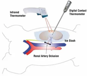
program was used for animal randomization. Coo-ling was obtained by placing 4ºC ice slush in 90% of total renal surface. The ice-free surface was used for temperature measurement. By using the digital contact thermometer (DT), we assured similar tempe-rature was obtained both in ice covered and ice-free areas. In both groups, renal temperature was simul-taneously measured with a DT and IRT (Figure-1). DT was used to measure not only surface but also deep kidney temperatures during the experiment in order to assure homogeneous kidney cooling. As a result, two temperature curves for DT and a third curve for the IRT were obtained in Groups 1 and 2.
Thermometer Characteristics and Specifications IRT: a portable infrared thermometer (Mi-nipa®, model MT-360) with laser pointer and LCD display was used for non-contact temperature mea-surement. Specifications according to manufacturer were: temperature precision of 2ºC within a tempera-ture variation of -30 to 100ºC, distance-to-spot ratio of 10:1, emissivity of 0.1 to 1.0 and response time of 250 ms. Emissivity was adjusted to 0.98. To include a renal surface diameter of at least 5 mm we perfor-med temperature measurement with the device in a 5 cm range from the kidney in a 90-degree angle, according to manufacturer instructions.
Figure 1 - Schematic Swine Model for simultaneous kidney temperature measurement with IRT and DT during renal ischemia procedures.
Ice Slush
Renal Artery Oclusion
Digital Contact Thermometer Infrared
DT: we used a waterproof digital contact thermometer (Incoterm®, model 9791.16.0.00) with a 1ºC measurement precision and tempera-ture window scale of -50 to 300ºC. The device rigid metal tip was inserted in the renal paren-chyma with maximum depth of 10 mm (surface DT temperature). Also, it was inserted deeply in renal parenchyma with a depth of 30 mm in a 45-degree angle to ensure parenchyma tempera-ture measurement (deep DT temperatempera-ture). Tempe-rature was measured until the sound alarm was heard according to manufacturer instructions.
Comparative and Statistic Analyses
Temperature values at each time point were expressed in mean, standard deviation and range for each thermometer in all groups. Tem-perature curves of IRT and DT were compared in Groups 1 and 2. We used the Kolmogorov-Smir-nov test to assure data normality. The Student-T test was used to compare mean values at each time point (T0, T2, T5, T10) between the different thermometers. We considered p < 0.05 to be sta-tistically significant.
RESULTS
Group 1 – Cold Ischemia (n = 10)
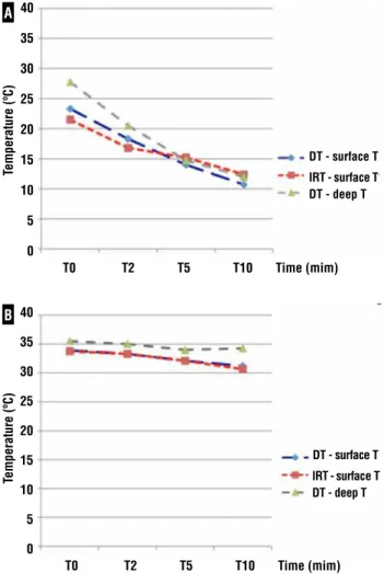
Mean swine temperature was 34.2 ± 1.7ºC (31-37ºC). Figure-2A shows temperature curves in renal cold ischemia group for the two thermo-meters. For the DT, mean surface temperature de-crease was 12.6 ± 4.1ºC (5-19ºC; Table-1). At T5 and T10, 8 and 9 of 10 animals had surface renal temperature ≤ 15ºC, respectively. All had kidney temperature below 20ºC already at T5. Mean deep temperature decrease was 15.8 ± 1.5ºC (15-18ºC). For the IRT, mean temperature decrease was 9.1 ± 3.8ºC (3-14ºC). At T5 and T10, 70% and 80% of animals had kidney temperature ≤ 15ºC, respec-tively. All had renal temperature below 20ºC al-ready at T5. There was no statistically significant difference between thermometers.
Group 2 – Warm Ischemia (n = 10)
Mean animal temperature was 34.9 ± 1.3ºC (33-36ºC). Temperature curves in renal warm ischemia group for both thermometers are
exposed on Table-2. Mean surface temperature decrease for DT was 2.7 ± 1.8ºC (0-4ºC; Figure--2B). Mean deep temperature decrease was 0.5 ± 1.0ºC (0-3ºC). For the IRT, mean temperature decrease was 3.1 ± 1.9ºC (0-6ºC). No statistically significant difference between thermometers was found at any time point.
DISCUSSION
Transitory interruption of kidney blood su-pply is a necessary step in particular for urologi-cal procedures such as partial nephrectomy, renal transplantation and renovascular surgery. Metho-ds to decrease cellular metabolism are required to
Figure 2 - Mean temperature decrease during cold (A) and warm (B) renal ischemia measured by digital and infrared thermometers.
T0 T2 T5 T10 Time (mim) 40
35
30
25
20
15
10
5
0
DT - surface T
IRT - surface T DT - deep T
Temperature (ºC)
T0 T2 T5 T10 Time (mim) 40
35
30
25
20
15
10
5
0
DT - surface T
IRT - surface T DT - deep T
Temperature (ºC)
A
prevent ischemic-reperfusion injury, being exter-nal reexter-nal cooling with ice slush the most accep-ted technique. The first authors to examine this matter were Wickham et al. in 1967 (1), followed by Ward who used a canine model and determi-ned 15ºC to be the ideal temperature to avoid re-nal damage caused by ischemia and reperfusion phenomenon (2). Since then, such figures have been adopted as goals every time renal cooling is required. Although effortless to execute in con-ventional surgeries, accurate ice slush placement during laparoscopic procedures is technically de-manding and less attractive. For that reason, new renal cooling methods have been created (3-10). Each method has its pros and cons, none being able to completely reproduce the gold-standard
open technique. That led many surgeons to per-form laparoscopic partial nephrectomy without renal vascular clamping. The temperature of re-nal tissue during warm ischemia has been poorly investigated. A maximum warm ischemia time of 30 minutes is supposed to avoid permanent renal damage (17,18). However, even in excel-lence laparoscopic centers a longer ischemia time is reported (19), in part explaining why la-paroscopic partial nephrectomy is underperfor-med worldwide (20).
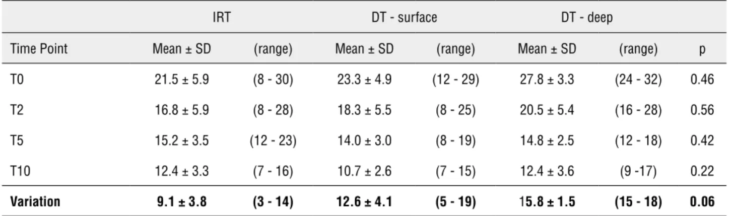
To date, we are the first to test IRT ac-curacy during renal surgery. IRT infer tempera-ture by using a portion of the thermal radiation emitted by the object of measurement. If a laser is used to help aim the thermometer at certain Table 1 - Comparative analysis of temperature decrease during cold renal ischemia procedure.
IRT DT - surface DT - deep
Time Point Mean ± SD (range) Mean± SD (range) Mean ±SD (range) p
T0 21.5 ± 5.9 (8 - 30) 23.3± 4.9 (12 - 29) 27.8 ± 3.3 (24 - 32) 0.46
T2 16.8 ± 5.9 (8 - 28) 18.3 ± 5.5 (8 - 25) 20.5± 5.4 (16 - 28) 0.56
T5 15.2 ±3.5 (12 - 23) 14.0± 3.0 (8 - 19) 14.8± 2.5 (12 - 18) 0.42
T10 12.4 ±3.3 (7 - 16) 10.7 ± 2.6 (7 - 15) 12.4 ± 3.6 (9 -17) 0.22
Variation 9.1 ± 3.8 (3 - 14) 12.6 ± 4.1 (5 - 19) 15.8 ± 1.5 (15 - 18) 0.06
IRT = Infrared thermometers; DT = Digital contact thermometer; SD = Standard deviation
Table 2 - Comparative analysis of temperature decrease during warm renal ischemia procedure.
IRT DT - surface DT - deep
Time Point Mean ±SD (range) Mean ± SD (range) Mean ± SD (range) p
T0 33.8 ± 1.0 (32 - 35) 33.9 ± 1.3 (32 - 36) 35.5 ± 1.3 (34 - 37) 0.77
T2 33.3 ± 1.3 (31 - 36) 33.3 ± 1.3 (31 - 36) 35.0 ± 0.8 (34 - 36) 1.0
T5 32.1 ± 1.4 (30 - 34) 32.1 ± 2.0 (29 - 36) 34.0 ± 1.2 (33 - 35) 1.0
T10 30.7 ± 2.0 (28 - 34) 31.2 ± 2.5 (29 - 37) 34.3 ± 1.0 (33 - 35) 0.65
Variation 3.1 ± 1.9 (0 - 6) 2.7 ± 1.8 (0 - 4) 0.5 ± 1.0 (0 - 3) 0.62
distance it is called laser non-contact thermome-ter. The basic design consists of a lens to focus the infrared thermal radiation on to a detector, which converts radiant power to an electrical signal that can be displayed in units of tempe-rature after being compensated for ambient tem-perature. Specifications of portable handheld sensors include ratings of temperature accuracy, distance-to-spot ratio (D:S) and emissivity. In ge-neral, measurement uncertainty remains around ± 2ºC/± 4ºF. D:S ratio is the relationship between object distance and diameter of the temperature measurement area. Finally, the sensor may have an adjustable emissivity setting, which can be set to measure the temperature of shiny and non--reflective surfaces. The value of emissivity for various materials can be looked up in published emissivity table. We adjusted emissivity to 0.98 because it is the closest to renal tissue.
Initial studies testing IRT accuracy and practicability were undertaken for febrile pa-tients evaluation. Results vary among series with some disappointing (13,14) and other promising reports (11,12). Our experiment showed excellent accuracy of the IRT compared to digital contact thermometer. We found no difference in tempe-rature values in each time point studied. Also, IRT proved to be reliable not only during small temperature variation but also for sizeable va-riations. IRT was very comfortable to manipulate during the experiment and we faced no problems following manufacturer instructions. In addition, it is a non-contact device and facilitates tempe-rature measurement without increasing the risk of tissue damage or contamination. This is a se-rious advantage when temperature measurement is required. The device may have a potential use in lap/robotic procedures and could be used to-gether with the camera or even built-in. Althou-gh initially planned, a cost analysis was not done because thermometers’ prices are very similar. Therefore, being an uncomplicated, innocuous and low-cost new technology, its incorporation in surgical routine is mainly dependent on its availability in an integrated system with lapa-roscopic cameras.
We could assess real effectiveness of ice slush renal cooling. The essential issue in
achie-ving hypothermia is to ensure that the hypo-thermia is uniform throughout the kidney sur-face, cortex and the medulla and is constant during the procedure. Desired renal temperatures bellow were already achieved after five minutes of renal cooling in all animals of our experiment. If cutoff value was 15ºC, at T5 and T10 70-80% and 80-90% of kidneys were adequately cooled, respectively. We confirmed data from other stu-dies proving ice slush to be an excellent fast and efficient method for renal cooling (10,17,18). Gill et al. achieved a nadir renal parenchymal tempe-rature of 5 to 19ºC by using ice slush during la-paroscopic partial nephrectomy in humans (21). Guerra et al. recently compared ice slush with retrograde ureteral cold saline perfusion during experimental renal cooling (10). In the ice slush group, they obtained a mean renal temperature decrease rate of 1.37ºC per minute in the medulla and 1.44ºC per minute in renal cortex during the first 20 minutes. After 5 and 10 minutes, mean renal temperature was 17ºC and 12ºC, respec-tively. Those findings are very similar to ours. Small differences may be related to animal size and weight. In humans, temperature decrease may be a little slower because of renal paren-chyma thickness.
Finally, monitoring of renal temperature during those procedures may have an even more impor-tant role when a clinical research with strict me-thodological standards is being carried on. For example, accurate renal cooling should be asses-sed every time a serum or histological analysis is being undertaken in such organs or individuals. In addition, and not less important, IRT may be appropriate for investigative laboratory studies assessing methods or effects of kidney cooling during open renal procedures.
CONCLUSIONS
IRT proved to be an accurate device for renal temperature monitoring during cold and warm ischemia procedures. The noncontact in-frared thermometer is a non-invasive precise tool witch allows effortless temperature measurement during kidney surgery. External ice slush cooling confirmed to be fast and effective at cooling in the pig model.
ABBREVIATIONS
IRT = Infrared thermometer
DT = Digital contact thermometer
D:S = Distance-to-spot ratio
CONFLICT OF INTEREST
None declared.
REFERENCES
1. Wickham JE, Hanley HG, Joekes AM: Regional renal hypo-thermia. Br J Urol. 1967; 39: 727-43.
2. Ward JP: Determination of the Optimum temperature for regional renal hypothermia during temporary renal isch-aemia. Br J Urol. 1975; 47: 17-24.
3. Beri A, Lattouf JB, Deambros O, Grüll M, Gschwendtner M, Ziegerhofer J, et al.: Partial nephrectomy using renal artery perfusion for cold ischemia: functional and oncologic out-comes. J Endourol. 2008; 22: 1285-90.
4. Janetschek G, Abdelmaksoud A, Bagheri F, Al-Zahrani H, Leeb K, Gschwendtner M: Laparoscopic partial nephrec-tomy in cold ischemia: renal artery perfusion. J Urol. 2004; 171: 68-71.
5. Landman J, Rehman J, Sundaram CP, Bhayani S, Monga M, Pattaras JG: Renal hypothermia achieved by retrograde intracavitary saline perfusion. J Endourol. 2002; 16: 445-9. 6. Crain DS, Spencer CR, Favata MA, Amling CL: Transureteral
saline perfusion to obtain renal hypothermia: potential ap-plication in laparoscopic partial nephrectomy. JSLS. 2004; 8: 217-22.
7. Rama Raju BV, Atm Prakash, Kapur M, Gulati SM: Acute renal ischaemia and the effect of local hypothermia on the histological changes: an experimental study. Indian J Med Res. 1969; 57: 925-31.
8. Webster TM, Moeckel GW, Herrell SD: Second prize: simple method for achieving renal parenchymal hypothermia for pure laparoscopic partial nephrectomy. J Endourol. 2005; 19: 1075-81.
9. Kijvikai K, Viprakasit DP, Milhoua P, Clark PE, Herrell SD: A simple, effective method to create laparoscopic renal pro-tective hypothermia with cold saline surface irrigation: clin-icalapplication and assessment. J Urol. 2010; 184: 1861-6. 10. Guerra R, Leonardi EP, Otsuka RA, Quitzan J, Kawano PR,
Yamamoto HA, et al.: Cold renal ischemia: comparison of efficacy between two techniques of cooling, in a swine model. J Endourol. 2010; 24: 445-9.
11. Nguyen AV, Cohen NJ, Lipman H, Brown CM, Molinari NA, Jackson WL, et al.: Comparison of 3 infrared thermal de-tection systems and self-report for mass fever screening. Emerg Infect Dis. 2010; 16: 1710-7.
12. Osio CE, Carnelli V: Comparative study of body temperature measured with a non-contact infrared thermometer versus conventional devices. The first Italian study on 90 pediatric patients. Minerva Pediatr. 2007; 59: 327-36.
13. Fortuna EL, Carney MM, Macy M, Stanley RM, Younger JG, Bradin SA: Accuracy of non-contact infrared thermometry versus rectal thermometry in young children evaluated in the emergencydepartment for fever. J Emerg Nurs. 2010; 36: 101-4.
14. Hausfater P, Zhao Y, Defrenne S, Bonnet P, Riou B: Cuta-neous infrared thermometry for detecting febrile patients. Emerg Infect Dis. 2008; 14: 1255-8.
15. Lapostolle F, Catineau J, Le Toumelin P, Proust C, Garrigue B, Galinski M, et al.: Intravenous fluid temperature man-agement by infrared thermometer. Am J Emerg Med. 2006; 24: 174-6.
16. Estrella EP: An infrared forehead thermometer for flap monitoring. J Plast Reconstr Aesthet Surg. 2009; 62: 736. 17. Ramani AP, Ryndin I, Lynch AC, Veetil RT: Current concepts
in achieving renal hypothermia during laparoscopic partial nephrectomy. BJU Int. 2006; 97: 342-4.
19. Gill IS, Kavoussi LR, Lane BR, Blute ML, Babineau D, Co-lombo JR Jr, et al.: Comparison of 1,800 laparoscopic and open partial nephrectomies for single renal tumors. J Urol. 2007; 178: 41-6.
20. Gerber GS, Stockton BR: Laparoscopic nephron-sparing surgery. J Endourol. 2007; 21: 458-9.
21. Gill IS, Abreu SC, Desai MM, Steinberg AP, Ramani AP, Ng C, et al.: Laparoscopic ice slush renal hypothermia for par-tial nephrectomy: the inipar-tial experience. J Urol. 2003; 170: 52-6.
22. Gill IS, Patil MB, Abreu AL, Ng C, Cai J, Berger A, et al.: Zero ischemia anatomical partial nephrectomy: a novel ap-proach. J Urol. 2012; 187: 807-14.
_____________________
Correspondence address: Dr. Giovanni Scala Marchini Division of Urology, Clinics Hospital, University of Sao Paulo Medical School