Key words:
Calcium Oxalate; Urine; Ethylene Glycol; Kidney Calculi
Int Braz J Urol. 2013; 39: 565-71
__________________
Submitted for publication: December 19, 2012
__________________
Accepted after revision: May 29, 2013
Purpose: To investigate the anti-urolithiatic effect of cow urine ark (medicinal distilled cow urine) on ethylene glycol (EG) induced renal calculi.
Materials and Methods: 36 male Wistar rats were randomly divided into 6 equal groups. Group I animals served as vehicle control and received distilled water for 28 days. Group II to VI animals received 1% v/v EG in distilled water for 28 days. Group II served as EG control. Group III and IV (preventive groups) received cow urine ark orally for 28 days in doses of 1 mL/kg and 2 mL/kg, respectively. Group V and VI (treatment groups) received 1 mL/kg and 2 mL/kg cow urine ark orally, respectively from 15th to 28th days. 24-hour urine samples were collected on day 0 and 28. Urine volume and oxalate levels were measured. On day 28, blood was collected for biochemical parameters. Animals were sacrificed and kidneys were harvested, weighed and histopathologically evaluated for calcium oxalate (CaOx) crystals. To calculate the percentage of inhibition of mineraliza-tion, simultaneous flow static in-vitro model was used.
Results: EG significantly increased urine oxalate, serum creatinine, blood urea level; kidney weight and CaOx deposits. Provision of cow urine ark resulted in significantly lower levels of urine oxalate, serum creatinine, blood urea and CaOx depositions as compared to Group II. (p value < 0.05) It also significantly restored kidney weight. (p value < 0.05) Cow urine ark inhibited 40% and 35% crystallization of CaOx and calcium phosphate, respectively.
Conclusion: Cow urine ark is effective in prevention and treatment of EG induced uroli-thiasis in Wistar rats.
INTRODUCTION
Urolithiasis is extremely painful and the third prevalent disorder of urinary system (1,2). Life time risk for renal calculi is 7-12% among general population (3). Alteration in crystalliza-tion condicrystalliza-tions of urine is responsible for stone formation (4). Genetic factor, diet, sedentary lifes-tyle, climate, and urinary tract infection are com-mon risk factors (5). The studies have shown that consumption of high dietary oxalate is a major
risk factor for stone formation (6). The most com-mon metabolic abnormality acom-mong the recurrent stone formers is hypercalciuria (7). In humans, al-most 80% calculi are composed of calcium oxa-late (CaOx) and calcium phosphate whereas uric acid, cystine and magnesium ammonium phosphate (struvite stone) are forming the rest of the calculi (8). Surgical treatment is the definitive management of renal calculi at present (9). Recurrent stone forma-tion is the major problem even after surgical remo-val of the stone. Recurrence rate is more than 50%
Anti-Urolithiatic Effect of Cow Urine Ark on Ethylene
Glycol-Induced Renal Calculi
_______________________________________________
A. B. Shukla, D. R. Mandavia, M. J. Barvaliya, S. N. Baxi, C. B. Tripathi
Dept. of Pharmacology (ABS, DRM, MJB, CBT) and Department of Pathology (SNB), Government Medical College and Sir Takhtasinhji General Hospital, Bhavnagar-364001 (Gujarat), India
ABSTRACT
ARTICLE
INFO
over 10 years duration of stone formation without medical treatment (10). It is more frequent in male as compared to female (70-81% vs. 47-60%) (11). Established medical treatment is lacking for pre-vention and treatment of renal calculi.
Variety of herbal drugs and alternative tre-atments has been tried for prevention and treat-ment of renal calculi but none of them got the commercial importance. From thousands of years in India, use of cow urine for cardiac and renal problems, weight loss, jaundice, anemia, diarrhea, stomach ache, piles, skin diseases, and vitiligo has been mentioned in ancient ayurvedic literature such as Charaka Samhita and Shushruta Samhi-ta (12). Cow urine is known to have diuretic and nephroprotective effect (13). However, scientific evidences are not available for anti-urolithiatic activity of cow urine. The present study was de-signed to evaluate the anti-urolithiatic activity of cow urine ark (medicinal distilled cow urine) in ethylene glycol (EG) induced renal calculi.
MATERIALS AND METHODS
Animals
All the experiments were performed after approval (IAEC no-21/2011; dated 13/09/2011) of Institutional Animal Ethics Committee of Gover-nment Medical College Bhavnagar; Gujarat, In-dia and in accordance with the guidelines for the Committee for Purpose of Control and Supervision of Experiments on Animals (CPCSEA). Healthy male Wistar albino rats weighting 250-350 g were procured from the central animal house of the ins-titute. They were housed in standard transparent polycarbonate cages and kept in a 12 hour light--dark cycle under controlled room temperature and humidity (24 ± 2 ºC; 65 ± 10%). Animals were fed with standard laboratory diet and tap drinking water. The animals were allowed to acclimatize for at least three days before the starting experiments. The animal handling was performed according to the Good Laboratory Practice (GLP) guidelines.
Drugs and Chemicals
Cow urine ark was purchased from Go-Vi-gyan Anushandhan Kendra, Devalapar, Nagpur, Maharashtra, India (US Patent No 6410 059/2002).
It is prepared by vaporization of cow urine and it has been used in human for many years in In-dia. Ethylene glycol (Fisher Scientific Co., Mum-bai, India), oxalate kit (Trinity Biotech, Ireland), sodium phosphate (Aldrich, India), sodium oxalate and calcium acetate (Alfa Aesar, Hyderabad, India) were used in the study.
The dose of cow urine ark was extrapola-ted from the human dose (10 mL/day) (14,15).
Study design and procedure
Thirty six male Wistar albino rats were as-signed randomly into six equal groups.
Group I (Vehicle control): received distilled water for 28 days;
Group II (EG control): received 1% v/v ethylene glycol (EG) in distilled water for 28 days;
Group III (preventive group): received 1% v/v EG in distilled water for 28 days + cow urine ark 1 mL/kg orally for 28 days;
Group IV (preventive group): received 1% v/v EG in distilled water for 28 days + cow urine ark 2 mL/kg orally for 28 days;
Group V (treatment group): received 1% v/v EG in distilled water for 28 days + cow urine ark 1 mL/kg orally from 15 to 28 days;
Group VI (treatment group): received 1% v/v EG in distilled water for 28 days + cow urine ark 2 mL/kg orally from 15 to 28 days;
Outcome measures
Urinary parameters
24 hour urine specimens were collected by keeping each rat in separate metabolic cage (B.I.K Industries, Mumbai, India) on day 0 and 28. Uri-ne volume was measured. Acidified uriUri-ne samples were kept in refrigeration and analyzed for urinary oxalate level using oxalate kit and spectrophoto-meter within 7 days of collection of samples (16).
Biochemical parameters
Histopathological parameters
All rats were sacrificed after blood col-lection and both kidneys were removed by midli-ne incisions. Both kidmidli-neys were weighed and kept in formaldehyde (10% v/v) for at least 24 hours. 5 mm sections were taken and processed in a series of graded acetone and xylene, embedded in paraffin wax. 5 µm thin sections were taken and stained with haematoxylin-eosin (H&E) and evaluated under op-tical light microscope. CaOx crystal depositions were calculated in 10 microscopic fields (159 x 10-9 m2
each) and other changes like necrosis and tubular dilatations were also noted.
In-vitro method
Determination of mineralization
Inhibition of calcium oxalate and cal-cium phosphate mineralization by cow urine ark was measured by simultaneous flow static model (S.S.M.) described by Farook et al. (17). For inhi-bition of calcium oxalate and calcium phosphate mineralization, procedures were carried out in two sets; one served as blank set and other as expe-rimental set. In blank set, 0.1 M sodium oxalate (25 mL), and 0.1 M calcium acetate (25 mL) were taken in two separate burettes whereas in experi-mental set cow urine ark (25 mL) was taken in a third burette. In both sets of experiments, chemi-cals were allowed to fall simultaneously, slowly and at steady pace into a 250 mL beaker. After 30 min., the mixture was kept in hot water bath for 10 min., cooled to room temperature and collected into a pre-weighed centrifuge tube. Centrifugation of mixture was done at 3000 rpm for 15 min. Su-pernatant fluid was discarded and precipitate (ppt) was obtained. All tubes with ppt were dried in a hot air oven at 120 ºC, cooled to room tempera-ture and weighed. Similar process was repeated using 0.1 M sodium phosphate (25 mL) and 0.1 M calcium acetate (25 mL) for inhibition of calcium phosphate mineralization. The percentage inhibi-tion of mineralizainhibi-tion was calculated by the follo-wing formula:
Statistical analysis
All parameters were expressed as mean ± standard error of mean (S.E.M.) except inhibition of calcium oxalate and calcium phosphate mine-ralization described in percentage. Biochemical pa-rameters between the groups were compared using Kruskal-Wallis test followed by Dunn’s multiple comparison test. Urinary parameters, kidney weight and CaOx crystal depositions were compared using ANOVA followed by Tukey-Kramer multiple com-parison test. The statistical calculations were done using GraphPad InStat, Demo version 3.06. P < 0.05 was considered as statistical significant.
RESULTS
Effect of cow urine ark on urinary oxalates and volume
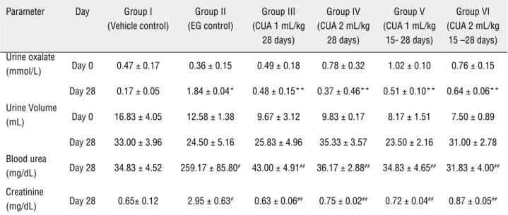
All the groups were related at baseline. Uri-ne volume and oxalate level among all the groups were not statistically significant on day 0. Hype-roxaluria was evident in the EG control group on 28th day as compared to the vehicle control group. Both prophylactic and treatment groups of cow urine ark had shown significant reduction in the urinary oxalate concentration on day 28 (p value < 0.05; Table-1). There was no significant difference in the urinary volume on day 28 betwe-en normal and experimbetwe-ental groups.
Effect of cow urine ark on renal function and kidney weight
Administration of EG resulted in significant abnormalities of serum creatinine and blood urea (Table-2). EG control group had shown 7 times hi-gher value of blood urea and 4 times hihi-gher value of serum creatinine as compared to vehicle treated group. Weight of the kidney also almost doubled in the EG treated group. Serum creatinine and blood urea were significantly reduced in all the cow urine ark treated rats as compared to EG treated rats (p value < 0.05; Table-1). These observed values in cow
Table 1 - Comparison of urinary and biochemical parameters between the groups at study end point.
Parameter Day Group I (Vehicle control)
Group II (EG control)
Group III (CUA 1 mL/kg
28 days)
Group IV (CUA 2 mL/kg
28 days)
Group V (CUA 1 mL/kg
15- 28 days)
Group VI (CUA 2 mL/kg
15 –28 days)
Urine oxalate
(mmol/L) Day 0 0.47 ± 0.17 0.36 ± 0.15 0.49 ± 0.18 0.78 ± 0.32 1.02 ± 0.10 0.76 ± 0.15
Day 28 0.17 ± 0.05 1.84 ± 0.04* 0.48 ± 0.15** 0.37 ± 0.46** 0.51 ± 0.10** 0.64 ± 0.06**
Urine Volume
(mL) Day 0 16.83 ± 4.05 12.58 ± 1.38 9.67 ± 3.12 9.83 ± 0.17 8.17 ± 1.51 7.50 ± 0.89
Day 28 33.00 ± 3.96 24.50 ± 5.16 25.83 ± 4.96 35.33 ± 3.57 23.50 ± 2.16 31.00 ± 2.78
Blood urea
(mg/dL) Day 28 34.83 ± 4.52 259.17 ± 85.80# 43.00 ± 4.91## 36.17 ± 2.88## 34.83 ± 4.65## 31.83 ± 4.00##
Creatinine
(mg/dL) Day 28 0.65± 0.12 2.95 ± 0.63# 0.63 ± 0.06## 0.75 ± 0.02## 0.72 ± 0.04## 0.87 ± 0.05##
(All values are expressed as mean ± SEM. n = 6 for all groups.)
*P value < 0.001 as compared Group I and **P value < 0.001 as compared Group II by one way ANOVA followed by Tukey-Kramer multiple comparison tests.
#P value < 0.05 as compared Group I;
##P value < 0.05 as compared Group II by Kruskal-Wallis test followed by Dunn’s multiple comparisons. EG- Ethylene glycol, CUA-Cow urine ark.
Table 2 - Comparison of body weight, kidney weight, CaOx crystals deposition.
Groups Baseline body weight (gm)
Mean Increment of body weight (gm)
Kidney weight (gm)
No. of CaOx crystals in 10 microscopic field (X 40)
Group I (Vehicle control ) 276.67 ± 7.82 15.00 ± 1.29 1.18 ± 0.05 0.333 ± 0.211
Group II (EG control) 302.50 ± 5.74 9.17 ± 1.54 2.05 ± 0.10# 17.66 ± 1.96###
Group III (CUA 1 mL/kg 28 days) 266.67 ± 16.67 12.50 ± 3.10 1.33 ± 0.07## 1.67 ± 0.33##
Group IV (CUA 2 mL/kg 28 days) 296.67 ± 16.87 13.33 ± 2.47 1.27 ± 0.08## 1.50 ± 0.34##
Group V (CUA 1 mL/kg 15- 28 days) 291.67 ± 11.95 13.33 ± 1.67 1.35 ± 0.09## 2.50 ± 0.43##
Group VI (CUA 2 mL/kg 15 - 28 days) 328.33 ± 8.72 12.50 ± 2.81 1.60 ± 0.06## 2.33 ± 0.33##
(All values are expressed as mean ± SEM. n = 6 for all groups.) #P value < 0.05 as compared group I;
##P value < 0.05 as compared group II;
urine ark treated animals were comparable to vehi-cle treated group. Administration of cow urine also significantly reduced the kidney weight (Table-2).
Effect of cow urine ark on body weight and mean increment of body weight
There was no significant difference in body weight in EG and cow urine ark treated animals on baseline. Mean increment of body weight during 28 days was not significantly affected in EG control group as well in all the cow urine ark treated groups at the completion of experiment (Table-2).
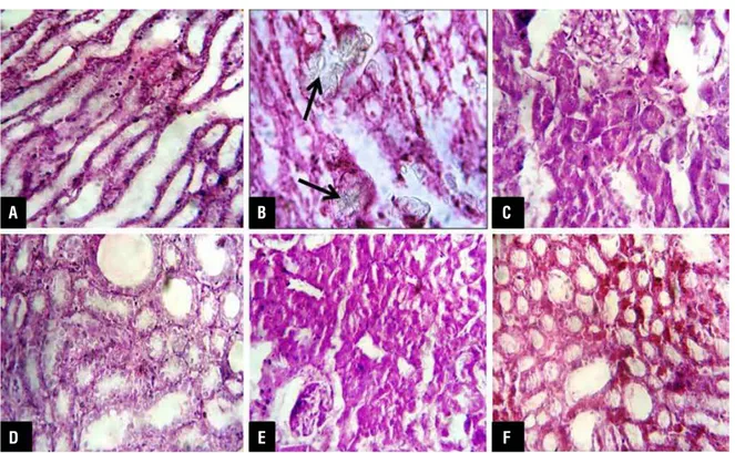
Effect of cow urine ark on renal oxalate crystals at histopathological evaluation
EG treated rats had significant CaOx crys-tal deposition in the all major three parts of kidney. These intratubular, interstitial crystals were found mostly in cortex and medulla associated with sig-nificant leukocyte infiltration, necrosis, hemorrhage and tubular dilatation. (Figure-1) EG control group showed the maximum CaOx crystals in renal paren-chyma and on papillary tips as compared to vehicle control group. These crystals were large polygonal
in shape, heterogeneous in distribution and pattern. There was marked reduction in number of crystal de-position in all the cow urine ark treated rats (p value < 0.05; Table-2). In these groups, tubular dilatation, leukocyte infiltrations were found less than in EG treated group and necrosis was not detected.
Effect of cow urine ark on In vitro mineralization Cow urine ark showed 40% and 35% inhi-bition of CaOx and calcium phosphate crystalliza-tion, respectively.
DISCUSSION
Among many in vivo models developed to evaluate anti-urolithiatic effect, EG induced calculi are widely used (18,19). Rat has been found a suita-ble and frequently used animal to induce CaOx de-position in kidney because of its close resemblance of urinary system to human. Selection of male rats was based on previous studies showing higher rate of crystal depositions in male as compared to fe-male rats (20,21).
Figure 1 - Histopathological images of kidney sections after H&E staining under light microscope (X 40) (A) Vehicle control group, (B) EG control group showing calcium oxalate crystals (arrows in figure), (C to F) Cow urine ark treated groups.
A
D
B
E
C
The basic mechanism behind EG induced calculi is hypercalciuria and hyperoxaluria lea-ding to CaOx crystal formation (22). In addition to development of oxalate crystal, it is also associa-ted with severe oxidative stress to renal tissue (23). Oxalate has been reported to induce lipid peroxi-dation and causes renal tissue damage by reacting with polyunsaturated fatty acids in cell membra-nes and by generation of reactive oxygen species like hydroxyl and superoxide ions (24). Due to re-nal papillary hypertrophy and crystal depositions, kidney weight is increased in EG treated rats (25). It increases blood urea and creatinine levels signi-ficantly due to impairment in renal function.
Up to the best of our knowledge, the pre-sent study is the first study that evaluate cow uri-ne ark as an antiurolithiatic agent. In the present study, cow urine ark had significantly reduced the excretion of oxalate and CaOx crystal depositions in kidney in EG treated rats. EG is metabolized into glycolaldehyde acid, glycolic acid, glyoxylic acid, and oxalic acid. The formed oxalic acid is largely excreted in urine as oxalate. It is precipi-tated by calcium ions to form CaOx crystals (26). Reduction in urinary oxalate and CaOx crystal de-position shows the efficiency of cow urine ark in prevention of stone formation. Inhibition of mi-neralization of calcium oxalate and calcium phos-phate also support the efficacy of cow urine ark in prevention of crystal formation and hence in prevention of stone formation (17). Cow urine ark reduces the oxalate formation from EG and also prevents its precipitation with calcium to form CaOx crystals. It may be useful to prevent the recurrence as it prevents crystallization in urine. We have designed treatment groups as 0.75 % v/v EG administration leads to persistent crystalluria by day 12 and crystal formation from 50-75% in 15 days (27,28). We want to simulate the clinical scenario of exposure and treatment concurrently. In these groups cow urine ark also showed signifi-cant improvement.
In the present study, cow urine ark signi-ficantly restored the kidney weight and impaired renal function (Table-2). Cow urine ark has sig-nificant antioxidant and superoxide scavenging properties (29). Antioxidant property of cow urine ark may have contributed in restoration of EG
in-duced renal dysfunction. Positive effect on kidney weight and renal function suggests its nephro-protective effect in renal calculi. Cow urine ark is known to have diuretic effect (13). In our study, we did not find its diuretic effect in EG treated rats as the urine volume was not affected. It is known that due to compromised renal function caused by EG, the mean increment of body weight reduces (30). Though we have found reduction in mean in-crement of body weight in EG control but it could not reach statistical significant difference proba-bly due to small sample size. Some oxalate stones may have an origin after bacterial infection. Cow urine is known to have anti-microbial effect whi-ch can also contribute to its antiurolithiatic effect (29). However, we did not evaluate this possible mechanism in our study.
The present study has given first insight to the effect of Cow urine ark in renal calculi, ho-wever it has several limitations: smaller sample size; inability to evaluate molecular mechanisms; urine substrates for composition of stone. Further studies can be planned with active ingredients of Cow urine ark in different models of renal calculi. Clinical studies are required to determine the same effects in renal calculi in human beings.
CONCLUSIONS
Cow urine ark showed significant antiu-rolithiatic effect and restoration of compromised renal function in preventive as well as treatment groups. It probably exerts its action by reducing oxalate excretion and crystallization inhibition. Further studies are recommended to know its me-chanism of action in different preclinical and cli-nical settings.
ACKNOWLEDGMENTS
We are sincerely thankful to Dr. Tejas K. Pa-tel, Assistant professor, Department of Pharmacolo-gy, GMERS Medical College, Gotri, Vadodara, Gu-jarat (India) for critical review of this manuscript.
CONFLICT OF INTEREST
REFERENCES
1. Rad AK, Hajzadeh MAR, Rajaei Z, Sadeghian MH, HN, Kesha-varzi Z: Preventive effect of Cynodon dactylon against ethylene glycol-induced nephrolithiasis in male rats. Avicenna Journal of Phytomedicine. 2011; 1: 14-23.
2. Laikangbam R, Damayanti Devi M: Inhibition of calcium oxalate crystal deposition on kidneys of urolithiatic rats by Hibiscus sab-dariffa L. extract. Urol Res. 2012; 40: 211-8.
3. Grases F, Costa-Bauza A, Prieto RM: Renal lithiasis and nutrition. Nutr J. 2006; 5: 23-9.
4. Ehud Gnessin, James E. Lingeman, Andrew P, Evan: Pathogen-esis of renal calculi. Turkish Journal of Urology. 2010; 36: 190-9. 5. Romero V, Akpinar H, Assimos DG: Kidney stones: a global pic-ture of prevalence, incidence, and associated risk factors. Rev Urol. 2010; 12: e86-96.
6. Finkielstein VA, Goldfarb DS: Strategies for preventing calcium oxalate stones. CMAJ. 2006; 174: 1407-9.
7. Arrabal-Polo MA, Arrabal-Martin M, Garrido-Gomez J: Calcium renal lithiasis: metabolic diagnosis and medical treatment. Sao Paulo Med J. 2013; 131: 46-53.
8. Leonetti F, Dussol B, Berthezene P, Thirion X, Berland Y: Dietary and urinary risk factors for stones in idiopathic calcium stone formers compared with healthy subjects. Nephrol Dial Trans-plant. 1998; 13: 617-22.
9. Miller NL, Lingeman JE: Management of kidney stones. BMJ. 2007; 334: 468-72.
10. Patel RK, Patel SB, Shah JG: Anti-Urolithiatic Activity of Ethanolic Extract of Seeds of Benincasa Hispida (Thumb). Pharmacology-online. 2011; 3: 586-91.
11. Manjula K, Rajendran K, Eevera T, Kumaran S: Effect of Costus igneus stem extract on calcium oxalate urolithiasis in albino rats. Urol Res. 2012; 40: 499-510.
12. Arunkumar S, Muthuselvam M, Rajendran R: Antimicrobial Ac-tivities of Cow Urine Distillate Against Some Clinical Pathogen. Global Journal of Pharmacology. 2010; 1: 41-4.
13. Gururaja MP, Joshi AB, Joshi H, Sathyanarayana D, Subrah-manyam EVS, Chandrashekhar KS: Antidiabetic potential of cow urine in streptozotocin induced diabetic rats. Asian Journal of Traditional Medicines. 2011; 6: 8-13.
14. Davale S: Panchgavya Ayurvedic Chikitsa (5th ed.). India. Go-Vigyan Anusandhan Kendra, Deolapar, Nagpur. 2009; pp.54. 15. Ghosh MN: Toxicity studies. In: Fundaments of experimental
Pharmacology (5th ed.), Hilton & company, Kolkatta. 2011; pp.167.
16. Li MG, Madappally:MM: Rapid enzymatic determination of uri-nary oxalate. Clin Chem. 1989; 35: 2330-3.
17. Farook NA, Dameem GA, Alhaji NM, Sathiya R, Muniyandi J, Sangeetha SJ: Inhibition of Mineralization of Urinary Stone Forming Minerals by Hills Area Fruit. E-Journal of Chemistry. 2004; 1: 137-41.
18. Atmani F, Slimani Y, Mimouni M, Aziz M, Hacht B, Ziyyat A: Effect of aqueous extract from Herniaria hirsuta L. on experi-mentally nephrolithiasic rats. J Ethnopharmacol. 2004; 95: 87-93.
19. Atmani F, Khan SR: Effects of an extract from Herniaria hirsuta on calcium oxalate crystallization in vitro. BJU Int. 2000; 85: 621-5.
20. Vyas B, Vyas R, Joshi S, Santani D: Antiurolithiatic Activity of Whole-Plant Hydroalcoholic Extract of Pergularia daemia in Rats. J Young Pharm. 2011; 3: 36-40.
21. Vermeulen CW: Experiments on causation of urinary calculi. In: Vermeulen CW, editor. Essays in experimental biology. Chi-cago: University of Chicago Press. 1962; pp. 253-69. 22. Karadi RV, Gadge NB, Alagawadi KR, Savadi RV: Effect of
Mo-ringa oleifera Lam. root-wood on ethylene glycol induced uro-lithiasis in rats. J Ethnopharmacol. 2006; 105: 306-11. 23. Atmani F, Slimani Y, Mimouni M, Hacht B: Prophylaxis of
cal-cium oxalate stones by Herniaria hirsuta on experimentally induced nephrolithiasis in rats. BJU Int. 2003; 92: 137-40. 24. Thamilselvan S, Hackett RL, Khan SR: Lipid peroxidation in
ethylene glycol induced hyperoxaluria and calcium oxalate nephrolithiasis. J Urol. 1997; 157: 1059-63.
25. Kuo RL, Lingeman JE, Evan AP, Paterson RF, Parks JH, Bled-soe SB, et al.: Urine calcium and volume predict coverage of renal papilla by Randall’s plaque. Kidney Int. 2003; 64: 2150-4. 26. Atef M, Al-Attar: Antilithiatic Influence of Spirulina on Ethylene
Glycol-Induced Nephrolithiasis in Male Rats. American Jour-nal of Biochemistry and Biotechnology 2010; 6: 25-31. 27. Khan SR: Animal models of kidney stone formation: an
analy-sis. World J Urol. 1997; 15: 236-43.
28. Jie Fan, Michael A. Glass and Paramjit S. Chandhoke: Impact of Ammonium Chloride Administration on a Rat Ethylene Gly-col Urolithiasis Model. Scanning Microscopy 1999; 13: 299-306.
29. Edwin J, Sheej E, Vaibhav T, Rajesh G, Emmanuel T: Antioxi-dant and Antimicrobial Activities of Cow Urine. Global Journal of Pharmacology 2008; 2: 20-2.
30. Bouanani S, Henchiri C, Migianu-Griffoni E, Aouf N, Lecouvey M: Pharmacological and toxicological effects of Paronychia argentea in experimental calcium oxalate nephrolithiasis in rats. J Ethnopharmacol. 2010; 129: 38-45.
_____________________
Correspondence address: