Authors
Luis Alberto Batista Peres1
Sarah Sella Langer1 Raysa Cristina Schmidt1 Roberto Arthur Bavaresco Nacke1 Paulo Victor Muller Francescon1
Rogério Cavalcante de Almeida1
Renan Macedo Coimbra1
Tailla Michelle Ribas1 Tiago Dahrug Barros1 Tiemi Matsuo2
1 Universidade Estadual do Oeste do Paraná – UNIOESTE
2 Universidade Estadual de Londrina
Submitted on: 10/31/2010 Approved on: 12/23/2010
Corresponding author: Luis Alberto Batista Peres R. São Paulo, 769- Ap 901- Centro
Cascavel – PR – Brazil CEP: 85801-020
E-mail: [email protected]
The present study was carried out at Universidade Estadual do Oeste do Para-ná – UNIOESTE, Cascavel, Paraná, Brazil.
The authors declare no conflict of interestinteresse.
A
BSTRACTMetabolic disorders are frequently ob-served in pediatric patients with renal lithiasis. Objectives: Study the metabolic and anatomical alterations and perform the chemical analysis of stones found in children with nephrolithiasis in our region. Methods: A retrospective study on 158 children with evidence of recent renal stone formation was performed. One hundred and nine children con-cluded the metabolic study. Laboratory investigation consisted in two samples of 24-hour urine for calcium, uric acid, citrate, oxalate, sodium and creatinine; qualitative cystinuria, urinary pH fol-lowing 12-hour fasting and water restric-tion, urine culture and chemical analysis when the stones were available. Renal imaging techniques included, at least, re-nal ultrasound and excretory urogram. Results: A cause for nephrolithiasis was identified in 96.3% of children. The main metabolic alteration was hyper-calciuria (73.4%). Chemical analysis of stones showed calcium oxalate in 90.9% of the cases. Anatomical alterations were found in 18.0% of the investigated cases and the most frequently found al-teration was pyelo-ureteral duplication (28.6%). Conclusions: Hypercalciuria was the most frequently found disorder and pyelo-ureteral duplication was the most common anatomical alteration; moreover, calcium oxalate was the most frequent chemical constituent. The pres-ent study showed the characteristics of pediatric patients with nephrolithiasis in our region.
Keywords: nephrolithiasis, child, meta-bolic diseases.
[J Bras Nefrol 2011;33(1): 34-38]©Elsevier Editora Ltda.
Nephrolithiasis in pediatric patients: metabolic and
anatomical investigation
I
NTRODUCTIONNephrolithiasis in pediatric patients is rela-tively rare. In different series of patients at all age ranges with renal lithiasis, the prev-alence in children varies from 2 to 2.7%.1,2 Recent studies have shown that the annual incidence is increasing in different popula-tions.3,4 At the diagnosis, most calculi in children were found in the kidneys, with remnants being found in ureters.5 Several factors can predispose children to develop nephrolithiasis and among them, meta-bolic and genitourinary abnormalities are particularly important; these are often as-sociated with diet, environmental factors and infectious causes. Nephrolithiasis is associated with considerable morbidity and has high recurrence rates.6
The knowledge on nephrolithiasis in children has increased in recent years. Most children with urinary lithiasis have underlying metabolic abnormalities, with hypercalciuria being the most preva-lent.7,8,9 Other metabolic risk factors vary in frequency according to the different se-ries.10,11 Some other metabolic alterations that have been described are hypocitratu-ria, hyperuricosuhypocitratu-ria, hyperoxaluhypocitratu-ria, renal tubular acidosis and cystinuria.12
M
ETHODSA retrospective study was carried out in 158 children treated at the Service of Nephrology of the General Outpatient Clinic of Hospital Universitário do Oeste do Paraná, who presented evidence of recent neph-rolithiasis, from 1995 to 2010. The inclusion criteria for this retrospective study included spontaneous, endoscopic or surgical elimination of calculi and/or radiological confirmation of their presence in the uri-nary tract in the previous six months. The 24-hour urinary data of patients with more than one sample were recorded, as well as family history, clinical pre-sentation, chemical analysis of calculi and imaging assessment.
The laboratory investigation included two or more 24-hour urine samples, including calcium, uric acid, citrate, sodium, creatinine and calcium oxalate mea-surements, and uric acid, creatinine and parathor-mone in blood. Qualitative cystinuria, urinary pH after 12-hour fasting and water restriction, urine cul-ture and chemical analysis of calculi were performed. The laboratory methods employed and the ref-erence values used for 24-hour urine samples were: calcium – atomic absorption spectrophotometry (< 4.0 mg/kg); uric acid - uricase enzymatic method (< 15 mg per kg); citrate - citrate-lyase enzymatic meth-od (> 320 mg); smeth-odium - selective ion methmeth-od (< 150 mEq); creatinine - alkaline picrate method (> 1.000 mg) and urinary volume - volumetric measurement in Becker by visual analysis. For plasma measure-ments, the methods used were: calcium - colori-metric method (8,5-10,5 mg/dL); uric acid - uricase colorimetric method (2.0 to 7.0 mg/dL); creatinine - alkaline picrate method (0.7 to 1.4 mg/dL) and parathormone - intact molecule assay. For isolated urine sample analyses, the methods employed were: qualitative cystinuria - sodium nitroprusside test and urinary pH - measured by using reagent test strips with a methyl red and bromothymol blue indicator system. A decreased urinary volume was considered when at least one of the samples had a 24-hour uri-nary volume < 15 mL/kg. The chemical analysis was carried out using the colorimetric method.
The patients were divided in two groups accord-ing to age, with a cutoff of ten years, with 49 pa-tients being younger and 60 older than ten years.
The Chi-square test and Fisher’s Exact test were used to compare the variables. A p value < 0.05 was considered statistically significant. This study was approved by the Human Subject Research Ethics Committee of UNIOESTE.
R
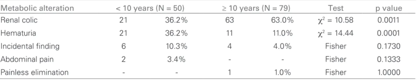
ESULTSThe most frequent clinical presentations were renal colic (65.2%) and hematuria (24.8%). In patients younger than 10 years, the most common clinical pre-sentation was hematuria and in the group aged above 10 years, renal colic was the most common presen-tation (p < 0.05). These data are shown in Table 1. A family history of nephrolithiasis was reported by 80% of the studied group.
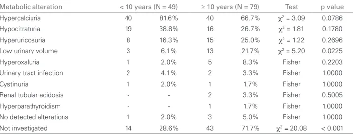
Of the 158 patients, 109 concluded the metabolic study. At least one alteration was found in 96.3% of them. Patient mean age was 11.4 ± 4.7 years (ranging from 4 months to 18 years) and 54.1% were males. The main metabolic alterations identified in the study group were: hypercalciuria (73.4%), hypocitraturia (32.1%) and uric acid hyperexcretion (21.1%). The differences found between the age and sex distribu-tions were not statistically significant (Tables 2 and 3, p > 0.05). There was a significant difference regarding the number of patients with low urinary volume and of those that were not investigated, being higher in the group older than 10 years (Table 3).
The chemical analysis of the calculi showed the presence of calcium oxalate in 90.9%, calcium car-bonate in 54.5%, uric acid in 18.2%, phosphorus and magnesium in 9.1% of the cases.
Anatomic alterations were observed in 18.1% of the cases, with pyeloureteral duplication being found in four patients (28.6%), renal cyst in three (21.4%), neurogenic bladder in two (14.3%) and pyelocali-ceal obstruction, extrarenal pelvis, horseshoe kidney, polycystic kidney disease and distal ureteral stenosis in one patient each.
D
ISCUSSIONThe present study presents the main metabolic risk factors, the clinical presentation, the anatomic altera-tions and the family history of pediatric patients with nephrolithiasis that were referred to our institution for metabolic assessment.
Nephrolithiasis can occur at any age. The litera-ture has shown that the mean age at diagnosis ranges from 7 to 10.3,13 In the present study, the mean age of patients was 11 years, with 40% of them being younger than 10 years at admission.
completed the metabolic assessment is similar. When the metabolic alterations were compared between the age groups < 10 and > 10 years, there were no sta-tistically significant differences. The epidemiology of nephrolithiasis in the pediatric population is not as well defined as in the adult population. In the first decade of life, nephrolithiasis was more prevalent in the male sex, being the opposite in the second decade of life, as it was more prevalent in the female sex.15
Hypercalciuria is associated with the development of nephrolithiasis. In children with hypercalciuria, the prevalence of familial nephrolithiasis is 46 to 49%.11,16 Idiopathic hypercalciuria has been identified as the most frequent metabolic risk factor, detected in 40% to 69% of the cases.17,19 A positive family his-tory seems to be the most important isolated risk fac-tor.20 A large number of genes have been suggested as responsible for the pathogenesis of hipercalciúria.21-26 In the present study, 89.6% of the patients with hy-percalciuria had a positive family history.
Other main metabolic disorders identified in the study group were hypocitraturia (32.1%) and hy-peruricosuria (21.1%). Until recently, hypocitraturia
was considered uncommon.10 Tefekli et al27 identified hypocitraturia as the most prevalent metabolic risk factor in children with nephrolithiasis (60.6%). Van Dervoort et al.4 also observed that hypocitraturia was the most commonly identified metabolic abnormal-ity, present in 52% of the children studied between 2003-2005. Hyperuricosuria has been detected in 16 to 54% of the children.28-30
The signs and symptoms of pediatric nephrolithia-sis are abdominal or flank pain, with or without he-maturia, urinary tract infection and isolated hematu-ria.29,31 The classic renal colic is rarer in children, who usually have more vague symptoms, such as flank pain or even painless hematuria.32 The most common clinical presentation in patients aged below 10 and above 10 years was hematuria and renal colic, respec-tively. These data are in agreement with the study by Alpav et al.31 Calcium oxalate is the main compo-nent of the analyzed calculi.28 In the present study, we found calcium oxalate in 90.9% of the analyzed calculi. Dursun et al.,29 in their study of 179 pediatric patients, observed abdominal and flank pain in 56% and macroscopic hematuria in 14% of the cases. The Table 1 CLINICALPRESENTATIONPERAGEDISTRIBUTION (ANALYSISOF 129 PATIENTS)
Metabolic alteration < 10 years (N = 50) ≥ 10 years (N = 79) Test p value
Renal colic 21 36.2% 63 63.0% χ2 = 10.58 0.0011
Hematuria 21 36.2% 11 11.0% χ2 = 14.44 0.0001
Incidental finding 6 10.3% 4 4.0% Fisher 0.1730
Abdominal pain 2 3.4% - - Fisher 0.1333
Painless elimination - - 1 1.0% Fisher 1.0000
χ2: Chi-square.
Table 2 METABOLICALTERATIONSDISTRIBUTEDBYSEX (ANALYSISOF 109 PATIENTS)
Metabolic alteration Female (N = 50) Male (N = 59) Test p value
Hypercalciuria 35 70.0% 45 76.2% χ2 = 0.55 0.4603
Hypocitraturia 20 40.0% 15 25.4% χ2 = 2.64 0.1043
Hyperuricosuria 8 16.0% 15 25.4% χ2 = 1.44 0.2295
Low urinary volume 9 18.0% 7 11.9% χ2 = 0.81 0.3671
Hyperoxaluria 1 2.0% 5 8.5% Fisher 0.2154
Urinary tract infection 3 6.0% 1 1.7% Fisher 0.3311
Cystinuria 1 2.0% 1 1.7% Fisher 1.0000
Renal tubular acidosis 1 2.0% 1 1.7% Fisher 1.0000
Hyperparathyroidism 1 2.0% 0 0 Fisher 0.4587
No detected alterations 4 8.0% 0 0 Fisher 0.4988
Not investigated 29 58.0% 20 33.9% χ2 = 9.13 0.0025
main anatomic alteration detected in the study group was ureteropelvic junction obstruction, and calcium oxalate was the most frequently identified chemical constituent.
Anatomic alterations were present in 18% of the cases in the present study, of which pyeloureteral du-plication was the most frequent one. Ureteropelvic junction obstruction was the most common abnor-mality in the study by Dursun et al.29 Alpav et al.31 found that vesicoureteral reflux was the most preva-lent abnormality, which would lead to urinary stasis and calculus formation.
C
ONCLUSIONSHypercalciuria was the most frequently found disor-der and pyeloureteral duplication was the most com-mon anatomical alteration; moreover, calcium oxalate was the most frequent chemical calculus constituent. There are no differences regarding the metabolic dis-orders between sexes and age ranges, but only regard-ing the clinical presentation. The present study was important as it analyzed the characteristics of pediat-ric patients with nephrolithiasis in our region.
R
EFERENCES1. Vahlensieck EW, Bach D, Hesse A. Incidence, prev-alence and mortality of urolithiasis in the German Federal Republic. Urol Res 1982; 10:161-4.
2. Borghi L, Ferretti PP, Elia GF et al. Epidemiological study of urinary tract stones in a Northern Italian City. Br J Urol 1990; 65:231-5.
3. Edvardsson V, Elidottir H, Indridason O, Palsson R. High incidence of kidney stones in Icelandic children. Pediatr Nephrol 2005; 20:940-4.
4. VanDervoort K, Wiesen J, Frank R et al. Urolithiasis in pediatric patients: a single center study of inci-dence, clinical presentation and outcome. J Urol 2007; 177:2300-5.
5. Gearhart JP, Herzberg GZ, Jeffs RD. Childhood uro-lithiasis: experiences and advances. Pediatrics 1991; 87:445-50.
6. Noe HN, Stapleton FB, Jerkins GR, Roy S. Clinical experience with pediatric urolithiasis. J Urol 1983; 129:1166-8.
7. Milliner DS, Murphy ME. Urolithiasis in pediatric patients. Mayo Clin Proc 1993; 68:241-8.
8. Srivastava T, Schwaderer A. Diagnosis and manage-ment of hypercalciuria in children. Curr Opin Pediatr 2009; 21:214-9.
9. Guven AG, Koyun M, Baysal YE et al. Urolithiasis in the first year of life. Pediatr Nephrol 2010; 25:129-34.
10. Stapleton FB. Childhood stones. Endocrinol Metab Clin North Am 2002; 31:1001-5.
11. Spivacow FR, Negri AL, del Valle EE, Calviño I, Fradinger E, Zanchetta JR. Metabolic risk factors in children with kidney stone disease. Pediatr Nephrol 2008; 23: 1129-33.
12. Worcester EM, Coe FL. Nephrolithiasis. Prim Care Clin Office Pract 2008; 35:369-91.
13. Ozokutan BH, Kucukaydın M, Gunduz Z,
Kabaklioglu M, Okur H, Turan C. Urolithiasis in childhood. Pediatr Surg Int 2000; 16:60-3.
14. Del Valle EE, Spivacow FR, Zanchetta JR. Metabolic evaluation at the time of the first renal lithiasis epi-sode. Medicina 1999; 59:417-22.
15. Novak TE, Lakshmanan Y, Trock BJ, Gearhart JP, Matlaga BR. Sex prevalence of pediatric kidney stone disease in the United States: an epidemiologic investi-gation. Urology 2009; 74:104-7.
16. Polito C, La Manna A, Cioce F, Villani J, Nappi B, Di Toro R. Clinical presentation and natural course of idiopathic hypercalciuria in children. Pediatr Nephrol 2000; 15:211-14.
Table 3 DISTRIBUTIONPERAGEANDMETABOLICALTERATIONS (ANALYSISOF 109 PATIENTS)
Metabolic alteration < 10 years (N = 49) ≥ 10 years (N = 79) Test p value
Hypercalciuria 40 81.6% 40 66.7% χ2 = 3.09 0.0786
Hypocitraturia 19 38.8% 16 26.7% χ2 = 1.81 0.1780
Hyperuricosuria 8 16.3% 15 25.0% χ2 = 1.22 0.2696
Low urinary volume 3 6.1% 13 21.7% χ2 = 5.20 0.0225
Hyperoxaluria 1 2.0% 5 8.3% Fisher 0.2203
Urinary tract infection 2 4.1% 2 3.3% Fisher 1.0000
Cystinuria 1 2.0% 1 1.7% Fisher 1.0000
Renal tubular acidosis - - 2 3.3% Fisher 0.5005
Hyperparathyroidism - - 1 1.7% Fisher 1.0000
No detected alterations 1 2.0% 3 5.0% Fisher 1.0000
Not investigated 14 28.6% 43 71.7% χ2 = 20.08 < 0.001
17. Basaklar AC, Kale N. Experience with childhood uroli-thiasis. Report of 196 cases. Br J Urol 1991; 67:203-5. 18. Stapleton FB, McKay CP, Noe HN. Urolithiasis in
children: the role of hypercalciuria. Pediatr Ann 1987; 16: 980-1.
19. Lieberman E. Importance of metabolic contributions to urolithiasis in pediatric patients. Mayo Clin Proc 1993; 68:313-15.
20. Curhan GC, Willett WC, Rimm EB, Stampfer MJ. Family history and risk of kidney stones. J Am Soc Nephrol 1997; 8:1568-73.
21. Loredo-Osti JC, Roslin NM, Tessier J, Fujiwara TM, Morgan K, Bonnardeaux A. Segregation of urine calci-um excretion in families ascertained for nephrolithiasis: evidence for a major gene. Kidney Int 2005; 68:966-71. 22. Vezzoli G, Soldati L, Gambaro G. Update on primary
hypercalciuria from a genetic perspective. J Urol 2008; 179:1676-82.
23. Reed BY, Heller HJ, Gitomer WL, Pak CY. Mapping a gene defect in absorptive hypercalciuria to chromosome 1q233-q24. J Clin Endocrinol Metab 1999; 84:3907-13. 24. Reed BY, Gitomer WL, Heller HJ et al. Identification
and characterization of a gene with base substitutions associated with the absorptive hypercalciuria pheno-type and low spinal bone density. J Clin Endocrinol Metab 2002; 87:1476-85.
25. Vezzoli G, Tanini A, Ferrucci L et al. Influence of calcium-sensing receptor gene on urinary calcium ex-cretion in stone-forming patients. J Am Soc Nephrol 2002; 13:2517-23.
26. Imamura K, Tonoki H, Wakui K et al. 4q33-qter dele-tion and absorptive hypercalciuria: report of two un-related girls. Am J Med Genet 1998; 78:52-4. 27. Tefekli A, Esen T, Ziylan O et al. Metabolic risk
fac-tors in pediatric and adult calcium oxalate urinary stone formers: is there any difference? Urol Int 2003; 70:273-77.
28. Rizvi SA, Sultan S, Zafar M Net al. Evaluation of chil-dren with urolithiasis. Indian J Urol 2007; 23:420-7. 29. Dursun I, Poyrazoglu HM, Dusunsel R et al. Pediatric
urolithiasis: an 8-year experience of single centre. Int Urol Nephrol 2008; 40:3-9.
30. Naseri M, Varasteh AR, Alamdaran SA. Metabolic factors associated with urinary calculi in children. Iran J Kidney Dis 2010; 4:32-8.
31. Alpay H, Ozen A, Gokce I, Biyikli N. Clinical and metabolic features of urolithiasis and microlithiasis in children. Pediatric Nephrol 2009; 24:2203-9.