182
J Bras Patol Med Lab, v. 52, n. 3, p. 182-188, June 2016 REVIEW ARTICLE
Bone marrow necrosis: literature review
Necrose de medula óssea: revisão da literatura
Tamara C. S. Cabral; Carolina M. Fernandes; Luis Alberto C. Lage; Maria Claudia Zerbini; Juliana Pereira
Universidade de São Paulo (USP), São Paulo, Brazil.
First submission on 16/09/15; last submission on 29/01/16; accepted for publication on 23/03/16; published on 20/06/16
ABSTRACT
Introduction: Bone marrow necrosis (BMN) is a rare pathologic entity that is commonly undiagnosed, and often associated with hematologic
diseases. Methodology: We conducted a literature review at PubMed using “bone marrow necrosis” as key words. Our search retrieved 25
articles written in English, and a further 65 case reports. Results and discussion: BMN pathophysiology is not well understood, but appears to be associated with vascular injuries that lead to oxygen and nutrient deprivation. Destructive tumor necrosis factor alpha (TNF-α) activity is also likely involved in the development of endothelial and bone marrow sinusoidal lesions. Diagnoses of BMN are commonly indicated by anemia, thrombocytopenia, high levels of lactic dehydrogenase and alkaline phosphatase, and the identiication of leukoerythroblastic reactions. Bone marrow (BM) aspirate and biopsy, and magnetic nuclear resonance imaging are the main diagnostic options. The only available treatments are those directed against the primary cause, with associated supportive care for what is ordinarily a rapidly lethal
state. Conclusion: The search for an underlying associated malignancy is important for the management of BMN.
Key words: bone marrow necrosis; tumor necrosis factor alpha; leucoerythroblastic; bone marrow aspirate; bone marrow biopsy.
INTRODUCTION
Bone marrow necrosis (BMN) is a rare clinical entity commonly deined as necrosis of the myeloid tissues and stroma evident in a substantial (> 50%) area of the bone marrow (BM) parenchyma, without cortical bone involvement. Since its initial description by Wade and Stevenson in 1941, in a patient suffering from sickle-cell disease, BMN has been associated with a variety of medical conditions. An uncertain etiology only adds to the complexity of diagnosing BMN, which remains controversial, and
is often only made post mortem(1-8).
Histological analyses of BMN cases show eosinophilic amorphous material with destruction of the normal BM architecture, together with fat-cell depletion(6, 8). Some authors advocate that the
BMN should only be diagnosed if the necrotic zone involves at least half of the specimen(3-17). As peripheral blood draws usually indicate
anemia, thrombocytopenia, and leukoerythroblastic pictures, the relevant differential diagnoses naturally encompass the causes of cytopenia such as aplastic anemia (AA), myeloibrosis, and avascular bone necrosis. Preservation of the BM architecture, with replacement
of hematopoietic tissue with adipocytes, rules out a BMN diagnosis in cases of AA. In addition, unlike BMN histopathology, avascular bone necrosis is evident by damage to cortical tissue alone(4, 8, 9).
Given the recent recognition of this lesion as a distinct clinical entity, and its complex diagnosis, we undertook a literature review of this topic to highlight the key clinical, etiologic, and pathophysiologic data.
METHODOLOGY
We conducted a literature review using PubMed and the search term “bone marrow necrosis”. Our criteria for article inclusion were: 1) an English language publication; 2) a publication date prior to June 2014; 3) open access text; 4) a deinition of BMN that agrees with that already described in this article, i.e. necrosis of the myeloid tissues and stroma in more than 50% of the specimen, with no cortical involvement. With these ilters in place, we recovered 25 articles and 65 case reports. Epidemiological descriptors of these data are provided in Table 1.
183
n 20 1 1 1 1 1 1 1 1 1 16 5 1 1 1 1 1 1 1 1 1 1 1 3 1 65
Age (years) 15-65 38 24 39 67 67 66 41 80 70 5-80 26-57 53 67 67 32 10 67 40 26 19 66 39 25-53 66 5-80
M:F 13:7 1:0 0:1 0:1 1:0 1:0 0:1 1:0 1:0 1:0 10:6 3:2 1:0 1:0 1:0 0:1 1:0 0:1 0:1 1:0 1:0 1:0 1:0 3:0 1:0 44:21
Etiology
AML 3 1 1 1 2 1 1 1 11
ALL 1 7 2 1 1 12
NHL 4 1 1 6
HL 4 3 1 8
CML 2 1 1 1 5 MDS/MPS 1 1
Solid malignancy 3 1 2 1 1 1 9
AFS 1 1 2
DIC 1 1 2
TTP 1 1
HbS 1 1 3 5 TB 2 1 2
Drugs 1 1 1 1 1 1 1 1 8
Unknown 2 2
Degree 28
I 3 1 4
II 5 5
III 12 1 1 1 1 1 2 1 19
Symptoms
Fever + + + + + + + + + + + + + + - + + + + +
Bone pain + + + + + + + + + + + + + - + + + + +
Jaundice + + + - +
Laboratory
Anemia + + + + + + + + + + + + + + + + + + + + + + +
Leucopenia + + + + + + + + + + + - - + + - - + +
Thrombocytopenia + + + + + + + + + + + + + - + + + + + + +
Leucoerythroblastic reaction
+ + + + + + - +
LDH + + + + + + + + - + - + + +
AF + + + + + + + - + - + + +
Prognosis 10d-22m (105,8d)
70d ¥ † ¥ ¥ ¥ ¥ ¥ ¥ - 20d - ¥ ¥ ¥ 120d - 6,5m ¥ ¥ ¥ 21d ¥ ¥ ¥ 2m
BMN: bone marrow necrosis; n: population of the study; M: male; F: female; AML: acute myeloid leukemia; ALL: acute lymphoblastic leukemia; NHL: non-Hodgkin lymphoma; HL: Hodgkin lymphoma; CML: chronic myeloid leukemia; MDS: myelodisplastic syndrome; MPS: myeloproliferative syndrome; AFS: antiphospholipid syndrome; DIC: disseminated intravascular coagulation; TTP: thrombotic thrombocytopenic purpura; HbS: sickle-cell disease; TB: tuberculosis; LDH: lactic dehydrogenase; AF: alkaline phosphatase; +: present; -: absent; †: premature death, without known time; ¥: survival not achieved.
Tamara C.
S.
Ca
bral; Car
olina M.
F
ernandes; Luis Alber
to C.
Lage; Maria Claudia Zerbini; Juliana Per
184
Bone marrow necrosis: literature review
EPIDEMIOLOGY
The incidence of bone marrow necrosis is extremely variable in the literature and ranges from 0.3% to 37% according to whether analyses were conducted in vivo or post mortem, the expertise of the pathologist, and the used diagnostic criteria(4, 6, 10, 12, 19, 23, 27).
Including the caveat that necrosis should be found in more than half of the specimen reduces incidence to 0.3%-12%(4). Using
the same criteria, Dunn et al. (1995)(28) and Pennaforte et al.
(1986)(29) determined remarkably similar incidences of 0.3% and
0.4%, for BMN detected in vivo.
ETIOLOGY
BMN has been associated with many diseases, and multiple contributory factors are not infrequent. Possible causes include hematological diseases such as sickle-cell disease, hemolytic uremic syndrome, graft-versus-host disease, megaloblastic anemia, malignancies, and disseminated intravascular coagulation. In addition, infectious diseases, anorexia nervosa, and drugs/biologics such as alpha-interferon (IFN-α), ludarabine, granulocyte-colony stimulating factor (G-CSF), tretinoin, diclofenac, and paracetamol, are occasionally linked to diagnoses of BMN. Idiopathic cases have also been described(5, 6, 8-10, 12, 14, 15, 17, 24, 25, 29, 30). Table 2 indicates the
causal features of 240 cases of BMN published in 2000 by Janssens
et al. (2000)(8).
Neoplasia underlies 90% of diagnosed cases of BMN; of these, hematological malignancies constitute 60% of cases, with BMN identiied at primary diagnosis of the neoplasia, or at relapse(6, 8).
Acute lymphoblastic leukemia is the most common cause in adults and children (18%)(8), with acute myeloid leukemia being second to
it, although no subtype (using the French-American-British criteria) predominates. Non-Hodgkin lymphomas (NHL) are also commonly involved (10%-15%), with chronic myeloproliferative neoplasms and myelodysplastic syndromes being less common. In chronic myeloid leukemia, BMN is associated with blast crises(21). Ordinarily,
a diagnosis of BMN can accompany the primary diagnosis of a neoplasia or the detection of its recurrence. However, BMN might also be diagnosed prior to, or following a diagnosis of neoplasia, or during subsequent treatments with chemotherapy or radiotherapy. Clearly, any suspicion of BMN should be accompanied by a rigorous
assessment of any underlying malignant disease(4, 6, 8, 12, 16).
The association of BMN with solid malignancy is less consistent. Gastric tumors are most frequently implicated, with other lesions involving prostate, colon, and lungs(4, 6, 8). Janssens et al. (2000)(8)
TABLE 2 − Disorders associated with BMN diagnosed in vivo
Disorder Nº of patients
Malignant disorders 218
Hematologic malignancies 145
Acute leukemia 98
Acute lymphoblastic leukemia 43 Acute myeloid leukemia 31 Acute leukemia (not speciied) 24 Myeloproliferative syndrome 13 Chronic myeloid leukemia 11 Essential thrombocythemia 1
Myeloibrosis 1
Lymphomas 35
Non-Hodgkin lymphoma 26 Chronic lymphocytic leukemia 2
Hairy cell leukemia 1
Multiple myeloma 1
Hodgkin disease 5
Solid tumors 72
Unspeciied 34
Primary tumor of unknown origin 12
Stomach 14
Lung 3
Breast 1
Ovary 3
Prostate 1
Carcinoid 1
Kaposi 1
Esophagus 1
Neuroblastoma 2
Medulloblastoma 1
Non-malignant disorders 22
Infections 17
Sepsis 12
Pneumonia 1
Mucormycosis 1
Q fever 1
Tuberculosis 2
Parvovirus 4
HIV 1
Drugs 8
Sulfasalazine 1
Interferon-alpha 3
All-transretinoic acid 4
Fludarabine 1
Granulocyte-colony stimulating factor 1
Sickle-cell disease 6
Others 12
Hyperparathyroidism 1
Anorexia nervosa 1 Hemolytic uremic syndrome 1 Antiphospholipid syndrome 3 Disseminated intravascular coagulation 4
Idiopathic 2
showed infection as an important non-neoplastic cause of BMN, especially if associated with other comorbidities such as sickle-cell disease. The most common infectious agents include
Escherichia coli, Streptococcus sp., Staphylococcus sp.,
Citrobacter freundii, Salmonella sp., and Pseudomonas aeruginosa. However, other more unusual agents have also been described. These include the Mucoromycotina fungi that cause mucormycosis, the bacterium Coxiella burnetii (Q fever),
B19 erythrovirus, and tuberculosis(6, 8, 11, 12, 20). Drug use is rarely
associated with BMN and, when implicated, usually occurs because of chemotherapeutic applications(8). It is important to
be aware that BMN may arise prior to clinical disease, making its diagnosis far from straightforward(6, 7).
PATHOPHYSIOLOGY
While the pathophysiology of BMN remains unclear and involves multiple mechanisms, the vascular damage that causes cell hypoxia appears to be a commonly accepted causal injury. BM iniltration by non-hematologic cells may also disrupt its microenvironment and provoke an unbalance in the delivery of oxygen and nutrients. The mechanical obstruction of BM sinusoids by sickle cells, or neoplastic cells, together with the vascular obstruction provoked by inlammatory products, or as a byproduct of certain drugs, irradiation, and endotoxins, also constitutes potential causal mechanisms(3, 4, 6-8, 10-13). The role of tumor necrosis
factor alpha (TNF-α) derived from macrophages and monocytes, which subsequently causes lesions in endothelial and bone marrow sinusoids, leading to BM infarct and necrosis, is also well known (Figure 1)(4, 11, 13). The release of superoxide free radicals
by activated granulocytes may also activate pro-thrombotic mechanisms that lead to BM endothelial cell commitment(19).
Following ischemic injury, necrotic cells are cleared by phagocytosis, with repair mechanisms resulting in vascular and ibroblast proliferation. As a result, normal bone marrow tissue should replenish the injured marrow microenvironment. However, marrow reconstruction with abnormal or atypical cells may occur. Likewise, prolonged injury and suboptimal tissue recovery can also result in ibrotic tissue deposition, which impairs tissue architecture and function(6, 8, 11).
DIAGNOSIS
The main clinical features associated with BMN are non-speciic, with the most common being bone pain that occurs in 80% of patients. This pain is severe, of acute onset, and generalized; when localized, the affected area is commonly the lumbar region. Pain management is dificult and frequently necessitates hospitalization(3, 6, 8, 20, 22). Fever, anemia, and jaundice
are seen in 55% to 70% of cases and are related to the cytokine release provoked by tissue necrosis or infection(3, 5, 6, 18, 26).
BMN is a rare cause of BM failure (0.5%-1%), with cytopenias being the main laboratory abnormality detected following the onset of bone pain(6, 8). Anemia and thrombocytopenia are seen in 90% and
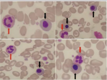
80% of cases, respectively. The white blood count is not stereotypically altered and may be normal or reduced. However, if the patient presents with leukocytosis, abnormal cells are commonly identiied. A leukoerythroblastic inding in the peripheral blood is present in 50% of cases (Figure 2)(5, 6, 8, 22, 26, 27). In addition, the patient can
present with elevated transaminases and uric acid; high levels of
FIGURE 1 − BMN pathophysiology: inflammation and plugs of sickle cells or neoplastic cells cause vascular obstruction and disrupt oxygen/nutrient delivery to hematopoietic tissue. BM infiltration (for example, by leukemic cells) can provoke microenvironment imbalance, as does cytotoxicity induced by irradiation, or drugs – either can cause vascular collapse –. TNF-α produced by monocytes and macrophages, and superoxide released by granulocytes, induce endothelial lesions with platelet aggregation, and vascular obstruction. All of these events can lead to BMN
BMN: bone marrow necrosis; TNF-α: tumor necrosis factor alpha; BM: bone marrow.
FIGURE 2 − A peripheral blood smear (100×): Leishman’s stain indicating leukoerythroblastic picture in a case of BMN. Black arrows show nucleated erythrocyte precursors, with red arrows indicating granulocytes
186
FIGURE 3 − BM biopsy in BMN: A) BM biopsy of a patient with Burkitt lymphoma showing severe necrosis (HE, 100×); B) BM coagulative necrosis with “ghost” neoplastic cells (HE, 200×); C) CD20 staining, 400×
BM: bone marrow; BMN: bone marrow necrosis; HE: hematoxilin and eosin.
lactic dehydrogenase and alkaline phosphatase are found in 41% and 51% of cases, respectively. There are no laboratory abnormalities associated speciically with BMN and, if suspected, its diagnosis should be supported by conirmatory data collated from a number of different approaches(6, 8, 22).
The main procedures needed to reach a diagnosis of BMN are BM aspiration, BM biopsy, and magnetic nuclear resonance (MNR) imaging. However, BM biopsy is the most illustrative and requires evaluation by an expert pathologist(16, 19). Although
BM aspirates are not commonly dry, their morphology may indicate a gelatinous degeneration, including cellular debris. The main indings observed from BM histology are amorphous and eosinophilic material dispersed in the extracellular spaces that should be quantiied to permit lesion classiication (see below). Rare cells with poorly delimited cytoplasmic membranes, irregular nuclei, and shrunken or vacuolated cytoplasm may be
also found(6, 8, 15, 18). In some instances, coagulative necrosis may
be observed with “ghost” cells that may reveal neoplastic features and even a diagnostic immune phenotype (Figure 3). Bone destruction with a lack of osteoblasts, osteoclasts, and osteocytes, together with empty osteocytic lacunae, may also occur in more advanced lesions(8).
The classiication of BMN is based upon the extension of BM involvement and ranges from grade I to III (Table 3). Bone scintigraphy using 99technetium may also prove to be useful
in reaching a diagnosis of BMN. Like MNR, it is a noninvasive technique and can also be used as a guide for the optimal sampling of a BM aspirate and/or biopsy. Areas with 99technetium uptake
higher than 2 cm are suggestive of BMN(8). By MNR, a diffuse and
TABLE 3 − Classification of BMN according to the extent
of BM involvement in a biopsy
BMN grade Classification Extent
I Slight < 20%
II Moderate 20%-50%
III Severe > 50%
BMN: bone marrow necrosis; BM: bone marrow.
FIGURE 4 − MNR in BMN 1: upper left to lower right – MNR sagittal T1, sagittal, coronal, and axial T2 with fat saturation. A patient with dermatomyositis diagnosed with BMN. Red arrows indicate areas of increased liquid content
MNR: magnetic nuclear resonance; BMN: bone marrow necrosis.
FIGURE 5 − MNR in BMN 2: left to right – MNR coronal STIR, axial T2 with fat saturation, T1 after contrast with fat saturation. Red arrows indicate areas of increased BM liquid
MNR: magnetic nuclear resonance; BMN: bone marrow necrosis; STIR: short-TI inversion recovery; BM: bone marrow.
extensive increase of liquid content, usually in the vertebral body and pelvic bones, may be seen (Figures 4 and 5)(3, 8, 11). The BMN
lesion is more extensive and diffuse compared to avascular bone injury, which involves more focal and periarticular necrosis.
A
C
B
TREATMENT
The approach to BMN involves treatment of the primary cause together with supportive care with antibiotics and blood transfusion. Most studies consider that a diagnosis of BMN implies a poor prognosis that, ordinarily, will rapidly lead to death. However, new studies indicate that survival may be prolonged if BMN is not associated with neoplasia(7, 8, 15). In cases associated with neoplasia,
overall survival remains poor, although a lack of data precludes more succinct outcome descriptions. Moreover, a rare but possible complication of BMN is the embolism of necrotic bone marrow tissue that then blocks vessels in multiple organs, to provoke infarct.
CONCLUSION
Based on this review, we conclude that BMN is associated with many diseases, and yet presents with no clear pathophysiology. In addition, clinical and laboratory characteristics are non-speciic and, in many cases, symptoms are related to the causal condition that is most commonly a hematological disease. Finally, the most important conclusion is that in all cases of BMN a malignant disease must be ruled in or out, given that malignancies are the most common cause of this lesion.
ACKNOWLEDGMENTS
We would like to thank Drs. Liliana Suganuma (physician – hematology and hemotherapy) and Bruno Trindade (medical resident – radiology), both from the Medical College of Universidade de São Paulo (USP), for images indicating the leukoerythroblastic reaction, and MNR data, respectively.
CONFLICT OF INTEREST
None of the authors have any conlict of interest regarding inancial, ethical, or any other personal issues concerning this article.
CONTRIBUTORS
All authors have materially contributed to this article in terms of their research, written contributions, revision, or selection of appropriate supporting material including igures.
All authors have approved the inal version of this article.
RESUMO
Introdução: Necrose de medula óssea (NMO) é uma entidade rara, frequentemente não diagnosticada e mais comumente associada a doenças hematológicas. Metodologia: Realizou-se revisão da literatura na base de dados do PubMed, utilizando o termo “necrose de medula óssea”. Foram encontrados 25 artigos em inglês e 65 relatos de caso. Resultados e discussão: A fisiopatologia da NMO não é bem elucidada e parece estar associada a lesão vascular com consequente hipóxia celular por desbalanço na oferta de oxigênio e nutrientes. O fator de necrose tumoral alfa (TNF-α) provavelmente também está implicado na lesão endotelial e nos sinusoides da medula óssea. Sugere-se o diagnóstico pela presença de anemia, trombocitopenia, reação leucoeritroblástica, níveis elevados de desidrogenase lática e fosfatase alcalina. Aspirado e biópsia de medula óssea e ressonância nuclear magnética são os principais exames diagnósticos. As únicas possibilidades terapêuticas são tratamento da causa de base e medidas suportivas. Conclusão: O ponto mais importante no manejo da NMO é a busca por condições neoplásicas associadas.
Unitermos: necrose de medula óssea; fator de necrose tumoral alfa; leucoeritroblástica; aspirado de medula óssea; biópsia de medula óssea.
REFERENCES
1. Wade L, Stevenson L. Necrosis of bone marrow with fat embolism in sickle cell anemia. Am J Pathol. 1941; 17: 47-54.
2. Jain D, Singh T, Kumar N. Hypocellular acute myeloid leukemia with bone marrow necrosis in young patients: two case reports. J Med Case Rep. 2009; 3: 27-31.
3. Campiotti L, Codari R, Appio L, et al. Bone marrow necrosis related to imatinib mesylate therapy for CML bilateral blast crisis. Leuk Res. 2007; 31: 1765-72.
4. Azuma T, Sakai I, Matsumoto T, et al. Leukemoid reaction in association with bone marrow necrosis due to metastatic prostate cancer. Intern Med. 2005; 44: 1093-6.
188
CORRESPONDING AUTHOR
Tamara Carvalho dos Santos Cabral
Rua Manoel Barreto, 324, apto 902; Graça; CEP: 40150-360; Salvador-BA, Brasil; e-mail: [email protected].
thrombotic thrombocytopenic purpura as a manifestation of occult colon cancer. Jpn J Clin Oncol. 2004; 34: 476-80.
6. Paydas S, Ergin M, Baslamisli F, et al. Bone marrow necrosis:
clinicopathologic analysis of 20 cases and review of the literature. Am J Hematol. 2002; 70: 300-5.
7. Bashawri L, Satti MB. Bone marrow necrosis: report of ive cases and review of the literature. Ann Saudi Med. 2000; 20: 78-82.
8. Janssens A, Offner FC, van Hove WZ. Bone marrow necrosis. Cancer. 2000; 88: 1769-80.
9. Chim CS, Lam CK, Wong KF, et al. Atypical blasts and bone marrow necrosis associated with near-triploid relapse of acute promyelocytic leukemia after arsenic trioxide treatment. Hum Pathol. 2002; 33: 849-51. 10. Markovic SN, Phyliky RL, Li CY. Pancytopenia due to bone marrow necrosis in acute myelogenous leukemia: role of reactive CD8 cells. Am J Hematol. 1998; 59: 74-8.
11. Lee Y, Hong YC, Yang CF, et al. Severe extensive bone marrow necrosis from military tuberculosis without granulomas and pulmonary presentation. J Chin Med Assoc. 2010; 73: 208-11.
12. Rossi D, Ramponi A, Franceschetti S, Stratta P, Gaidano G. Bone marrow necrosis complicating post-transplant lymphoproliferative disorder: resolution with rituximab. Leuk Res. 2008; 32: 829-34. 13. Kumakura S, Ishikura H, Kobayashi S. Bone marrow necrosis and Lambert-Eaton syndrome associated with interferon alpha treatment. N Engl J Med. 1998; 338: 196-203.
14. Katayama Y. Bone marrow necrosis in a patient with acute myeloblastic leukemia during administration of G-CSF and rapid hematologic recovery after allotransplantation of peripheral blood stem cells. Am J Hematol. 1998; 57: 238-40.
15. Al-Gwaiz LA. Bone marrow necrosis. Ann Saudi Med. 1997; 17: 374-6. 16. Invernizzi R, D’Alessio A, Iannone AM, Pecci A, Bernuzzi S, Castelloal A. Bone marrow necrosis in acute lymphoblastic leukemia. Haematologica. 1995; 80: 572-3.
17. Godeau B, Galacteros F, Schaeffer A, et al. Aplastic crisis due to extensive bone marrow necrosis and human parvovirus infection in sickle cell disease. JAMA. 1991; 91: 557-8.
18. Ianotto JC, Tempescul A, Eveillard JR, Charles F, Berthou C. Non-lethal bone marrow necrosis under irst line arsenic trioxide therapy in acute promyelocytic leukaemia. Acta Haematol. 2010; 123: 146-7.
19. Khoshnaw NS, Al-Rawi HA, Nore BF. Precursor T-cell acute lymphoblastic leukemia presenting with bone marrow necrosis: a case report. J Med Case Rep. 2012; 6: 349-55.
20. Aras Y, Akakaya MO, Unal SN, Bilgic B, Unal OF. Bone marrow necrosis secondary do imatinib usage, mimicking spinal metastasis on magnetic resonance and FDG-PET/CT. J Neurosurg Spin. 2012; 16: 57-60. 21. Khalil YK, Pistoria MJ, Wright RE. Catastrophic antiphospholipid antibody syndrome with bone marrow necrosis: a rare complication. J Rheumatol. 2011; 38: 2279-81.
22. Aydogdu I, Erkurt MA, Ozhan O, et al. Reversible bone marrow necrosis in a patient due to overdosage of diclofenac sodium. Am J Hematol. 2006; 81: 298-305.
23. Ozkan A, Ozkalemkas F, Ali R, Ozkocaman V, Ozelik T. Severe bone marrow necrosis without suggestive features. Am J Hematol. 2006; 81: 382-8. 24. Vermeersch P, Zachee P, Brusselmans C. Acute myeloid leukemia with bone marrow necrosis and Charcot Leyden crystals. Am J Hematol. 2007; 82: 1029.
25. Rossi P, Curiel M, Demoux AL et al. Bone marrow necrosis and sickle
cell crisis associated with double heterozygosity for HbS and HbOARAB. Am J Hematol. 2010; 86: 309-10.
26. Adamski J, Hanna CA, Reddy VB, Litovsky SH, Evans CA, Marques MB. Multiorgan failure and bone marrow necrosis in three adults with sickle
cell β+-thalassemia. Am J Hematol. 2012; 87: 621-4.
27. Noguchi M, Oshimi K. Extensive bone marrow necrosis and symptomatic hypercalcemia in B cell blastic transformation of chronic myeloid leukemia: report of a case and review of the literature. Acta Haematol. 2007; 118: 111-6.
28. Dunn P, Shin LY, Liaw S, Sun C. Bone marrow necrosis in 38 cancer patients. J Formos Med Assoc. 1985; 92: 1107-10.
29. Pennaforte JL, Dufour M, Etienne JC, Schvartz H, Caulet T. Diagnostic problems in bone marrow necrosis: report of a case revealing acute leukemia. Arch Anat Cytol Pathol. 1986; 34: 111-6.
30. Brown CH. Bone marrow necrosis: a study of seventy cases. John Hopkins Med. 1972; 131: 189-203.