against Multiresistant Bacteria without Inducing Strong
Resistance
Edwin J. A. Veldhuizen1*, Ellen C. Brouwer2, Viktoria A. F. Schneider1, Ad C. Fluit2
1Department of Infectious Diseases and Immunology, Division of Molecular Host Defence, Faculty of Veterinary Medicine, Utrecht University, Utrecht, The Netherlands,
2Department of Medical Microbiology, University Medical Center Utrecht, Utrecht, The Netherlands
Abstract
The increased prevalence of multidrug-resistant (MDR) bacteria in combination with the relatively limited development of new antibiotics presents a serious threat to public health. In chicken, especially Extended-Spectrum ß-Lactamase (ESBL) carrying Enterobacteriaceae are often asymptomatically present but can infect humans. Due to their broad range antimicrobial activity cathelicidins and other host defence peptides, are considered to be an attractive alternative to conventional antibiotics. In this study, the antimicrobial activity of three chicken cathelicidins against a broad array of multidrug resistant bacteria was determined. All three peptides showed high antibacterial activity independent of the presence of MDR characteristics. Induction experiments using S. aureus and K. pneumoniae showed that although an increase in resistance was initially observed, susceptibility towards chicken cathelicidins remained high and no major resistance was developed. The combined results underline the potential of chicken cathelicidins as a new alternative to antibiotics.
Citation:Veldhuizen EJA, Brouwer EC, Schneider VAF, Fluit AC (2013) Chicken Cathelicidins Display Antimicrobial Activity against Multiresistant Bacteria without Inducing Strong Resistance. PLoS ONE 8(4): e61964. doi:10.1371/journal.pone.0061964
Editor:Axel Cloeckaert, Institut National de la Recherche Agronomique, France
ReceivedJanuary 23, 2013;AcceptedMarch 18, 2013;PublishedApril 22, 2013
Copyright:ß2013 Veldhuizen et al. This is an open-access article distributed under the terms of the Creative Commons Attribution License, which permits unrestricted use, distribution, and reproduction in any medium, provided the original author and source are credited.
Funding:The study was partly funded by a SEED grant from the Infection & Immunity Center Utrecht of the University Medical Center Utrecht and University of Utrecht. The funders had no role in study design, data collection and analysis, decision to publish, or preparation of the manuscript. No additional external funding received for this study.
Competing Interests:The authors have declared that no competing interests exist.
* E-mail: [email protected]
Introduction
The world-wide increase in antibiotic resistance has severely reduced the current treatment options for infectious diseases. This issue is particularly serious in the case of Extended-Spectrum ß-Lactamase (ESBL) carrying Enterobacteriaceae. ESBLs confer resistance to third-generation cephalosporins, a class of antibiotics that is often used for empiric therapy. The growing levels of third-generation cephalosporin resistance leave only carbapenems as reliable treatment option, however, resistance against these antibiotics is also increasingly reported [1]. Similarly, methicillin-resistantStaphylococcus aureus(MRSA) remain a problem with rising numbers of community-acquired MRSA and the global spread of livestock-associated MRSA, in particular ST398 [2,3]. Alternative treatment options are urgently required to address the dangers posed by these drug-resistant pathogens. Cathelicidins are a class of antimicrobial peptides that may provide this alternative. Chicken cathelicidins are particularly interesting in this respect, considering the fact that the chicken is a major non-symptomatic carrier of multiresistant bacteria.
Cathelicidins are Host Defence Peptides (HDP) that play an important role in the innate immune system. They exhibit broad range antimicrobial activity against both Gram-negative and Gram-positive bacteria, as well as against fungi and parasites. To date, most study has focused on the antibacterial mode of action of these peptides. So-called lytic peptides, such as the human cathelicidin LL-37, bind to bacterial membranes and either form
pores or lead to destabilization of the membrane, eventually leading to lysis of the bacteria. Other cathelicidins, such as porcine PR-39 cross the bacterial membrane to access intracellular targets leading to inhibition of protein and DNA synthesis [4,5]. In addition to antimicrobial activity, many HDPs possess immuno-modulatory activities including lipopolysaccharide (LPS) binding, induction of cytokine production and chemotaxis [6]. This multiplicity of functions of the HDPs adds to their potential for use as alternative for antibiotics.
these cells upon degranulation of these cells. The localization of CATH-3 has not been determined but is assumed to be similar to CATH-1 and -2 [13]. Contrary to this localization in immune cells, CATH-B1 is exclusively produced in the epithelial cells surrounding the M-cells in the bursa of Fabricius, suggesting that this peptide has a local role in forming a defense layer to protect the bursa from infection [7], although low levels of CATH-B1 RNA were also found in other tissues [14]. Despite the differences in localization, sequence and structure of the chicken cathelicidin subset, no significant functional differences between them have been identified.
To investigate the antimicrobial potential of chicken CATH-1-3, the minimal inhibitory concentration against a large set of clinically relevant bacterial species and strains was investigated. In addition, induction of resistance against cathelicidins was deter-mined in three bacterial species.
Materials and Methods
Bacterial Isolates
In total 39 clinical bacterial isolates belonging to 25 Gram-negative and Gram-positive species were used in this study. The minimal inhibitory concentrations (MICs) of the isolates for different sets of antibiotics were determined according to the CSLI guidelines (Table S1) [15,16]. The selection included methicillin-resistantS. aureus, vancomycin-resistant enterococci, ESBL-positive
Escherichia. coli and carbapenemase-positive Klebsiella pneumoniae. MICs against the three isolates used in resistance development (see below) for the three chicken CATHs were determined using the same method.
Peptides
The mature peptides of chicken CATH-1, -2 and -3 were synthesized by Caslo laboratory APS, Lyngby, Denmark. All three peptides were purified to .95% purity using HPLC, and mass spectrometry analysis indicated that the mass of the peptides were within 1 dalton of the theoretical value.
Antimicrobial Activity Assays
-Colony count assays. Antimicrobial activity of the three
chicken cathelicidins was tested against two MRSA, five ESBL-positive strains and a vancomycin-resistant Enterococcus faecium. Bacteria were maintained in Tryptic Soy Broth (TSB, Oxoid Limited, Hampshire, UK) at 37uC and grown to mid-logarithmic phase before testing. Colony count assays were performed to test the activity of CATHs as described previously [17]. In short, bacteria were pelleted and resuspended in 10 mM sodium phosphate buffer pH 7.0 containing 1/100 TSB and diluted to 26106CFU/ml. A 25ml aliquot of CATH peptide solution was mixed with 25ml of bacterial culture and incubated for 3 h at
37uC. Subsequently, the cultures were diluted 50–5000 fold and spread plated on Tryptic Soy Agar plates and after 24 h at 37uC counted for surviving bacteria. The plates containing less than 10 colonies at the lowest sample dilution was defined as the minimal bactericidal concentration (MBC,.3 log reduction in CFU/ml).
-Spot-test. CATH-1, -2, and -3 were screened against 39
bacterial strains from different Gram-negative and Gram-positive species. The bacteria were cultivated overnight at 37uC, a suspension of bacteria (56105CFU/ml) in distilled water was
prepared and spread over Mu¨ller-Hinton (MH) agar plates. After the plates were dried, 20ml of the CATH peptide solution (32mM) was spotted. After overnight culture at 37uC the plates were examined for growth inhibition by measuring the diameter of the zones and noting the presence or absence of colonies within the spot area.
Induction of CATH Resistance inS. aureusandK. pneumoniae
S. aureus S0385, K. pneumoniae 03C006, and K. pneumoniae
NCTC13443 were cultured for 10 days in MH broth in the presence of CATH-1, -2, or -3, in a series of concentrations ranging from 0 to 40mM. Daily, 50ml samples displaying 80% growth compared with growth in MH broth only, were used to inoculate a new dilution series (0.5 ml). At day 10 broth dilution assays were performed to determine MIC values. Specifically, MICs were measured by sampling from the overnight cultures with highest peptide concentration that still showed detectable growth. These cultures were diluted till 26106CFU/ml after which a 25ml aliquot was incubated with 25ml CATH (0–80mM) for 3 h. Two-hundred ml MH broth was then added and incubation was continued for an additional 21 h. The concentra-tion at which no visible bacterial growth was present was taken as the MIC value.
Results
Antimicrobial Activity Tests
-Colony count assays. MBCs for CATH-1-3 were
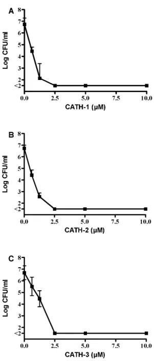
deter-mined using colony count assays against two MRSA, five ESBL-positive strains and a vancomycin-resistantE. faecium. The data for the activity of all three CATHs against ESBL-positiveE. coli38.34 is shown in Figure 1. At a concentration of 0.6mM an approximately 2 log decrease in bacterial counts is observed for all three peptides. A higher concentration of peptide (2.5mM) led to complete killing of all bacteria. MBC values using this method against the other strains tested are shown in Table 2 and indicate that no significant differences in MBC values were observed between the three peptides, and similar MBC values were observed for all bacteria tested. MIC values for the peptides determined by broth dilution assays in MH broth of the three isolates used in resistance development were in the same order of magnitude (0.63–1.25mM).
-Spot-test. CATH-1, -2, -3 showed antibacterial activity
against all 39 Gram-negative and Gram-positive bacterial strains belonging to 25 different species (Table 3). Peptide addition to a spread layer of Gram-positive bacteria resulted in clear inhibition zones for all bacteria tested. However, varying numbers of colonies were observed in the clear zone for most Gram-negative species. Exceptions were the Acinetobacter spp, Stenotrophomonas maltophilia, and oneK. pneumoniaestrain. The presence of colonies in the clear zone was strongest for CATH-1 where 13 of 18 tested Gram-negative strains (72%) showed colonies in the clear zone and lowest for CATH-3 where this effect was only present in 2 of the 18 (11%) strains. No difference was observed for ESBL- or Table 1.Amino acid sequence of mature chicken
cathelicidins.
Amino acid sequence AA Charge
CATH-1 RVKRVWPLVIRTVIAGYNLYRAIKKK 26 +8
CATH-2 RFGRFLRKIRRFRPKVTITIQGSARF 26 +9
CATH-3 RVKRFWPLVPVAINTVAAGINLYKAIRRK 29 +7
CATH-B1 PIRNWWIRIWEWLNGIRKRLRQRSPFYVR GHLNVTSTPQP
40 +7
carbapenemase-positive strains. For some bacteria, colonies from the clearance zone were grown overnight (in the absence of CATH) and used again for a spot test using the same peptide, to determine if total resistance was acquired. Without exception these experiments resulted in a similar clearance zone diameter and a comparable number of colonies in the clearance zone (data not shown) indicating the presence of heterogeneous resistance in these bacterial strains.
Induction of CATH Resistance inS. aureusandK. pneumoniae
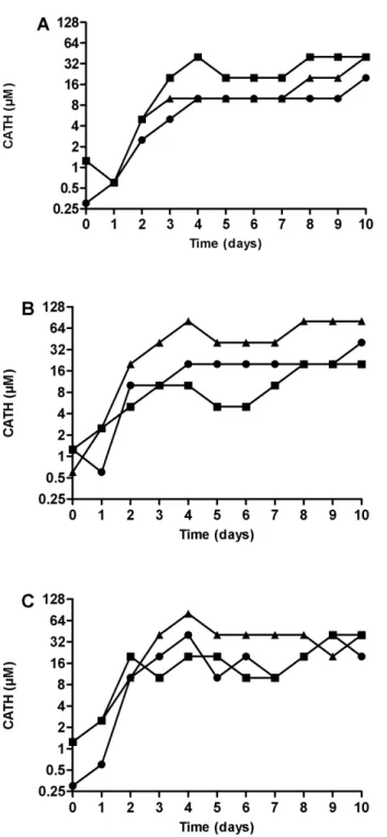
As a representative Gram-positive bacteria,S. aureusS0385 was used, whereas for Gram-negative bacteria twoK. pneumoniaestrains were selected: one strain that showed colonies in the clear zone in the spot test (K. pneumoniae NCTC13443) and one that did not show any colony growth in this zone (K. pneumoniae03C006). All isolates reached maximum levels of reduced susceptibility within 4 days of the start of the experiment (Figure 2). No clear differences in the level of reduced susceptibility between the three CATHs were observed.
After day 10 of the induction with each CATH the MICs for each isolate from the dilution showing at least 80% growth were determined using broth dilution assays and colony count assays. Interestingly, the MIC values were much lower than the tolerated levels of CATHs during the resistance development experiment. In addition, although the MICs were higher for bacteria with reduced sensitivity than bacteria that did not have reduced sensitivity induced, the actual increase is still relatively small: on average 2–4 fold in the broth dilution assay (Table 4). These results were confirmed for S. aureus S0385 using colony count assays where even smaller, and in some cases no differences, were observed between the bacteria grown in the presence or absence of CATHs (data not shown).
Discussion
In this investigation we determined the antimicrobial activity of chicken CATHs 1–3 against ESBL-positive Enterobacteriaceae, MRSA, and other bacterial species. The fourth chicken cathe-licidin CATH- B1 was left out of our studies due to its considerable bigger size and restricted localization, which lowers the potential of this peptide as alternative to antibiotics. Whether chicken CATHs are lytic to bacteria or possess a mode of action involving binding to intracellular targets is as of yet unknown. Interestingly, our results show no differences in MIC and MBC values for the three chicken CATHs tested, despite their considerable differences in amino acid sequence and structure, especially between CATH-2 and the other two CATH peptides. In addition, MBC values for all multiresistant strains tested were comparable to
non-multire-Figure 1. Antibacterial activity of CATH-1-3 against E. coli 38.34.E. coli(16106CFU/ml) were incubated with CATH-1-3 for 3 h.
Surviving bacteria were determined using colony count assays. All experiments were performed at least in triplicate. A: CATH-1; B: CATH-2; C: CATH-3.
doi:10.1371/journal.pone.0061964.g001
Table 2.Antibacterial activity of chicken cathelicidins against multiresistant bacteria.
Bacterial strain MBC (mM)
* CATH-1 CATH-2 CATH-3
Escherichia coli38.34 a 1.25–2.5 1.25–2.5 2.5
Escherichia coli38.16 b 1.25–2.5 1.25–2.5 2.5–5
Staphylococcus aureusS0385 c 1.25 1.25–2.5 1.25–5
Staphylococcus aureusWKZ2 d 1.25–2.5 2.5 2.5
Klebsiella pneumoniaeNCTC-13443 e 1.25–2.5 1.25–2.5 1.25–5
Klebsiella pneumoniaeATCC-BAA-1705 f 1.25–2.5 1.25–2.5 1.25–2.5
Pseudomonas aeruginosaVW178 g 0.6–1.25 1.25 1.25–2.5
Enterococcus faeciumE155 h 0.6–1.25 1.25 1.25
*: a) CTX-M-1 ESBL positive from chicken, b) TEM-52 -ESBL positive from chicken, c) methicillin resistant, livestock-associated ST398, d) methicillin resistant, clinical isolate, e) NDM-1 carbapenemase positive, f) KPC carbapenemase positive, g) cystic fibrosis patients, h) vancomycin resistant. Bacteria (16106CFU/ml) were incubated with CATH-1-3 for 3 h. Surviving
bacteria were determined using colony count assays. All experiments were performed at least in triplicate.
sistant strains [10]. This indicates that the antibacterial action of chicken CATHs is unrelated to the mechanism of action of classical antibiotics.
Remarkably, in the spot test heterogeneous resistance was present among the Gram-negative species tested, but absent among Gram-positive tested bacteria. Although it was not observed in this study, the phenomenon of heterogeneous Table 3.Antimicrobial activity of chicken cathelicidins against a broad range of bacterial strains using spot-test.
CATH-1 CATH-2 CATH-3
Species isolate nr. * zone Ø (mm)colonies zone Ø (mm)colonies zone Ø (mm)colonies
Enterococcus faecalis 11E098 10 – 10 – 10 –
Enterococcus faecalis 15A374 9 – 9 – 9 –
Enterococcus faecium E155 a 10 – 10 – 10 –
Enterococcus faecium 16D030 9 – 9 – 9 –
Enterococcus faecium 15A623 10 – 10 – 10 –
Staphylococcus aureus S0385 b 10 – 11 – 11 –
Staphylococcus aureus 03A194 10 – 10 – 10 –
Staphylococcus epidermidis 08A1057 12 – 12 – 12 –
Staphylococcus epidermidis 08A1071 12 – 12 – 12 –
Staphylococcus haemolyticus 10A630 11 – 11 – 11 –
Staphylococcus intermedius 09D123 11 – 11 – 11 –
Staphylococcus lugdunensis 10A302 10 – 10 – 10 –
Staphylococcusspecies 19A337 11 – 11 – 11 –
Streptococcus bovis 12A090 10 – 8 – 8 –
Streptococcus pyogenes 05D015 12 – 11 – 11 –
Streptococcus pyogenes 23M092 11 – 11 – 11 –
Streptococcus agalactiae 05A396 10 – 10 – 10 –
Streptococcus mitis 01A162 10 – 10 – 10 –
Streptococcus pneumonia 14B186 11 – 11 – 11 –
Streptococcus salivarius 14A071 10 – 10 – 10 –
Streptococcus sanguis 08A557 11 – 11 – 11 –
Acinetobacter baumannii 06A330 10 – 10 – 10 –
Acinetobacter calcoaceticus 15A600 10 – 10 – 10 –
Citrobacter diversus 08A083 11 11 – 0 –
Citrobacter freundii 15C098 10 + 10 – 0 –
Enterobacter aerogenes 20A063 11 + 11 – 11 +++
Escherichia coli 01A280 11 +++ 11 ++ 0 –
Escherichia coli 23E068 10 + 10 – 10 –
Escherichia coli 38.34 c 10 +++ 11 – 0 –
Escherichia coli 38.16 d 0 +++ 10 ++ 0 –
Klebsiella pneumoniae 03C006 10 – 10 – 0 –
Klebsiella pneumoniae NCTC-13443 e 10 +++ 11 – 0 –
Klebsiella pneumoniae BAA-1705 f 11 ++ 10 + 0 –
Pseudomonas aeruginosa VW178 g 10 + 10 + 0 –
Pseudomonas aeruginosa 04A191 10 +++ 10 +++ 0 –
Pseudomonas aeruginosa 13A066 10 + 10 + 10 ++
Salmonella enteritidis 19A060 0 +++ 10 – 0 –
Salmonella typhimurium 10A629 0 +++ 10 ++ 0 –
Stenotrophomonas maltophilia 16C077 11 – 11 – 10 –
All strains were human isolates unless otherwise noted.
*: a) vancomycin resistant, b) methicillin resistant, livestock associated ST398, c) CTX-M-1 ESBL positive from chicken, d) TEM-52 -ESBL positive from chicken, e) NDM-1 carbapenemase positive, f) KPC carbapenemase positive, g) cystic fibrosis patients. Bacteria were cultivated overnight at 37uC, a suspension of bacteria (56105CFU/ml) in distilled water was prepared and spread over Mu¨ller-Hinton agar plates. After the plates were dried, 20ml of a CATH solution (32mM) was spotted, plates were
cultured at 37uC and examined for growth inhibition by measuring the diameter of the zones and noting the presence or absence of colonies within the spot area. The table represents single or duplicate screening experiments.
resistance has been described for Staphylococcus aureus, where vancomycin and methicillin resistance can be heterogeneously present. In only approximately one in a million cells in a population, resistance is expressed. The exact mechanisms are still
unknown, but for methicillin, next to the loss of a regulator protein, a chromosomal mutation appears to be necessary to obtain homogeneously expressed resistance [18]. An analogous phenomenon may occur here. Due to higher (or lower) than usual expression of resistance, e.g. due to a regulator, in only a few cells in a population, resistant colonies survive on a plate. In induction experiments these cells are increasingly selected and a mutation may further enhance the resistance level. When the culture is no longer exposed, revertants will take over due to a fitness advantage and resistance levels drop to wild-type values.
In the induction experiments with K. pneumoniaeand S. aureus, reduced sensitivity to all three CATHs tested,was obtained within a few days. The mechanism(s) explaining these observations are not known because resistance to cationic antimicrobial peptides, including cathelicidins, is only partially understood. Different peptides and different bacterial species or groups appear to have different resistance mechanisms [19]. In S. aureus at least five different mechanisms exist including production of proteases and HDP inactivating proteins, alteration of membrane fluidity and membrane charge and expression of multidrug pumps [20]. Several of these mechanisms have also been shown to be induced in the presence of HDPs through the activation of bacterial two-and three-component systems. In Gram-negative bacteria similar mechanisms exist, e.g.,phoPQregulated genes [21–23]. An efflux pump has been implicated in Neisseria gonorrhoeae [24], a phoPQ
regulated protease inSalmonella entericaserovar Typhimurium [21], and a protease in enterohemorrhagicE. coli[25]. However, these mechanisms mostly seem to involve an intrinsic resistance to cathelicidins, because knock-outs of the proposed genes result in reduced resistance compared to wild-type.
In the 10 day resistance induction experiment, overnight growth was used as read out parameter but this has only limited value since a very small number of surviving bacteria could potentially grow out to a full density overnight culture. Indeed growth curves of bacteria in the presence of sub-MIC concentration of CATH show that the peptide increases the time till exponential growth phase is reached (data not shown). Obviously, in vivo this delay could be enough for other immune factors to effectively eradicate the remaining bacteria. In addition, the actual MIC determined by colony count assays and broth dilution assays were much lower than the concentration where overnight growth was observed, and only slightly higher compared to non-induced bacteria. The larger volumes used during the induction experiment compared to broth dilution assays might partially explain this difference. If heteroge-neous resistance is present, the chance of having resistant bacteria is higher at larger volumes. More importantly, the number of bacteria transferred to the next generation (,107CFU/ml) is 10 Figure 2. Induction of CATH-1,-2, and-3 resistance inS. aureus
S0385 (panel A),K. pneumoniaeNCTC-13443 (panel B) andK. pneumoniae 03C006 (panel C). Bacteria were grown o/n in the presence of 0–80mM peptide. The sample containing the highest CATH
concentration showing.80% bacterial growth compared to a control without CATH is shown. Subsequently, this bacterial culture was subcultured into new medium containing 0–80mM CATH peptide. This
procedure was repeated for 9 consecutive days. Circles: CATH-1; squares: CATH-2; triangles: CATH-3. Shown is the peptide tolerance (80% growth) over 10 days in a single induction experiment. doi:10.1371/journal.pone.0061964.g002
Table 4.Antibacterial activity of chicken cathelicidins before and after induction of resistance.
MIC (mM)
CATH-1 CATH-2 CATH-3
Bacterial strain Day 0 Day 10 Day 0 Day 10 Day 0 Day 10
Staphylococcus aureus
S0385
0.3 1.25 1.25 5 1.25 1.25
Klebsiella pneumoniae 03C006
0.6 2.5 1.25 10 1.25 1.25
Klebsiella pneumonia
NCTC-13443
1.25 2.5 1.25 2.5 0.6 5
fold higher than used in the broth dilution assays, again increasing chances of transferring resistant bacteria. Finally, in experiments in our group using fluorescently labeled peptides, it was observed that peptides instantly localize to microbial membranes and that the peptide was often heterogeneously distributed among cells. This heterogeneous distribution would lead to bacterial cells receiving sub-MIC concentrations of peptide, enabling them to eventually grow out to a proper overnight culture. Overall, CATH resistance as determined by MIC values only increases slightly and this small increase is achieved within a few days and does not seem to develop further upon prolonged incubation periods. This indicates that, at least in our experimental set-up, no major resistance mechanisms leading to loss of susceptibility towards CATHs are induced, contrary to the resistance development described for more classic antibiotics.
In summary, our experiments show an antimicrobial activity of CATH-1, -2, and -3 against both negative and Gram-positive bacteria, independent of the presence of resistance mechanism towards classic antibiotics. No clear differences in activity were observed between the three CATHs. Heterogeneous resistance was noted in Gram-negative species in spot assays, but
induction of resistance towards chicken CATHs was low and leveled off after 3–4 days indicating that development of major resistance is unlikely to occur.
Supporting Information
Table S1 Susceptibility of bacterial strains used in this study for a large set of antibiotics. MICs were determined according to CSLI guidelines.
(XLSX)
Acknowledgments
The authors wish to thank Prof. Dr. Henk P. Haagsman and Dr. Nathaniel I. Martin for critically reading of the manuscript.
Author Contributions
Conceived and designed the experiments: EV EB AF. Performed the experiments: EV EB VS. Analyzed the data: EV EB AF. Contributed reagents/materials/analysis tools: EV AF. Wrote the paper: EV AF.
References
1. Woodford N, Turton JF, Livermore DM (2011) Multiresistant Gram-negative bacteria: The role of high-risk clones in the dissemination of antibiotic resistance. FEMS Microbiol Rev 35: 736–755.
2. Gould IM, David MZ, Esposito S, Garau J, Lina G, et al. (2012) New insights into meticillin-resistantStaphylococcus aureus(MRSA) pathogenesis, treatment and resistance. Int J of Antimicrob Agents 39: 96–104.
3. Fluit AC (2012) Livestock-associatedStaphylococcus aureus. Clin Microbiol Infect 18: 735–744.
4. Boman HG, Agerberth B, Boman A (1993) Mechanisms of action onEscherichia coliof cecropin P1 and PR-39, two antibacterial peptides from pig intestine. Infect Immun 61: 2978–2984.
5. Shi J, Ross CR, Chengappa MM, Sylte MJ, McVey DS, et al. (1996) Antibacterial activity of a synthetic peptide (PR-26) derived from PR-39, a proline-arginine-rich neutrophil antimicrobial peptide. Antimicrob Agents Chem 40: 115–121.
6. Kai-Larsen Y, Agerberth B (2008) The role of the multifunctional peptide LL-37 in host defense. Front Biosci : 13: 3760–3767.
7. Goitsuka R, Chen CL, Benyon L, Asano Y, Kitamura D, et al. (2007) Chicken cathelicidin-B1, an antimicrobial guardian at the mucosal M cell gateway. Proc Natl Acad Sci USA 104: 15063–15068.
8. Xiao Y, Cai Y, Bommineni YR, Fernando SC, Prakash O, et al. (2006) Identification and functional characterization of three chicken cathelicidins with potent antimicrobial activity. J Biol Chem 281: 2858–2867.
9. van Dijk A, Veldhuizen EJA, van Asten AJ, Haagsman HP (2005) CMAP27, a novel chicken cathelicidin-like antimicrobial protein. Vet Immunol Immuno-pathol 106: 321–327.
10. van Dijk A, Molhoek EM, Veldhuizen EJA, Bokhoven JL, Wagendorp E, et al. (2009) Identification of chicken cathelicidin-2 core elements involved in antibacterial and immunomodulatory activities. Mol Immunol 46: 2465–2473. 11. Bommineni YR, Dai H, Gong YX, Soulages JL, Fernando SC, et al. (2007) Fowlicidin-3 is an alpha-helical cationic host defense peptide with potent antibacterial and lipopolysaccharide-neutralizing activities. FEBS J 274: 418– 428.
12. Xiao Y, Herrera AI, Bommineni YR, Soulages JL, Prakash O, et al. (2009) The central kink region of fowlicidin-2, an alpha-helical host defense peptide, is critically involved in bacterial killing and endotoxin neutralization. J Innate Immun 1: 268–280.
13. van Dijk A, Molhoek EM, Bikker FJ, Yu PL, Veldhuizen EJA, et al. (2011) Avian cathelicidins: Paradigms for the development of anti-infectives. Vet Microbiol 153: 27–36.
14. Achanta M, Sunkara LT, Dai G, Bommineni YR, Jiang W, et al. (2012) Tissue expression and developmental regulation of chicken cathelicidin antimicrobial peptides. J Anim Sci and Biotechnol 3: 15.
15. Wayne P (2009) M07-A8. Methods for dilution antimicrobial susceptibility tests for bacteria that grow aerobically; approved standard: Eighth edition. Clinical and Laboratory Standards Institute.
16. Wayne P (2012) M100-S22. Performance standards for antimicrobial suscepti-bility testing: 22nd informational supplement. Clinical and Laboratory Standards Institute.
17. Veldhuizen EJA, Rijnders M, Claassen EA, van Dijk A, Haagsman HP (2008) Porcine beta-defensin 2 displays broad antimicrobial activity against pathogenic intestinal bacteria. Mol Immunol 45: 386–394.
18. Kondo N, Kuwahara-Arai K, Kuroda-Murakami H, Tateda-Suzuki E, Hiramatsu K (2001) Eagle-type methicillin resistance: New phenotype of high methicillin resistance undermec regulator gene control. Antimicrob Agents Chemother 45: 815–824.
19. Peschel A (2002) How do bacteria resist human antimicrobial peptides? Trends Microbiol 10: 179–186.
20. Koprivnjak T, Peschel A (2011) Bacterial resistance mechanisms against host defense peptides. Cell Mol Life Sci 68: 2243–2254.
21. Guo L, Lim KB, Poduje CM, Daniel M, Gunn JS, et al. (1998) Lipid A acylation and bacterial resistance against vertebrate antimicrobial peptides. Cell 95: 189– 198.
22. Guina T, Yi EC, Wang H, Hackett M, Miller SI (2000) A PhoP-regulated outer membrane protease of Salmonella enterica serovar Typhimurium promotes resistance to alpha-helical antimicrobial peptides. J Bacteriol 182: 4077–4086. 23. Belden WJ, Miller SI (1994) Further characterization of the PhoP regulon:
Identification of new PhoP-activated virulence loci. Infect Immun 62: 5095– 5101.
24. Shafer WM, Qu X, Waring AJ, Lehrer RI (1998) Modulation ofNeisseria gonorrhoeaesusceptibility to vertebrate antibacterial peptides due to a member of the resistance/nodulation/division efflux pump family. Proc Natl Acad Sci USA 95: 1829–1833.
25. Thomassin JL, Brannon JR, Gibbs BF, Gruenheid S, Le Moual H (2012) OmpT outer membrane proteases of enterohemorrhagic and enteropathogenic