Received : 28.03.2014 Accepted : 19.06.2014
Correspondence:Arbil AÇIKALIN
Çukurova Üniversitesi Tıp Fakültesi, Patoloji Anabilim Dalı, 01130, Sarıçam, ADANA, TURKEY
E-mail: [email protected] Phone: +90 532 470 21 20 (Turk Patoloji Derg 2014, 30:184-188)
ABSTRACT
Objective: Frozen section is an accurate diagnostic tool with some unavoidable pitfalls in gynecologic tumors. We aimed to evaluate the diagnostic value of frozen section, and to detect the factors causing erroneous diagnosis in ovarian tumors.
Material and Method: Frozen section and parafin section reports of 282 patients with ovarian neoplasms diagnosed between July 2006 and January 2013 in our institute were re-analyzed. Results were grouped into benign, borderline (for epithelial tumors) and malignant categories and compared between frozen section and parafin section diagnosis, statistically.
Results: Overall diagnostic accuracy of frozen section was 96.5%. Sensitivity of frozen section for benign, borderline and malignant tumors were 97.5%, 95.8%, and 95.6% and the related specificities were 97.5%, 97.6%, and 100%, respectively. We found the lowest positive predictive value in borderline group (79.3%), all of them with mucinous type epithelium. Second frequent discordant tumor type was immature teratoma.
Conclusion: Apart from the limitations of frozen section, pathologists should be aware of the pitfalls of technique and tumor types and tend to sample from the solid areas particularly in mucinous tumors and teratomas to avoid inappropriate surgery.
Key Words: Frozen section, Ovarian neoplasms, Quality control
ÖZ
Amaç: Jinekolojik tümörler için frozen kesit inceleme bazı tanısal tuzaklara karşın yeterli bir tanısal yöntemdir. Bu çalışmada, over tümörlerinde frozen incelemenin tanısal değerini ve tanısal yanlışlıkların nedenlerini tespit etmeyi amaçladık.
Gereç ve Yöntem: Kurumumuzda Haziran 2006 ve Ocak 2013 yılları arasında over tümörü nedeni ile frozen inceleme yapılmış olan toplam 282 hastaya ait sonuçlar yeniden değerlendirildi. Tanılar benign, borderline (epitelyal tümörler için) ve malign olmak üzere üç alt gruba ayrıldı. Frozen ve parafin kesit sonuçları istatistiksel olarak karşılaştırıldı.
Bulgular: Frozen incelemenin genel tanısal doğruluk oranı %96,5 idi. Benign, borderline ve malign tümörler için frozen incelemenin duyarlığı sırasıyla %97,5, %95,8 ve %95,6 idi. Özgüllük ise aynı sırayla, %97,5, %97,6 ve %100 idi. En düşük pozitif prediktif değer borderline grubunda saptandı (%79,3) ve hataya neden olan tüm olgular müsinöz tip epitelyal tümörler idi. Tanı uyumsuzluğu bulunan ikinci tümör grubu ise immatür teratom idi.
Sonuç: Frozen incelemenin kısıtlılıkları yanısıra, patoloğun frozen incelemenin teknik veya tümör tipi ile ilgili tuzaklarını bilmesi, özel-likle müsinöz tümörler ve teratom olgularında solid görünen alan-lardan örneklenmesi yetersiz veya gereksiz operasyon yapılmasını önlemek adına yararlı olacaktır.
Anahtar Sözcükler: Frozen inceleme, Over neoplazileri, Kalite kontrol
Intraoperative Frozen Section in Ovarian Neoplasms;
A Tertiary Center Experience
Over Tümörlerinde İntraoperatif Frozen İncelemenin Tanısal Yeri;
Tek Merkez Deneyimi
Arbil AÇIKALIN1, Goncagül TORUN1, Emine BAĞIR1, Fatma BAYRAM1, Handan ZEREN1, Ümran GÜLEÇ2, Ahmet Barış GÜZEL2, Derya GÜMÜRDÜLÜ1
Departments of 1Pathology and 2Obstetrics and Gynecology, Çukurova University, Faculty of Medicine, ADANA, TURKEY
INTRODUCTION
Ovarian cancer is the most common lethal gynecologic cancer and the seventh most common cause of death from cancer in women (4.2% of deaths) worldwide (1, 2). Surgical management of ovarian neoplasms difers according to their subtypes. It is therefore important for the surgeon to determine the category of ovarian neoplasm preoperatively. Preoperative imaging methods such as ultrasonography or serologic evaluations have been used for this aim but they
MATERIALS and METHOD
In this retrospective study, 282 ovarian neoplasms (42.8% of all gynecologic FSs) with intraoperative FS reports diagnosed in our institute between July 2006 and January 2013 (6.5 years) were re-analyzed. Parain section diagnoses with non-tumoral ovarian lesions (massive ovarian edema, hemorrhagic necrosis, benign cysts, infections) were not included.
All fresh gross specimen were examined by a resident and a pathologist or particularly gynecopathologist, in terms of localization, size, color, content, heterogeneity, iniltration pattern of the tumor and condition of the ovarian capsule. One to four sections depending on the size and heterogeneity of the tumor were sampled in a cryostat and sections were stained by hematoxylin-eosin. Slides were evaluated and reported to the surgeon by the pathologist. Final PS diagnosis reported by an experienced gynecopathologist was accepted as accurate diagnose. In present study, we re-analyzed the FS and PS reports of cases. FS reports were categorized as benign, borderline (BL)(for epithelial tumors) and malignant. Benign and malignant groups included all epithelial, sex-cord stromal and germ cell tumors. BL group included only epithelial tumors. Results were compared for each category. Discordant cases were discussed depending on the clinical efects of the discordant FS diagnosis. Overall accuracy meant concordance between FS and PS results regardless of subtypes of the tumors. Sensitivity and
speciicity, positive and negative predictive values of FS were determined statistically. Statistical tests were based on benign/BL/malignant categorization.
RESULTS
he mean age of all cases was 44.9 (min: 12, max: 80) years. A total 282 ovarian neoplasms consisting of: 235 (83.3%) epithelial tumors (85 benign, 24 BL, 126 malignant), 39 (13.8%) sex-cord stromal tumors, and 8 (2.8%) germ cell tumors were reviewed. All cases were diagnosed as benign, borderline (for epithelial tumors) or malignant on FS. Sub-categorization, in terms of histologic type, primary or metastasis was not possible in some cases of FS.
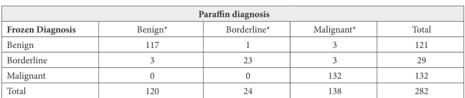
Overall diagnostic accuracy of FS was 96.5%. Sensitivity, speciicity, positive and negative predictive values of FS for benign, BL and malignant tumors are summarized in Table I.
Ten (3.5%) out of 282 cases were discordant based on benign/BL/malignant sub-categorisation, which also caused over- or under-surgery (Table II). Histopathologic diagnoses of PSs in 10 discordant cases were as follows: 3 benign mucinous tumors, 1 mucinous BL tumor, 3 malignant mucinous tumors and 3 immature teratomas. Immature teratomas were diagnosed as mature teratomas in FS, due to the sampling error. Two cases, 28 and 37 years old ages were grade 1 immature teratoma with 16 and 21 cm diameter tumor, subsequently. hree solid areas sampled during FS
Table I: Statistical values of benign, borderline and malignant tumors
Statistical result (%) Benign* Borderline* Malignant*
Sensitivity 97.5 95.8 95.6
Speciicity 97.5 97.6 100
Positive predictive value 96.7 79.3 100
Negative predictive value 98.1 99.6 96
* Benign and malignant tumors included epithelial, sex-cord stromal, germ cell tumors and metastasis, whereas borderline group included only epithelial tumors.
Table II: Comparison of frozen and parain diagnosis of all patients with respect to potential of malignancy
Parain diagnosis
Frozen Diagnosis Benign* Borderline* Malignant* Total
Benign 117 1 3 121
Borderline 3 23 3 29
Malignant 0 0 132 132
Total 120 24 138 282
did not include immature areas. On permanent sections, 3 slides for initial and 1 slide for the latter case included Grade 1 immature neuronal component among 20 parain samples plus 3 frozen samples (total 23 slides) for each. he third discordant case was 47 years old with a 10 cm diameter teratoma. Only 1 slide among 17 samples (14 parain+ 3 frozen) included Grade 2 immature neuronal area. Gross appearances of all these 3 cases were like a mature teratoma with chondroid, cystic, with keratinized material, and focal leshy gray-yellow areas. Although focal, representative areas were detected mainly on samples from solid ields. he other 7 discordant cases were all mucinous epithelial tumors. hree benign mucinous tumors were over-diagnosed as BL mucinous tumor on FS. One mucinous BL tumor was under-diagnosed as benign on FS. hree mucinous carcinomas were also under-diagnosed as BL. In terms of histologic subtypes; benign serous tumors (56 cases), Brenner tumors (2 cases), serous BL tumors (18 cases), endometrioid adenocarcinomas (3 cases), metastasis (35 cases), ibroma/ibrotechomas (29 cases), granulosa cell tumors (9 cases), mature cystic teratomas (5 cases) were all correctly diagnosed in FS. here was some discordance with respect to histologic subtype among the malignant tumors (Table III). hey were all reported to be malignant during FS, however histopathologic subtypes were diferent on PS report in 6 cases. Among these cases three were major mistakes, which caused under-/over-treatment due to FS diagnose. Two of them were 48 and 69 years old high-grade serous carcinoma cases, which were misdiagnosed as metastatic carcinoma during FS. hese tumors were bilateral and showed capsular involvement grossly and with additive role of the freezing artefact microscopically, yielded us to think about metastasis. he third case (45 years old) was a surprise diagnosis with malignant mesothelioma metastasis, which was misdiagnosed as mucinous carcinoma on FS, and the patient was over-treated.
he only case with small cell carcinoma; hypercalcemic type was diagnosed as malignant mesenchymal tumor in FS. his was a rare tumor, which we could not recognize
during FS. Other two misdiagnosed cases were one serous carcinoma and one clear cell carcinoma, which were subtyped as mucinous carcinoma and serous carcinoma on FS, respectively. hese latter 3 cases were minor mistakes, which did not afect the surgical procedure.
here were 8 clear cell carcinoma cases (3 pure, 5 mixed with serous carcinoma) in the present study. hree cases (37.5%) were misdiagnosed as serous (1 of the pure clear) or “malignant tumour; primary or metastatic carcinoma cannot be decided” (2 of the 5 mixed carcinomas) on FS. Diagnosis of metastasize was principally based on clinical information, per-operative indings purposive for primary tumor (in case of unknown primary), bilaterality of the tumor, nodular iniltration pattern, involvement of ovarian capsule. Almost all metastatic tumors (94.3%) were correlated with PS diagnoses except for 2 cases with malign mesothelioma and adenocarcinoma metastasis. Distribution of histopathologic subtypes of metastatic tumors was as follows: 18/35 (51.4%) adenocarcinomas, 10/35 (28.6%) ductal and 2/35 (5.7%) lobular carcinomas of breast, 4/35 (11.4%) signet ring cell carcinoma, and 1/35 (2.9%) malignant mesothelioma.
DISCUSSION
FS diagnosis of ovarian neoplasms is a stressful application for the pathologist considering the expectation of reproduction in young or middle aged women. Clinical, laboratory and radiologic information are important in FS evaluation as well as in all pathology area. However, ovarian FSs have an advantage of observation of the totally excised specimen mostly. his is very useful if the pathologist examines the gross specimen himself or herself with knowing the gross approaching to the ovarian tumors. Of course there are some tricks originating from FS technique or the tumor itself in which our hands are tied. Pathologist’s role is to minimize the false positive and false negative rates by being aware of these handicaps and make the surgeon informed about them. In most institutes pathologists have been studying in particular sections. We sometimes see the disadvantages of this
Table III: Discordant cases with respect to histopathologic subtypes
Frozen diagnose Parain diagnose Number of cases
Mucinous carcinoma (2 cases) Serous carcinoma (high grade) 1
Malign mesothelioma metastasis 1
Metastatic carcinoma Serous carcinoma (high grade) 2
Serous carcinoma, high grade Clear cell carcinoma 1
he possible reasons for discordance between FS and PS in BL mucinous tumors are associated with characteristics of these tumors, such as larger sizes, and presence of benign, BL and malignant components in the same tumor, in contrast to serous tumors (12). he samples should be taken from the thickest and solid areas of the cystic neoplasms. Immature teratoma was the second discordant tumor type with 3 cases in our study. he diiculty of this tumor has been stressed by some authors in the literature (8). In these cases, immature focuses were detected in parain section samples, which were not represented in FS as in present study. Being aware of possibility of immature areas should lead the pathologist to sample as more as possible from leshy, gray colored areas.
Metastatic tumors especially the mucinous carcinomas may be challenging in FS if there is inadequate clinical information and communication with the surgeon. Additionally tumor size less than 10 cm, bilaterality of the tumor, involvement of ovarian surface and nodular iniltration pattern are diagnostic clues for metastasize. In conclusion, there are unavoidable limitations of FS of ovarian neoplasms originating from techniques, time and sampling narrowness, characteristics of tumor itself especially mucinous tumors and teratomas as present in this study. To avoid insuicient and unnecessary operations, preferably an experienced gynecopathologist should examine the gross specimen by himself or herself, be aware of the tricks and inform the surgeon to be cautious about the limitations of FS.
REFERENCES
1. Bige O, Demir A, Saygili U, Gode F, Uslu T, Koyuncuoglu M. Frozen section diagnoses of 578 ovarian tumors made by pathologists with and without expertise on gynecologic pathology. Gynecol Oncol. 2011;123:43-6.
2. Rosenthal AN, Menon U, Jacobs IJ. Screening for ovarian cancer. Clin Obstet Gynecol. 2006;49:433-47.
3. Yarandi F, Etekhar Z, Izadi-Mood N, Shojaei H. Accuracy of intraoperative frozen section in the diagnosis of ovarian tumors. Aust N Z J Obstet Gynaecol. 2008;48:438-41.
4. Rakhshan A, Zham H, Kazempour M. Accuracy of frozen section diagnosis in ovarian masses: Experience at a tertiary oncology center. Archiv Gynecol Obstet. 2009;280:223-8.
5. Subbian A, Devi UK, Bafna UD. Accuracy rate of frozen section studies in ovarian cancers: A regional cancer institute experience. Indian J Cancer. 2013;50:302-5.
6. Toneva F, Wright H, Razvi K. Accuracy of frozen section in the diagnosis of ovarian tumours. J Obstetrics Gynaecol. 2012;32:479-82.
procedure especially in periods when the gynecopathologist is away. he pathologist of other sections may not be familiar with diagnostic clues and FS may become diicult. In spite of these disadvantages overall diagnostic accuracy of FS was reported to be reliable in large series ranging from 84.25% to 97.1%, which was 96.5% in our study (1, 3-7). We think that attendance of the gynecopathologist to gross examination is a very important factor to increase the diagnostic accuracy of FS. Sensitivity and speciicity of the FS in terms of benign and malignant tumors is also reported to be higher than 90% in the same studies.
Gross examination of the size, color, presence of solid/ cystic/papillary components, content of the cysts, necrosis, hemorrhage, capsule involvement, and iniltration pattern of the tumor (nodular, difuse) will be helpful to reduce the number of tumors in the diferential diagnosis. However, there are some tricks on microscopic evaluation. Freezing artifact usually dazzles the true cytologic features. Usubutun et al. have reported a false negative case of Krukenberg tumor diagnosed as ibrotechoma at FS (8, 9). Additional causative technical artifacts are massive hemorrhage and/or necrosis in the specimen. Samples should be selected apart from these changes as much as possible.
Ovarian BL tumors, especially mucinous types are the most stressful neoplasms in FS. Unsurprisingly, BL tumors show the lowest diagnostic values in the literature as well as in our study (sensitivity: 95.8%, ppv: 79.3%). In a pooled analysis of 1104 women with BL tumors showed an overall accuracy of 67.1%, a sensitivity of 82.1% and ppv of 78.7% (10). hese cases were either under-diagnosed or over-diagnosed. In present study majority of the discordant cases (7/10) were completely mucinous tumors. hree benign cases were over-diagnosed as BL tumor at FS. Two of them were 27 and 32 years old ages were not radically operated because of their fertility-sparing willing. However, third over-diagnosed patient at 48 years old, who was informed by the surgeon about the possible extent of the surgery up to the FS diagnoses, underwent total abdominal hysterectomy and bilateral salpingo-oopherectomy and appendectomy procedure. he patient was over-treated hence the PS report was mucinous cystadenoma.
7. Ilvan S, Ramazanoglu R, Ulker Akyildiz E, Calay Z, Bese T, Oruc N. he accuracy of frozen section (intraoperative consultation) in the diagnosis of ovarian masses. Gynecol Oncol. 2005;97:395-9. 8. Usubutun A, Altinok G, Kucukali T. he value of intraoperative
consultation (frozen section) in the diagnosis of ovarian neoplasms. Acta Obstet Gynecol Scand. 1998;77:1013-6.
9. Young RH. From krukenberg to today: he ever present problems posed by metastatic tumors in the ovary: part I. Historical perspective, general principles, mucinous tumors including the krukenberg tumor. Adv Anat Pathol. 2006;13:205-27.
10. Song T, Choi CH, Kim HJ, Kim MK, Kim TJ, Lee JW, Bae DS Kim BG. Accuracy of frozen section diagnosis of borderline ovarian tumors. Gynecol Oncol. 2011;122:127-31.
11. Shih KK, Garg K, Soslow RA, Chi DS, Abu-Rustum NR, Barakat RR. Accuracy of frozen section diagnosis of ovarian borderline tumor. Gynecol Oncol. 2011;123:517-21.