M a i l i n g A d d r e s s : S i l v i o H e n r i q u e B a r b e r a t o • R u a S a i n t H i l a i r e , 1 2 2 / 2 0 3 , 8 0 2 4 0 - 1 4 0 – C u r i t i b a , P R - B r a z i l
E-mail: [email protected] Received on 07/02/05 • Accepted on 08/29/05
Influence of Preload Reduction on Tei Index and
Other Doppler Echocardiographic Parameters of Left
Ventricular Function
Silvio Henrique Barberato e Roberto Pecoits Filho
Centro de Ciências Biológicas e da Saúde e Pontifícia Universidade Católica do Paraná - Curitiba, PR - Brazil
O
BJECTIVESTo assess the influence of preload reduction by hemodialysis on Doppler Tei Index of myocardial performance and other parameters of cardiac function.
M
ETHODSThe Tei index and left ventricular (LV) systolic and diastolic function parameters were estimated, before and after a single hemodialysis session. Only subjects who were in sinus rhythm, without history of coronary artery disease, and no evidence of cardiac valve disease and pericardial effusion were included in the study.
R
ESULTSFifteen patients (8 men, mean age 53 ± 14 years) completed the study. After an ultrafi ltration of 2,2 ± 1,1 liters, peak mitral E velocity decreased (p< 0,05) and A velocity remained unchanged (p = ns), resulting in reduction of E/A ratio (p< 0,01). The Tei index increased (from 0,57 ± 0,07 to 0,65 ± 0,09, p< 0,01) because of signifi cant prolongations in isovolumetric relaxation time (from 101 ± 14 to 113 ± 17 ms, p< 0,01) and ejection time (from 271 ± 22 to 252 ± 22, p< 0,05). The isovolumetric contraction time did not vary (p = ns). There was no change in diastolic tissue Doppler parameters, while systolic velocities increased (p< 0,05).
C
ONCLUSIONThe Tei index was affected by hemodialysis-induced preload alterations, as well as other mitral infl ow Doppler-derived parameters. The diastolic parameters of mitral annulus Doppler tissue were independent of preload, while systolic velocities suggested improved systolic function.
K
EY WORDSPatients with chronic renal failure (CRF) on maintenance hemodialysis (HD) experience a series of metabolic and hemodynamic abnormalities that predispose to anatomic
and functional change in myocardial performance1.
Thus, left ventricular (LV) hypertrophy, a geometric
change independently predictive of mortality2,3, is usually
accompanied by diastolic dysfunction. Left ventricular
systolic dysfunction seems to be less frequent4,5, although
it also adds prognostic value6.
Tei et al described a Doppler echocardiographic myocardial performance index combining time intervals related to systolic and diastolic function that refl ects global
cardiac function7. The Tei index is defi ned as the sum of
isovolumetric contraction time (IVCT) with isovolumetric relaxation time (IVRT) divided by left ventricular ejection time (ET). Numerous studies have demonstrated its clinical value as a sensitive indicator of the severity of
myocardial dysfunction8-10 and also a prognostic predictor
in several heart conditions11-14. Theoretically, this index
presents a series of advantages that can be effectively used to evaluate cardiac function of CRF patients on hemodialysis. It is easily obtained and reproducible,
regardless of ventricular cavity geometry15; in addition,
it is not affected by heart rate and blood pressure8.
However, human16,17 and animal18,19 studies have shown
the sensitivity of the index to loading manipulations. During hemodialysis, there is a sudden decrease in plasma volume secondary to ultrafi ltration, thereby constituting an interesting clinical model of preload reduction.
A number of studies have analyzed the infl uence of preload reduction by hemodialysis in isolated Doppler echocardiographic indices of systolic or diastolic function, evaluated by several parameters, such as ejection fraction20 and other contractility indices21, mitral infl ow
velocities and intervals by pulsed Doppler22-25, mitral
annular velocities by tissue Doppler26-28, and left atrial
volume29. Nevertheless, few data exist about preload
infl uence on the Tei index in a clinical condition of rapid changes in blood volume. This study aims at evaluating the effect of acute preload reduction mediated by a single hemodialysis session on the Tei index, as well as on other systolic and diastolic function indices provided by Doppler echocardiography, investigating whether these parameters depend on intravascular volume.
M
ETHODSNineteen CRF patients referred for transthoracic echocardiography (pre-renal transplantation assessment) were studied. These patients had been on maintenance hemodialysis for at least one month at the Nephrology Service of our institution (four-hour sessions, three times a week). Inclusion criteria were clinically stable patients in sinus rhythm, with no history of coronary artery disease (evaluated by medical chart review and detailed medical history), nor evidence of signifi cant valvular heart disease (any degree of mitral or aortic stenosis; more than mild
mitral or aortic insuffi ciency) or pericardial effusion. The project was approved by the institution’s Ethics Committee, and all patients gave a written informed consent to participate in the study.
Dry weight (volume to be removed by ultrafi ltration) was estimated based on clinical signs of hydration and blood pressure behavior during the session, together
with electrical bioimpedance30. Hemodialysis machines
used were Altra Touch (Althin, Miami, Florida, FL, USA) equipped with cellulose-acetate dialyzers regulated to a blood fl ow rate of 200mL/min and a dialysate fl ow rate between 300 and 400 mL/min.
Systolic and diastolic blood pressure, heart rate, height, and body weight were measured before and after HD. Ultrafi ltrate was estimated by the difference between pre- and post-hemodialysis body weight, considering 1
kg = 1 liter, as in previous HD studies24-26. Body surface
area was calculated according to Mosteller’s simplifi ed
formula (0.20247 x weight0,425 x height0,725)31. The body
mass index was calculated by dividing weight in kilograms by height in meters squared.
Echocardiograms were performed immediately before and approximately 30 minutes after the HD session by a single level-3 cardiologist-echocardiographer blind to clinical data, in compliance with guidelines established by the American College of Cardiology/American
Heart Association task-force32. All examinations were
conducted using an HDL 3000 echocardiograph (ATL-Philips Ultrasound Systems, Bothell, Washington, EUA) equipped with a 2.5-MHz transducer, with patients in left lateral decubitus. The usual sections were used to allow a thorough M-mode, two-dimensional, and Doppler echocardiography study (pulsed, continuous, color, and tissue) before and after the session. The following parameters were derived from M-mode measurements: left atrial anteroposterior diameter, interventricular septal and posterior wall thickness during diastole, LV end-diastolic and end-systolic dimensions. Left atrium was considered enlarged when it was > 40 mm, and LV was considered dilated when end-diastolic diameter was > 55 mm. Left ventricular mass was calculated using Devereux’s formula
according to the Penn convention33 and indexed to body
surface area. Left ventricular hypertrophy was diagnosed when the LV mass index was greater than 134 and 110
g/m2 of body surface for men and women, respectively.
Left ventricular fractional shortening was calculated from M-mode-derived diameters, as well as ejection
fraction by the cube method.35. Left ventricular systolic
dysfunction was diagnosed when ejection fraction was < 65%. Mitral infl ow velocities were measured in the apical four-chamber view with pulsed the Doppler sample
placed between the leafl et tips of the mitral valve36; at
a = mitral closure-to-opening interval (time interval from cessation to onset of mitral infl ow); and b = ET (aortic
fl ow ejection time, obtained at the LV outfl ow tract)8.
Isovolumetric contraction time (IVCT) was determined from the following subtraction: a – (IVRT + ET).
Mitral annular velocities measured by tissue Doppler were recorded in the apical four-chamber view, with a 2- to -4 mm sample volume placed at the junction
of the LV lateral wall with the mitral annulus37. For
optimal recording of low-velocity and high-amplitude myocardial signals, both gain and fi lter were set as low
as possible36. Both early (E’) and late (A’) diastolic mitral
annular velocities were obtained, in addition to E’/A’ and E/E’ ratios. Systolic mitral annular velocity (S) was also recorded for longitudinal contractile function. All Doppler echocardiographic measurements represented an average of three heart cycles.
Left ventricular diastolic function was categorized based on the interpretation of both mitral infl ow Doppler and tissue Doppler indices in four patterns: normal (grade 0), abnormal relaxation (grade I), pseudonormal (grade 2),
and restrictive (grade 3)38. With the E/A ratio < 1, it was
classifi ed as grade I; while with the E/A ratio > 2 it was considered grade 3. In the discrimination between true normal and pseudonormalized pattern, the concomitant presence of E’/A’ ratio < 1 and E/E’ ratio > 10 was used
to defi ne increased LV fi lling pressure37,39. An S-wave
lower than 9 cm/s was considered abnormal39.
Continuous variables were tested for the type of distribution, and their results were expressed as mean and standard deviation (parametric distribution) or median (non-parametric distribution). Categorical variables were expressed as percentages. Paired Student’s t test was used for comparisons before and after hemodialysis. Statistical analyses were performed using JMP 5.0 software (SAS Institute Inc, USA), and the signifi cance level was set at 0.05. Intraobserver variability was calculated in seven patients (7.5 ± 2 days after the fi rst measurement) and expressed in percentage for the primary parameters (absolute difference between two measurements divided by the mean value of both observations).
R
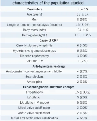
ESULTSFifteen patients completed the study (two were excluded due to sinus tachycardia, one because of aortic stenosis, and one because of moderate mitral regurgitation). No patient showed abnormal LV segmental contractility at rest. Table 1 shows clinical characteristics and anatomic changes diagnosed by echocardiogram in the patients studied. At baseline (pre-HD), eight patients showed abnormal relaxation (grade I diastolic dysfunction), six met criteria for mitral infl ow pseudonormalization through concomitant analysis by mitral annular tissue Doppler (grade II diastolic dysfunction), and one showed E/A ratio 2.5 (grade III diastolic dysfunction). Three patients
had LV systolic dysfunction detected by ejection fraction calculation, while lower systolic mitral annular velocity was shown in four by tissue Doppler imaging
After an average loss of 2.2 ± 1.1 kg through ultrafi ltration, expressive change was found in LV diastolic dimension (5.1 ± 0.6 to 4.7 ± 0.6 cm, p < 0.001) and systolic dimension (3.4 ± 0.5 to 2.9 ± 0.4 cm, p < 0.001), pointing to a preload reduction. No signifi cant changes were found in heart rate (83 ± 12 to 80 ± 13 beats/minute, p = 0.4), systolic blood pressure (163 ± 28 to 158 ± 24 mmHg, p = 0.3), and diastolic blood pressure (87 ± 14 to 87 ± 17 mmHg, p = 0.98) after dialysis. No patient experienced intradialytic hypotension requiring therapy discontinuation or change.
Table 2 shows the means of several Doppler echoardiographic indices, including the Tei index and its components, before and after HD session, as well as percentage changes from baseline conditions. After ultrafi ltration, mitral fl ow E-wave decreased (94 ± 22 to 78 ± 26 cm/s, p < 0.05), but mitral fl ow A-wave remained unchanged (100 ± 34 to 103 ± 30 cm/s, p = 0.6), resulting in a signifi cant decrease in the E/A ratio (1.1 ± 0.5 to 0.8 ± 0.3, p < 0.01). The Tei index increased (0.57 ± 0.07 to 0.65 ± 0.09, p < 0.01) due to IVRT prolongation (101 ± 14 to 113 ± 17 ms, p < 0.01) and ET shortening (271 ± 22 to 252 ± 22, p< 0.05). The IVCT remained unaltered (53 ± 9 to 50 ± 13 ms, p = 0.3).
Tissue Doppler-derived diastolic parameters showed no signifi cant change after hemodialysis: E’ (8.5 ± 0.9 to 8.0 ± 2 cm/s, p = 0.3), A’ (12.4 ± 2 to 11.9 ± 2 cm/s,
Table 1 - Clinical and echocardiographic characteristics of the population studied
Parameters n = 15
Age (years) 53 ± 14
Men 8 (53%)
Length of time on hemodialysis (months) 15 (3-96) Body mass index 24 ± 6 Hemoglobin (g/dL) 10.5 ± 2.5
Cause of CRF
Chronic glomerulonephritis 6 (40%) Hypertensive glomerulosclerosis 5 (33%) Diabetic nephropathy 3 (20%) SAH and DM 1 (7%)
Anti-hypertensive drugs
Angiotensin II-converting enzyme inhibitor 4 (27%) Beta-blockers 2 (13%) Amlodipine 2 (13%)
Echocardiographic anatomic changes
p = 0.5), E’/A’ (0.73 ± 0.2 to 0.70 ± 0.2, p = 0.7) e E/E’ (11 ± 2.6 to 10 ± 4.9, p = 0.8). However, the S-wave increased (10.5 ± 2 to11.4 ± 2 cm/s, p<0.05), as well as shortening percentage and ejection fraction (34 ± 5 to 37 ± 3%, p < 0.05; and 71 ± 7% to 75 ± 4%, p < 0.01, respectively). Intraobserver variability (expressed in percentage) for the study’s main variables was: IVCT + IVRT = 0.8 ± 1.1; ET = 2.2 ± 2.5; Tei index = 8 ± 2; E = 1.4 ± 1; A = 0.1 ± 0.8; E’ = 0.4 ± 0.9; A’ = 0 ± 0.4.
D
ISCUSSIONHemodialysis patients constitute an interesting group for assessing reduced preload effects on cardiac function parameters by Doppler echocardiogram. This study investigated the effects triggered by HD on the Tei index, which refl ects global myocardial function, and on systolic and diastolic function indices separately, in a group of CRF patients waiting for renal transplantation. Fluid removal resulted in a mean weight reduction of 2.2 Kg (or 2.2 liters of body water), causing a decline in intravascular volume and a drop in preload, as may be inferred by reduced LV
dimensions25. The decrease in circulating plasma volume
led to changes in the Tei index, demonstrating that it is affected by load conditions.
Diastolic indices derived from mitral infl ow, including E-wave, E/A ratio, and IVRT varied signifi cantly, similar to other studies using HD as clinical model of preload
reduction24-26,29. It is known that pulsed Doppler-derived
velocities are extremely volume-dependent40, and the
rapid drop in fi lling pressure caused by HD may expose to
a pseudonormalization of mitral fl ow27,41. Criteria for mitral
fl ow pseudonormalization were followed in six patients before HD, fi ve of whom showed abnormal relaxation on standard Doppler after hemodialysis. We thus confi rm that HD does not affect left ventricular diastolic function adversely, but rather induces changes that depend on the sensitivity of mitral Doppler parameters to preload variations, sometimes “unmasking” a preexistent diastolic dysfunction.
As this study demonstrates, the role of mitral annular tissue Doppler as a method to assess the relatively independent diastolic function of preload should be
underscored37, as it was also demonstrated in this study.
No change in E’ and A’ velocity was found with the average amount removed by ultrafi ltration in our group, a fi nding
similar to that reported in previous publications27,29. Other
authors who used high-fl ow HD and/or greater blood
volume loss28, or who included myocardial ischemia
patients26, observed variations in mitral annular velocities
after HD. It seems reasonable to say that E’ is little affected by HD, compared to E. The HD effect on E’ may be ignored as long as no excess fl uid is removed.
Pulsed-wave Doppler-derived systolic indices showed that IVCT remained unchanged and ET shortened signifi cantly. Because of the combined changes in mitral fl ow intervals (prolonged IVRT and shortened ET), there was signifi cant Tei index variation, which increased with preload reduction. An alternative analysis could propose a decrease in myocardial performance after the HD session due to systolic function deterioration, as may be suggested by the reduced ventricular ejection fraction.
Some arguments may be presented against this hypothesis. Firstly, we sought to exclude factors that could Table 2 – Doppler echocardiographic parameters of diastolic function and the Tei index: pre-HD, post-HD, and
modifi cation in percentage
Pre-HD Post-HD Modifi cation (%) P value
Mitral infl ow Doppler
IVRT 101 ± 14 113 ± 17 12 < 0.01
IVCT 53 ± 9 50 ± 13 -4.5 0.3
ET (ms) 271 ± 22 252 ± 22 -7 < 0.05 Tei index 0.57 ± 0.07 0.65 ± 0.09 14 < 0.01 E (cm/s) 94 ± 22 78 ± 26 -17 < 0.05
A (cm/s) 100 ± 34 103 ± 30 2 0.6
E/A 1.1 ± 0.5 0.8 ± 0.3 -16 < 0.01
Mitral annular TDI
E’ (cm/s) 8.5 ± 0.9 8.0 ± 2 -5.1 0.3
A’ (cm/s) 12.4 ± 2 11.9 ± 2 -2 0.5
S (cm/s) 10.5 ± 2 11.4 ± 2 8 < 0.05 E’/A’ 0.73 ± 0.2 0.70 ± 0.2 -5.8 0.7
E/E’ 11 ± 2.6 10 ± 4.9 -8.6 0.8
M-mode
PS (%) 34 ± 5 37 ± 3 11.2 < 0.05
EJ (%) 71 ± 7 75 ± 4 6.9 < 0.01
lead to an acute decrease in myocardial performance, such as valvular heart disease, pericardial disease, or myocardial ischemia. Although the presence of coronary heart disease was not checked by means of other non-invasive or non-invasive tests, the degree of plasma volume depletion during our experiment was probably within certain “physiological” ranges, since it was not followed by signifi cant change in heart rate and blood pressure. The occurrence of arterial hypotension could have led to refl ex tachycardia, subsequent ischemia, and a decrease in LV function parameters.
Secondly, the assumption that decreased ET may indicate a decline in systolic function is contradicted by the increase observed in ejection fraction (radial myocardial contractile function), and especially in S-wave velocity measured by mitral annular tissue Doppler (longitudinal contractile function). The ET shortening mechanism is likely to be related to the decrease in blood volume after HD, which reduces end-diastolic
volume and LV stroke volume21. It is known that HD may
induce a dissociation between changes in ventricular
fi lling and in contractile state20. Thus, despite the ET
abbreviation secondary to a decrease in left ventricular fi lling, contractile function apparently improved, probably due to uremic toxin removal. This effect is achieved by an isolated ultrafi ltration that antagonizes the Frank-Starling mechanism and does not remove the toxins that
depress myocardial function20. Therefore, while contractile
function indices improved, there was a paradoxical prolongation of the Tei index.
These fi ndings corroborate the known sensitivity of systolic intervals to preload variations, including ejection time21,42. The HD effects on LV systolic performance
vary and seem to depend partly on ventricular volumes and contractile function prior to dialysis. Alpert et al suggested that LV systolic function only improves after HD session in patients with previous systolic dysfunction, and that is does not change signifi cantly in individuals
with normal function43. It is worth noting that this study
used mean velocity of circumferential fi ber shortening (M-mode-derived) as the index for systolic function, rather than ejection fraction. On the other hand, a small study (ten patients) using the same method to evaluate a pediatric HD population suggested a real improvement
in myocardial contractile function in that group44.
In a more recent study performed on 128 chronic renal disease patients with normal baseline systolic function, no change was found in percent shortening and
ejection fraction with HD28, but the primary purpose of
the article was to study diastolic function, and presence of coronary artery disease was not an exclusion criterion. Our group showed improvement both in ejection fraction and systolic annular velocity measured by tissue Doppler. With regard to ejection fraction, this improvement could be suggested by the preload infl uence itself on the index, as demonstrated previously. Alternatively, the inclusion of three patients with reduced systolic dysfunction at baseline, a subgroup in which mean ejection fraction
increase after dialysis was 17%, could theoretically have created a bias for the overall result of the group. However, there was also a signifi cant and consistent increase in the S-wave, which is in keeping with a previous publication that pointed to improvement in myocardial systolic
velocities of CRF patients after HD45.
It seems reasonable to infer that the S-wave increase should represent a real improvement in contractility of longitudinal myocardial fi bers, since tissue Doppler is relatively preload-independent, and the amount of induced-volume variation in our group failed to produce changes in mitral annular diastolic velocities. The possibility of studying not only radial myocardial contractility but also longitudinal systolic function using novel echocardiographic techniques, such as tissue Doppler and myocardial strain, opens a new path to research aimed at understanding heart performance behavior under load variation.
A similar study performed by Koga et al also sought to evaluate the impact of HD-induced preload alterations
on the Tei index46. Evaluating 32 patients with mean age
of 72 ± 9, on HD for 40 ± 35 months and showing normal ejection fraction, these authors found a signifi cant decrease in E velocity and E/A ratio, associated with an increase in the Tei index (due to IVRT + IVCT prolongation and ET shortening). It is interesting to note that, in evaluating two groups according to the amount of weight
loss, whether ≥ 1.5 kg or <1.5 kg, only the fi rst group
showed changes in the Tei index. Analysis of the group as a whole revealed no changes in LV dimension and volume (weight loss 1.8 ± 0.7 kg), and ejection fraction remained unaltered after hemodialysis.
Our population consisted only of patients with myocardial hypertrophy and diastolic dysfunction, in addition to three patients with systolic dysfunction. This may explain baseline differences in the Tei index of the
populations evaluated (0.57 ± 0.07 vs 0.42 ± 0.16 in
the Japanese study). In our group, weight loss was greater ((2.2 ± 1.1 kg), allowing a more expressive reduction in circulating volume and thus relieving volume overload and resulting in different ejection fraction response. Therefore, the Tei index showed to be affected by preload reduction in a mixed sample as well, in patients with systolic and diastolic dysfunction, as had already been demonstrated in the population with normal systolic function in the Japanese study. This variation seems to depend on the volume removed, regardless of baseline systolic function.
has been present in other dialysis studies as well26,27.
Moreover, the fact that all the examinations were performed by a single echocardiographer contributed to reduce record variability.
Our results showed that the Tei index is affected by reduction in circulating blood volume induced by HD, raising its value, which could be misinterpreted as a decrease in myocardial performance. We concluded that, as with other parameters derived from mitral infl ow Doppler, this index may be affected by preload changes mediated by HD. Among the other parameters evaluated, diastolic mitral annular velocities measured by tissue Doppler proved to be unaffected by the volume
loss obtained in this study, and systolic velocities behavior suggested improved LV contractile function after the procedure. When evaluating the Tei index’s absolute value in a patient on maintenance HD in order to estimate global myocardial performance, one should interpret it with care, correlating it temporally with the dialysis session. Regardless of its sensitivity to preload, its prognostic value needs to be tested in this group of patients, considering its high predictive accuracy of
mortality in other clinical scenarios11-14.
Potencial Confl ict of Interest
No potential confl ict of interest relevant to this article was reported.
R
EFERENCES1. Alpert MA. Cardiac performance and morphology in end-stage renal disease. Am J Med Sci. 2003; 325: 168-78.
2. Foley RN, Parfrey PS, Harnett JD, Kent GM, Murray DC, Barre PE. The prognostic importance of left ventricular geometry in uremic cardiomyopathy. J Am Soc Nephrol. 1995; 5: 2024-31.
3. London GM. Cardiovascular disease in chronic renal failure: pathophysiologic aspects. Semin Dial. 2003; 16: 85-94. 4. Sarnak MJ. Cardiovascular complications in chronic kidney disease.
Am J Kidney Dis. 2003; 41: 11-7.
5. London GM. Left ventricular alterations and end-stage renal disease. Nephrol Dial Transplant. 2002; 17 Suppl 1: 29-36.
6. Sarnak MJ, Levey AS, Schoolwerth AC, et al. Kidney disease as a risk factor for development of cardiovascular disease: a statement from the American Heart Association Councils on Kidney in Cardiovascular Disease, High Blood Pressure Research, Clinical Cardiology, and Epidemiology and Prevention. Circulation 2003; 108: 2154-69. 7. Tei C. New non-invasive index for combined systolic and diastolic
ventricular function. J Cardiol. 1995; 26: 135-6.
8. Tei C, Ling LH, Hodge DO, et al. New index of combined systolic and diastolic myocardial performance: a simple and reproducible measure of cardiac function--a study in normals and dilated cardiomyopathy. J Cardiol. 1995; 26: 357-66.
9. Bruch C, Schmermund A, Marin D, et al. Tei-index in patients with mild-to-moderate congestive heart failure. Eur Heart J. 2000; 21: 1888-95.
10. Harjai KJ, Scott L, Vivekananthan K, Nunez E, Edupuganti R. The Tei index: a new prognostic index for patients with symptomatic heart failure. J Am Soc Echocardiogr. 2002; 15: 864-8.
11. Tei C, Dujardin KS, Hodge DO, Kyle RA, Tajik AJ, Seward JB. Doppler index combining systolic and diastolic myocardial performance: clinical value in cardiac amyloidosis. J Am Coll Cardiol. 1996; 28: 658-64.
12. Dujardin KS, Tei C, Yeo TC, Hodge DO, Rossi A, Seward JB. Prognostic value of a Doppler index combining systolic and diastolic performance in idiopathic-dilated cardiomyopathy. Am J Cardiol. 1998; 82: 1071-6.
13. Yeo TC, Dujardin KS, Tei C, Mahoney DW, McGoon MD, Seward JB. Value of a Doppler-derived index combining systolic and diastolic time intervals in predicting outcome in primary pulmonary hypertension. Am J Cardiol. 1998; 81: 1157-61.
14. Poulsen SH, Jensen SE, Tei C, Seward JB, Egstrup K. Value of the Doppler index of myocardial performance in the early phase of acute myocardial infarction. J Am Soc Echocardiogr. 2000; 13: 723-30.
15. Tei C, Nishimura RA, Seward JB, Tajik AJ. Noninvasive Doppler-derived myocardial performance index: correlation with simultaneous measurements of cardiac catheterization measurements. J Am Soc Echocardiogr. 1997; 10: 169-78.
16. Lutz JT, Giebler R, Peters J. The ‘TEI-index’ is preload dependent and can be measured by transoesophageal echocardiography during mechanical ventilation. Eur J Anaesthesiol. 2003; 20: 872-7. 17. Moller JE, Poulsen SH, Egstrup K. Effect of preload alternations on
a new Doppler echocardiographic index of combined systolic and diastolic performance. J Am Soc Echocardiogr. 1999; 12: 1065-72. 18. Lavine SJ. Effect of heart rate and preload on index of myocardial performance in the normal and abnormal left ventricle. J Am Soc Echocardiogr. 2005; 18: 133-41.
19. Cheung MM, Smallhorn JF, Redington AN, Vogel M. The effects of changes in loading conditions and modulation of inotropic state on the myocardial performance index: comparison with conductance catheter measurements. Eur Heart J. 2004; 25: 2238-42.
20. Nixon JV, Mitchell JH, McPhaul Jr JJ, Henrich WL. Effect of hemodialysis on left ventricular function. Dissociation of changes in fi lling volume and in contractile state. J Clin Invest. 1983; 71: 377-84.
21. Gilmartin JJ, Duffy BS, Finnegan P, McCready N. Non invasive study of left ventricular function in chronic renal failure before and after hemodialysis. Clin Nephrol. 1983; 20: 55-60.
22. Rozich JD, Smith B, Thomas JD, Zile MR, Kaiser J, Mann DL. Dialysis-induced alterations in left ventricular fi lling: mechanisms and clinical signifi cance. Am J Kidney Dis. 1991; 17: 277-85.
23. Sadler DB, Brown J, Nurse H, Roberts J. Impact of hemodialysis on left and right ventricular Doppler diastolic fi lling indices. Am J Med Sci. 1992; 304: 83-90.
24. Gupta S, Dev V, Kumar MV, Dash SC. Left ventricular diastolic function in end-stage renal disease and the impact of hemodialysis. Am J Cardiol. 1993; 71: 1427-30.
25. Chakko S, Girgis I, Contreras G, Perez G, Kessler KM, Myerburg RJ. Effects of hemodialysis on left ventricular diastolic fi lling. Am J Cardiol. 1997; 79: 106-8.
26. Agmon Y, Oh JK, McCarthy JT, Khandheria BK, Bailey KR, Seward JB. Effect of volume reduction on mitral annular diastolic velocities in hemodialysis patients. Am J Cardiol. 2000; 85: 665-8, A11. 27. Graham RJ, Gelman JS, Donelan L, Mottram PM, Peverill RE. Effect
of preload reduction by haemodialysis on new indices of diastolic function. Clin Sci (Lond). 2003; 105: 499-506.
function during hemodialysis. Am J Kidney Dis. 2004; 43: 1040-6. 29. Barberato SH, Mantilla DE, Misocami MA, et al. Effect of preload
reduction by hemodialysis on left atrial volume and echocardiographic Doppler parameters in patients with end-stage renal disease. Am J Cardiol. 2004; 94: 1208-10.
30. Pecoits-Filho R, Goncalves S, Barberato SH, et al. Impact of residual renal function on volume status in chronic renal failure. Blood Purif. 2004; 22: 285-92.
31. Mosteller RD. Simplifi ed calculation of body-surface area. N Engl J Med. 1987; 317: 1098.
32. Quinones MA, Douglas PS, Foster E, et al. American College of Cardiology/American Heart Association clinical competence statement on echocardiography: a report of the American College of Cardiology/American Heart Association/American College of Physicians - American Society of Internal Medicine Task Force on Clinical Competence. Circulation. 2003; 107: 1068-89.
33. Devereux RB, Reichek N. Echocardiographic determination of left ventricular mass in man. Anatomic validation of the method. Circulation. 1977; 55: 613-8.
34. Devereux RB. Detection of left ventricular hypertrophy by M-mode echocardiography. Anatomic validation, standardization, and comparison to other methods. Hypertension. 1987; 9: II9-26. 35. Pombo JF, Troy BL, Russell Jr RO. Left ventricular volumes and ejection
fraction by echocardiography. Circulation. 1971; 43: 480-90. 36. Quinones MA, Otto CM, Stoddard M, Waggoner A, Zoghbi WA.
Recommendations for quantifi cation of Doppler echocardiography: a report from the Doppler Quantifi cation Task Force of the Nomenclature and Standards Committee of the American Society of Echocardiography. J Am Soc Echocardiogr. 2002; 15: 167-84.
37. Nagueh SF, Middleton KJ, Kopelen HA, Zoghbi WA, Quinones MA. Doppler tissue imaging: a noninvasive technique for evaluation of left
ventricular relaxation and estimation of fi lling pressures. J Am Coll Cardiol. 1997; 30: 1527-33.
38. Nishimura RA, Tajik AJ. Evaluation of diastolic fi lling of left ventricle in health and disease: Doppler echocardiography is the clinician’s Rosetta Stone. J Am Coll Cardiol. 1997; 30: 8-18.
39. Khouri SJ, Maly GT, Suh DD, Walsh TE. A practical approach to the echocardiographic evaluation of diastolic function. J Am Soc Echocardiogr. 2004; 17: 290-7.
40. Choong CY, Herrmann HC, Weyman AE, Fifer MA. Preload dependence of Doppler-derived indexes of left ventricular diastolic function in humans. J Am Coll Cardiol. 1987; 10: 800-8.
41. Ie EH, Vletter WB, ten Cate FJ, et al. Preload dependence of new Doppler techniques limits their utility for left ventricular diastolic function assessment in hemodialysis patients. J Am Soc Nephrol. 2003; 14: 1858-62.
42. Gillebert TC, Van de Veire N, De Buyzere ML, De Sutter J. Time intervals and global cardiac function. Use and limitations. Eur Heart J. 2004; 25: 2185-6.
43. Alpert MA, Van Stone J, Twardowski ZJ, et al. Comparative cardiac effects of hemodialysis and continuous ambulatory peritoneal dialysis. Clin Cardiol. 1986; 9: 52-60.
44. O’Regan S, Villemand D, Revillon L, Robitaille P, Ducharme G, Davignon A. Effects of hemodialysis on myocardial function in pediatric patients. Nephron. 1980; 25: 214-8.
45. Hayashi SY, Brodin LA, Alvestrand A, et al. Improvement of cardiac function after haemodialysis. Quantitative evaluation by colour tissue velocity imaging. Nephrol Dial Transplant. 2004; 19: 1497-506. 46. Koga S, Ikeda S, Matsunaga K, et al. Infl uence of hemodialysis on