159
LETTERS
1. Horigome H, Nomura T, Saso K, et al. Coexistence of primary biliary cirrhosis and myasthenia gravis: a case study. Hepatogastroenterology 2000;47:125-127.
2. Finsterer J, Höflich S. Successful low-dose azathioprine for myasthenia gravis despite hepatopathy from primary sclerosing cholangitis: a case report. J Med Case Reports 2010;4:356.
3. Karlsen TH, Schrumpf E, Boberg KM. Primary sclerosing cholangitis. Best Pract Res Clin Gastroenterol 2010;24:655-666.
4. Sinakos E, Lindor K. Treatment options for primary sclerosing cholangitis. Expert Rev Gastroenterol Hepatol 2010;4:473-488.
5. Takiguchi J, Ohira H, Rai T, et al. Autoimmune hepatitis overlapping with primary sclerosing cholangitis. Intern Med 2002;41:696-700.
References
he presented report shows that in case of MG and collat-eral PSC, liver-toxic immunosuppression with AZT is tolerable and efective even over a longer period of time if the dosage is adapted according to the impaired liver functions. Low-dose AZT has a suicient therapeutic efect on MG without dete-rioration of PSC. Possibly, AZT was beneicial even for PSC, since the patient exclusively required ursodesoxycholic acid, PSC did not progress so far, and AZT has also been recom-mended as a treatment of PSC5. Immunosuppressive agents
other than AZT were considered, but they were rejected be-cause AZT is the only approved immunosuppressive drug for MG and since other agents are potentially liver-toxic as well. Continuation of steroids was also considered since they may have a beneicial efect on PSC5 and on MG, but they were
lastly stopped for their side efects, their inefectivity on the AchR-antibody titers, and since PSC did not deteriorate after their discontinuation.
Rare case of carotid artery occlusion due to
thrombosis of a giant cerebral aneurysm.
The role of cerebral revascularization.
Caso raro de oclusão da artéria carótida devido à trombose de um aneurisma cerebral
gigante. O papel da revascularização cerebral.
Rafael de Oliveira Sillero1, Valter José Sillero Filho2, Gislaine Priscila Momm Zimmermann3
1MD, Neurosurgeon-in-chief, Neurosurgery Unit, Regional Hospital of São José, São José SC, Brazil;
2MD, Neurosurgeon, Neurosurgery Unit, Regional Hospital of São José, São José SC, Brazil;
3MD, Ophthalmologist, Regional Hospital of São José, São José SC, Brazil.
Correspondence: Rafael de Oliveira Sillero; Unidade de Neurocirurgia, Hospital Regional de São José; Rua Adolfo Donato da Silva s/n; 88103-901 São José SC - Brasil; E-mail: [email protected]
Conflict of interest: There is no conflict of interest to declare.
Received 16 August 2011; Received in final for 06 September 2011; Accepted 13 September 2011
Spontaneous thrombosis of a giant cerebral aneu-rysm is a recognized phenomenon, however it becomes rare when the thrombosed aneurysm is associated with the occlusion of its parent artery1,2. The best
manage-ment strategy is not defined yet. Theoretically, it should
be directed to alleviating mass effect related symptoms caused by the aneurysm itself and to preventing cerebral ischemia.
We describe a case of carotid artery occlusion and dis-cuss the role of cerebral revascularization.
liver function parameters remained constantly elevated but in a tolerable range, without sudden increases, as previously described (Fig 1). AchR-antibodies had their ups and downs, however they also remained stable until the last follow-up in March, 2011 (Fig 2). Since the detection of PSC, the patient has never complained about liver symptoms. Total bilirubine did not exceed a maximal value of 3.0 mg/dL during ive years. Since the last myasthenic crisis, she remained clinically stable, with a persistent slight ptosis on the right side.
PSC is a chronic, inlammatory disease of the bile ducts, complicated by fatigue, pruritus, ibrotic biliary strictures, liver cirrhosis, and liver failure necessitating liver transplantation, cholangiocarcinoma, or colorectal cancer3. he etiology of PSC
is unknown but autoimmune mechanisms and genetic factors seem to play a pathogenetic role3. Although there is no causative
160
LETTERS
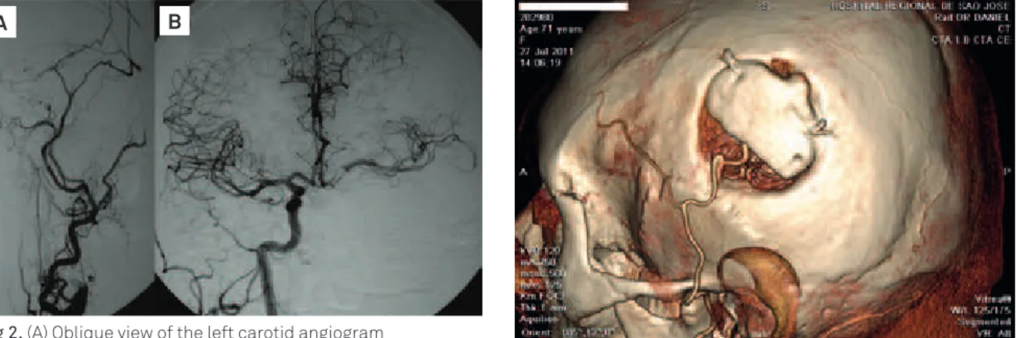
Fig 2. (A) Oblique view of the left carotid angiogram demonstrating occlusion (*) of the proximal internal carotid
artery in the neck. (B) Frontal view of the right carotid angiogram revealing good cross filling in the left side through the anterior communicating artery.
A B
Fig 1. (A) The computed tomography scan showing a high-density parasellar round lesion extending to the left middle cranial fossa. It was first misdiagnosed as a brain tumor. (B) Axial T2 weighted image revealing the giant thrombosed aneurysm.
A B
Fig 3. Postoperative computed tomography angiography showing a patent left-side superficial temporal artery-middle cerebral artery (STA-MCA) bypass.
CASE REPORT
A 69-year-old woman had sufered from left painful oph-thalmoplegia with a sudden onset. She had consulted her ophthalmologist, who suspected a left cavernous sinus syn-drome. A computed tomography of the head was obtained, and the patient was referred to our service with a diagnosis of brain tumor.
On admission, the patient was conscious and the left cavernous sinus syndrome was confirmed. The initial computed tomography (CT) scan showed a high-density parasellar round lesion extending to the left middle cra-nial fossa. Magnetic resonance imaging revealed a giant thrombosed aneurysm of the left cavernous internal ca-rotid artery (Fig 1). Further investigation with a four-ves-sel cerebral angiography (Fig 2) obtained 10 days after the onset of presentation revealed the finding of left internal carotid artery (ICA) occlusion with good cross filling in
the left-side circulation through the anterior communicat-ing artery. The patient was treated conservatively and her symptoms gradually improved. She was discharged on an-tiplatelet treatment.
At the 3-month follow-up consultation, the patient was complaining of some brief episodes of language disturbance related to verbal expression.
After demonstration of cerebral flow asymmetry by a single photon emission computed tomography (SPECT) study, the patient underwent extra-intracranial bypass surgery using the technique described by Yasargil3 to
anastomose the left superficial temporal artery to the middle cerebral artery. Postoperative course was un-eventful and control cerebral angiography confirmed by-pass patency.
At follow-up consultation 3-years later, the patient was asymptomatic and CT angiography (Fig 3) showed that the bypass remained patent.
DISCUSSION
ce-161
LETTERS
1. Sato K, Fujiwara S, Yoshimoto T, Onuma T. Two cases of spontaneous internal carotid artery occlusion due to giant intracranial carotid artery aneurysm. Stroke 1990;21:1506-1509.
2. Whittle IR, Williams DB, Halmagyi GM, Besser M. Spontaneous thrombosis of a giant intracranial aneurysm and ipsilateral internal
carotid artery. Case report. J Neurosurg 1982;56:287-289.
3. Yasargil MG. Microsurgery applied to neurosurgery. Stuttgart: Georg Thieme; 1969:60-81.
4. Vajkoczy P. Revival of extra-intracranial bypass surgery. Curr Opin Neurol 2009;22:90-95.
References
rebral fluid flow study criteria of reduced cerebrovascular reserve capacity4.
A report has been published in which aneurysmotomy and thrombectomy of a thrombosed giant intracavernous carotid aneurysm were performed and produced mass ef-fect relief with symptomatic improvement2. Similarly, our
patient had remission of mass effect symptoms, but with conservative management.