Patent Foramen Ovale and Ischemic Stroke in Young People:
Statistical Association or Causal Relation?
Edson Marcio Negrão
, Ivar Viana Brandi
2, Simone Vilela Nunes
, Daniel Gurgel Fernandes Távora
, Mauro Nakayama
,
Paulo Sergio Siebra Beraldo
Hospital SARAH - Brasília, DF – Belo Horizonte, MG2 – Fortaleza, CE
Summary
Objective: To determine if there are evidences of a causal relation between patent foramen ovale (PFO) x cryptogenic ischemic stroke (IS) in the young population and to analyze this relation in terms of causal criteria.
Methods: A total of 168 young patients with IS was retrospectively evaluated and divided into two groups: cryptogenic and with a defined cause. As a routine procedure, the patients underwent investigation of the PFO by means of transesophageal echocardiogram and/or transcranial Doppler sonography, both of them associated with the bubble test. Multivariate analysis was performed after demonstration of univariate statistical association between PFO x IS.
Results: After multivariate analysis, the association between PFO x cryptogenic IS was still statistically significant with odds ratio (adjusted25 &, 7KH WRWDO QXPEHU RI FHUHEUDO OHVLRQV DOVR SUHVHQWHG D VLJQLILFDQW association with cryptogenic IS (adjusted25 &,7KHDVVRFLDWLRQEHWZHHQ3)2DQGFU\SWRJHQLF,6 met all the causality criteria.
Conclusion: The causal relation between PFO and cryptogenic IS in the young population is highly probable. This fact should be considered in the therapeutic decision.
Key words: Patent foramen ovale, atrial septal defects, cerebrovascular accident, transesophageal echocardiography.
0DLOLQJDGGUHVV(GVRQ0DUFLR1HJUmR
SQN 212, bloco E ap.311, Asa Norte - 70864-050 – Brasília, DF E-mail: [email protected]
Manuscript received July 20, 2006; revised manuscript received July 20, 2006; accepted August 17, 2006.
Introduction
Ischemic stroke (IS) is the main cause of morbidity in the Brazilian population1. Although it is more prevalent in higher
age groups, its socioeconomic impact is greater when it affects young patients. In this population, the annual incidence of ischemic events is estimated between 6 and 26:100,0002.
At tertiary centers, up to 12% of patients admitted are aged under 45 years3. In the young population, despite extensive
investigation, up to 40% of cases do not have a clear etiology and are called cryptogenic4. In the last two decades
a significant portion of such cases has been attributed to paradoxical embolism and the main example is the patent foramen ovale (PFO). The statistical association between ischemic cerebrovascular events and PFO has already been widely documented in univariate analyses, including those carried out by our group5,6. This current study intends to
assess if the association between PFO and IS presents a causal relation or if there is just a statistical relation. In addition to a multivariate analysis, we also evaluated this association considering the well known causality criteria7.
Methods
We retrospectively included all the patients with diagnosis
of IS aged 15 to 45 years who had been consecutively admitted at the centers located in Brasilia, Salvador, Sao Luis and Fortaleza that are part of Rede Sarah de Hospitais de Reabilitação (Sarah Network of Rehabilitation Hospitals) between January 2003 and November 2004. In addition to clinical and neurological evaluation, the patients underwent basic hematological tests and study of thrombophilias. Patients also underwent computed tomography and/or nuclear magnetic resonance of the brain, Doppler examination of vertebral and carotid arteries, as well as transthoracic echocardiogram. When applicable, patients also underwent brain resonance angiography and/or cerebral arteriography.
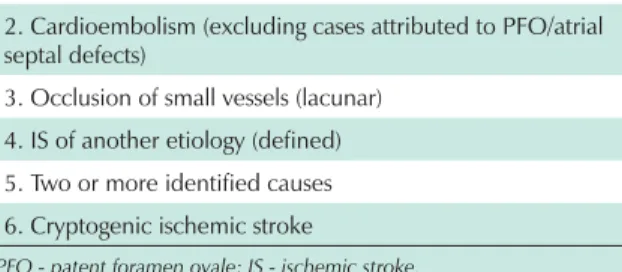
The cases were classified as cryptogenic or non-cryptogenic after analysis carried out by a neurologist (Ivar Viana Brandi or Simone Vilela Nunes), according to the TOAST criteria adapted for this study (Table 1)8. Cases with evaluation considered
patients were excluded due to incomplete evaluation. Two patients did not cooperate with the transesophageal echocardiogram and also did not undergo transcranial Doppler sonography. One patient presented a positive bubble test in transcranial Doppler but did not cooperate with the transesophageal echocardiogram. Therefore, 168 patients were considered for univariate analysis; 55% of them were females. The mean age was 33.9 years ± 9.3 (SD). The prevalence of traditional risk factors did not differ among patients with cryptogenic and non-cryptogenic IS, and the patients presented dyslipidemia (40%), smoking habit (32.5%), systemic arterial hypertension (35%) and diabetes mellitus (5%).
Ninety-eight patients (58.4%) underwent transesophageal echocardiogram and transcranial Doppler sonography; 34 (20.2%) patients underwent only the first procedure and 36 (21.4%) patients were only submitted to transcranial Doppler sonography.
Eighty-eight cases were considered cryptogenic (52%). According to modified TOAST classification, cases of defined etiology were those involving cardioembolism (28%), atherosclerosis of a great artery (17%), occlusion of a small vessel (11%) and IS of a different etiology (established) or with two or more causes (44%). The prevalence of PFO in the whole sample was 28% (47/168; 95% CI: 21.3 – 35.4). However, the prevalence of PFO was 15% (12/80) when only the defined etiology cases were considered. In the subgroup of IS cases classified as cryptogenic, PFO was present in 40% of patients (35/88). This difference was significant and represented a raw odds ratio of 3.7 (95% CI: 1.8 - 7.9).
In the multivariate analysis of 20 patients, some variables did not have the required responses; hence they were excluded from the study. The characteristics of these patients were not distinct from those of the 148 patients who remained in the analysis.
Based on the univariate analysis, three variables were considered in the logistic regression: PFO, total number of brain lesions and hyperhomocysteinemia. From the total of variables considered in the final model, only two were associated with the type of IS: presence of PFO (adjustedOR = 3.3; 95% CI: 1.5–7.4) and the total number of cerebral lesions (adjusted OR = 0.4; 95% CI: 0.2-0.9) (Table 3). The same model was tested, this time replacing PFO by shunt intensity, and it showed that cryptogenic IS was associated with more intense shunts (Figure 1). We tested the interactions among the interest variables without a change in the final model.
Discussion
By means of a multivariate analysis, the current study showed that young patients (younger than 45 years) with IS of undetermined cause may have their etiology related to the presence of a patent foramen ovale. Specifically, we confirmed that this association was more important in the cases with more intense shunts. In addition to an association, these results point to a causal relation between PFO and IS. Once this hypothesis is confirmed, the etiological possibilities for the cases of IS and transient ischemic attack should be broadened and they should include the cases of patent foramen ovale.
respective neurology teams of each participating hospital. The test was considered positive when at least one early hyperintense signal was observed (up to 10 seconds after infusion of macrobubbles)10.
In the transesophageal echocardiogram, the bubble test was defined as positive when at least three macrobubbles were visualized inside the left atrium up to the fifth cardiac cycle after maximum opacification of the right atrium. The shunt was considered small when up to nine macrobubbles were present in the left atrium; it was moderate when there were between 10 and 29 macrobubbles and marked shunt was defined with more than 30 macrobubbles in the left atrium.
Patients with a positive bubble test in the transcranial Doppler sonography who, for some reason, did not undergo transesophageal echocardiogram were excluded from the study due to the impossibility to determine the cause of shunt. Patients who underwent only transcranial Doppler sonography but had a negative bubble test were classified as non-patent foramen ovale due to the high negative predictive value of this exam for the diagnosis of right to left shunt11.
Statistical analysis - The data collected were stored in an Excel spreadsheet (Microsoft Excel 2003) and differential analysis was conducted using Epinfo (version 6.04d). All the statistical processing of the multivariate analysis was conducted in SPSS (version 13).
The candidate variables to be tested as independent in the final model were considered on the basis of clinical evidence, information available in the literature and univariate analysis, and in this case the decision criterion had a p value < 0.15. Univariate analysis examined contingency tables and the statistical association was checked for p < 0.05. Table 2 shows the details of all the variables considered.
The multivariate method used was multiple logistic regression, where the dependent variable was the type of IS dichotomized between cryptogenic and non-cryptogenic.12
For selection of the variables that remained in the logistic regression model a backward elimination process was used, and all variables with p < 0.05 remained in the final model.
Patients whose variables did not have the required responses were excluded from the study.
Results
A total of 175 patients was admitted to this study. Four Table 1 – Modified TOAST classification of ischemic stroke subtypes8
1. Atherosclerosis of great vessels
2. Cardioembolism (excluding cases attributed to PFO/atrial septal defects)
3. Occlusion of small vessels (lacunar)
4. IS of another etiology (defined)
5. Two or more identified causes
6. Cryptogenic ischemic stroke
In this context, in addition to diagnosis of PFO, the transesophageal echocardiogram also allows the description of several morphological and functional characteristics of the interatrial septum that enable its classification as a risk for IS13-16. In this study we found that the more intense shunt
presented a significant association with IS of undefined cause, even when it was adjusted for other independent variables. Two aspects should be emphasized: the first is the fact that the dose-effect relation meets the causality criteria7, which points
to the etiological relation of PFO in the cases of cryptogenic IS if others criteria are taken into account. The second fact has therapeutic implications showing that the shunt intensity should be one of the main factors in the risk stratification for indication of PFO closure. Actually, several articles have already considered this possibility; however, they included other factors that we have not considered in this current analysis, such as the concomitant presence of interatrial septum aneurism and prominent Eustachian valve. In addition to PFO, only one other variable was an independent predictor
for cryptogenic IS or IS of defined cause, and this was the total number of brain lesions. When three or more lesions were identified in the brain by means of imaging exams (computed tomography and/or magnetic resonance imaging – MRI) there was an increased 64% chance that the IS had a defined cause (adjusted OR = 0.36; 95% CI: 0.15-0.86). It is important to emphasize that the total number of lesions has two non-excluding interpretations. It may represent the presence of multiple lesions of the same ischemic event and/or lesions of temporally distinct events. Since our patients were investigated at a late stage of IS, we could not distinguish these two possibilities. In any case, we can infer that in cases of IS in which there is a suspition of PFO, the recurrence and/or multiple lesions are relatively low, at least when compared with more frequent cardioembolic causes, such as atrial fibrillation and Chagasic cardiopathy. It is noteworthy that the expressive number of 118 out of 168 patients (70%) underwent the brain imaging study with MRI, which is considered the best method for this type of analysis.
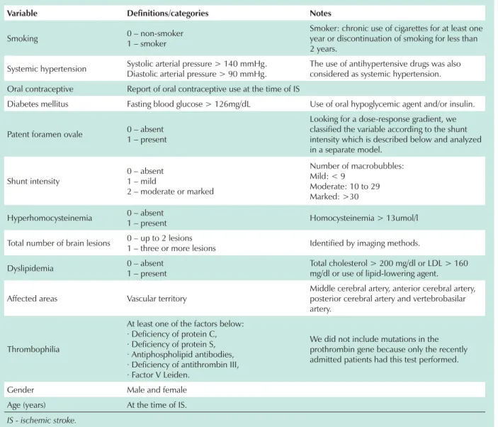
Table 2 – Details of variables considered in the analysis
Variable Definitions/categories 1RWHV
Smoking 0 – non-smoker
1 – smoker
Smoker: chronic use of cigarettes for at least one year or discontinuation of smoking for less than 2 years.
Systemic hypertension Systolic arterial pressure > 140 mmHg.
Diastolic arterial pressure > 90 mmHg.
The use of antihypertensive drugs was also considered as systemic hypertension.
Oral contraceptive Report of oral contraceptive use at the time of IS
Diabetes mellitus Fasting blood glucose > 126mg/dL Use of oral hypoglycemic agent and/or insulin.
Patent foramen ovale 0 – absent
1 – present
Looking for a dose-response gradient, we classified the variable according to the shunt intensity which is described below and analyzed in a separate model.
Shunt intensity
0 – absent 1 – mild
2 – moderate or marked
Number of macrobubbles: Mild: < 9
Moderate: 10 to 29 Marked: >30
Hyperhomocysteinemia 0 – absent
1 – present Homocysteinemia > 13umol/l
Total number of brain lesions 0 – up to 2 lesions
1 – three or more lesions Identified by imaging methods.
Dyslipidemia 0 – absent
1 – present
Total cholesterol > 200 mg/dl or LDL > 160 mg/dl or use of lipid-lowering agent.
Affected areas Vascular territory
Middle cerebral artery, anterior cerebral artery, posterior cerebral artery and vertebrobasilar artery.
Thrombophilia
At least one of the factors below: · Deficiency of protein C, · Deficiency of protein S, · Antiphospholipid antibodies, · Deficiency of antithrombin III, · Factor V Leiden.
We did not include mutations in the prothrombin gene because only the recently admitted patients had this test performed.
Gender Male and female
Age (years) At the time of IS.
7DEOH²8QLYDULDWHDQGPXOWLYDULDWHDQDO\VLVZLWKWKHLUUHVSHFWLYHRGGVUDWLR25DQGFRQILGHQFHLQWHUYDO&,GHSHQGHQW
variable is cryptogenic or non-cryptogenic IS
Variable Bivariate Final model
n OR &, p OR &, p
Patent foramen ovale No 121 1,00 - - - 1.00 - -
-Yes 47 3.74 2.16 6.48 0.001 3.32 1.49 7.38 0.003
Total number of lesions 0 to 2 129 1.00 - - - 1.00
3 or more 31 0.44 0.24 0.79 0.045 0.36 0.15 0.86 0.022
Hyperhomocysteinemia No 95 1.00 - - -
-Yes 60 0.50 0.31 0.81 0.038 - - -
-Dyslipidemia No 98 1.00 - - -
-Yes 65 0.60 0.38 0.96 0.118 - - -
-Age (years) Continuing 168 0.99 0.97 1.02 0.609 - - -
-Gender Male 75 1.00 - - -
-Female 93 0.93 0.60 1.46 0.824 - - -
-Smoking Never 99 1.00 - - -
-Yes 61 0.90 0.56 1.43 0.738 - - -
-Systemic hypertension No 108 1.00 - - -
-Yes 59 0.93 0.58 1.48 0.811 - - -
-Thrombophlia No 131 1.00 - - -
-Yes 27 1.19 0.64 2.21 0.683 - - -
-Diabetes mellitus No 159 1.00 - - -
-Yes 9 0.44 0.15 1.24 0.251 - - -
-Affected area 1 145 1.00 - - -
-2 or more 6 0.45 0.13 1.62 0.370 - - -
-Oral contraceptive No 58 1.00 - - -
-Yes 19 1.71 0.78 3.75 0.321 - - -
-Fig. 1 -Adjusted odds ratio (OR) of shunt intensity and type of IS (cryptogenic or defined cause).
1.0
4.8
1.4
0 2 4 6 8 10 12 14
None Mild Moderate / Marked
Shunt
A
d
ju
s
te
d
O
R
Although it has not been pointed as an independent variable in the logistic regression, the univariate analysis showed that hyperhomocysteinemia was associated with the type of IS (OR = 0.5; 95% CI: 0.31 – 0.81; P= 0.038). Several studies, including one study carried out in our Institution17,
showed the participation of hyperhomocysteinemia in cerebral ischemic events as an independent risk factor. In this specific investigation, the association observed – at least in the
univariate analysis – was in the sense of an association between IS of defined cause and high serum levels of homocystein. The fact that this variable did not remain in the final model must be cautiously interpreted, given the limitations inherent to studies with multivariate analysis18, in addition to the fact
that this variable possibly does not behave differently in IS of defined and undefined cause.
As to thrombophilia, we did not detect any association with cryptogenic IS, both in the univariate and multivariate analyses, i.e., 18.2% of patients with cryptogenic IS had some type of thrombophilia compared with 14.5% of patients with IS of undefined cause. Thrombophilias can be classified as predominantly arterial or venous. In the context of the current analysis we only evaluated venous thrombophilias, since they could be potentially related with paradoxical embolism. Although there is evidence of the association between mutation of the prothrombin gene (venous thrombophilia) and cryptogenic IS19,20, this measurement was not available to us
and the type of IS is an open question in the literature, with several conflicting studies. Our results pointed to non-existence of this association.
The other variables tested (age, gender, dyslipidemia, arterial hypertension, diabetes mellitus, oral contraceptives and smoking), even in the univariate analysis, were not associated with the type of IS. On the other hand, all these variables are considered as traditional risk factors for cerebrovascular diseases. In this sense, we should emphasize two aspects that can justify our results. First, the fact that we had a series of young patients aged under 45 years, in whom the prevalence of those factors is relatively low. The second aspect is the fact that the design of the current study does not allow us to draw any conclusions about the risk factors for IS, since all individuals affected presented this diagnosis. In fact, what we are evaluating are the factors associated with the type of event: with or without a defined cause. In this sense, we only found the presence of PFO (including the shunt intensity) and the number of brain lesions to be associated with the type of IS.
With these considerations as a background, we understand that the best way to conduct the discussions involving the possibility of a causal relationship should follow principles established for previously equivalent situations. In this scenario we reestablish the well accepted causality criteria7,20 and
emphasize the evidences available in the literature about a possible association between PFO and IS. Therefore, we prepared Table 4 where we try to answer the nine causality criteria in the present context. Considering the knowledge accumulated up to this moment, we believe that the best answer to this question will possibly come with the release of currently ongoing randomized clinical trials comparing the effectiveness of PFO closure versus clinical treatment and the recurrence of IS. Until then, we can only ponder on the basis of the available evidence about a probable causal relation and its repercussions in therapeutic decisions.
Potential Conflict of Interest
No potential conflict of interest relevant to this article was reported.
7DEOH²(YDOXDWLRQFULWHULDRIDFDXVDOUHODWLRQ7
Criteria Main question Patent foramen ovale x IS
Time sequence Did exposure happen
before the outcome?
It would be hardly probable to imagine that PFO would appear after an ischemic cerebrovascular event. It is an anatomic structure that is present since intrauterine life.
Strength of association How strong is the effect?
Both the present study and, in particular, a meta-analysis involving 9 patients with IS of cryptogenic origin and of defined cause, in individuals younger than
55 years old21, showed an odds ratio that is greater than 3.0.
Consistency of association
Have others detected the
same effect? Yes; there are meta-analyses about this topic
21/22.
Biological gradient Is there a dose-response
relation?
Possibly yes, because important shunts have a higher chance to be related with
cryptogenic IS and recurrence compared with small shunts23;24. The multivariate
analysis in this current study also showed this gradient.
Specificity of associationDoes exposure lead solely
to the outcome?
No. There are several other clinical conditions that are being associated with
persistence of PFO, such as migraine22;25-27, transient global amnesia28;29, diver´s
decompression disease30;31, platypnea-orthodeoxia syndrome32;33. All of them
would have the passage of microthrombi through the PFO and/or vasoactive substances not retained or metabolized by the lungs as the pathophysiological basis.
Biological feasibility Does the association make
sense?
There are published studies showing the presence of thrombi circulating
through the foramen ovale34;35.
Consistency with previous knowledge
Is the association consistent with the available evidence?
Yes. Several epidemiological studies, many of them cited here, point to this direction. Although the pressure in the right atrium is lower than the pressure in the left atrium, in several physiological situations (posture, inhalation, coughing,
Valsalva maneuver)36 there is an inversion of this gradient favoring right/left
shunt in the presence of a patent foramen ovale. On the other hand, we know that patients with pulmonary artery hypertension and PFO waiting for a
transplantation have a longer survival37.
Experimental evidence Is there a randomized
clinical trial about this topic?
There are other ongoing studies with this design comparing the percutaneous
closure and clinical treatment (CLOSURE-I, RESPECT and CARDIA)38.
Analogy Is the association similar to
others?
Yes. There are data in the literature showing the association of pulmonary
arteriovenous fistulae causing cerebral ischemia39;40 and PFO with ischemia of
References
1. Ministério da Saúde. Anuário Estatístico de Saúde no Brasil. Brasília, 2001. 2. Bartz PJ, Cetta F, Cabalka AK, Reeder GS, Squarcia U, Agnetti A, et al.
Paradoxical emboli in children and young adults: role of atrial septal defect and patent foramen ovale device closure. Mayo Clin Proc. 2006; 81(5): 615-8.
3. Kizer JR, Devereux RB. Clinical practice: patent foramen ovale in young adults with unexplained stroke. N Engl J Med. 2005; 353 (22): 2361-72. 4. Sacco RL, Ellenberg JH, Mohr JP, Tatemichi TK, Hier DB, Price TR, et al. Infarcts
of undetermined cause: the NINCDS Stroke Data Bank. Ann Neurol. 1989; 25 (4): 382-90.
5. Negrao EM, Brandi IV, Nunes SV, Beraldo PS. Abnormalities of interatrial septum and ischemic stroke in young people. Arq Neuropsiquiatr. 2005; 63 (4): 1047-53.
6. Lechat P, Mas JL, Lascault G, Loron P, Theard M, Klimczac M, et al. Prevalence of patent foramen ovale in patients with stroke. N Engl J Med. 1988; 318 (18): 1148-52.
7. Grimes DA, Schulz KF. Bias and causal associations in observational research. Lancet. 2002; 359 (9302): 248-52.
8. Adams HP Jr., Bendixen BH, Kappelle LJ, Biller J, Love BB, Gordon DL, et al. Classification of subtype of acute ischemic stroke. Definitions for use in a multicenter clinical trial. TOAST. Trial of Org 10172 in Acute Stroke Treatment. Stroke. 1993; 24 (1): 35-41.
9. Lupetin AR, Davis DA, Beckman I, Dash N. Transcranial Doppler sonography. Part 1. Principles, technique, and normal appearances. Radiographics. 1995; 15 (1): 179-91.
10. Jauss M, Zanette E. Detection of right-to-left shunt with ultrasound contrast agent and transcranial Doppler sonography. Cerebrovasc Dis. 2000; 10 (6): 490-6.
11. Sloan MA, Alexandrov AV, Tegeler CH, Spencer MP, Caplan LR, Feldmann E, et al. Assessment: transcranial Doppler ultrasonography: report of the Therapeutics and Technology Assessment Subcommittee of the American Academy of Neurology. Neurology. 2004; 62 (9): 1468-81.
12. Hosmer PM, Lemeshow S. Applied logistic regression. New York: Wiley; 1989.
13. Mesa D, Franco M, Suarez DL, Munoz J, Rus C, Delgado M, et al. Prevalence of patent foramen ovale in young patients with cerebral ischemic accident of unknown origin. Rev Esp Cardiol. 2003; 56 (7): 662-8.
14. Schuchlenz HW, Weihs W, Horner S, Quehenberger F. The association between the diameter of a patent foramen ovale and the risk of embolic cerebrovascular events. Am J Med. 2000; 109 (6): 456-62.
15. Schuchlenz HW, Saurer G, Weihs W, Rehak P. Persisting eustachian valve in adults: relation to patent foramen ovale and cerebrovascular events. J Am Soc Echocardiogr. 2004; 17 (3): 231-3.
16. Steiner MM, Di Tullio MR, Rundek T, Gan R, Chen X, Liguori C, et al. Patent foramen ovale size and embolic brain imaging findings among patients with ischemic stroke. Stroke. 1998; 29 (5): 944-8.
17. Harboe-Goncalves L, Vaz LS, Buzzi M. Association between plasmatic levels of homocysteine and stroke: a transversal analytic study. Arq Neuropsiquiatr. 2005; 63 (1): 97-103.
18. Concato J, Feinstein AR, Holford TR. The risk of determining risk with multivariable models. Ann Intern Med. 1993; 118 (3): 201-10.
19. Pezzini A, Del Zotto E, Magoni M, Costa A, Archetti S, Grassi M, et al. Inherited thrombophilic disorders in young adults with ischemic stroke and patent foramen ovale. Stroke. 2003; 34 (1): 28-33.
20. Karttunen V, Hiltunen L, Rasi V, Vahtera E, Hillbom M. Factor V Leiden and prothrombin gene mutation may predispose to paradoxical embolism in subjects with patent foramen ovale. Blood Coagul Fibrinolysis. 2003; 14 (3): 261-8.
21. Overell JR, Bone I, Lees KR. Interatrial septal abnormalities and stroke: a
meta-analysis of case-control studies. Neurology. 2000; 55 (8): 1172-9. 22. Orgera MA, O’Malley PG, Taylor AJ. Secondary prevention of cerebral
ischemia in patent foramen ovale: systematic review and meta-analysis. South Med J. 2001; 94 (7): 699-703.
23. Stone DA, Godard J, Corretti MC, Kittner SJ, Sample C, Price TR, et al. Patent foramen ovale: association between the degree of shunt by contrast transesophageal echocardiography and the risk of future ischemic neurologic events. Am Heart J. 1996; 131 (1): 158-61.
24. Homma S, Di Tullio MR, Sacco RL, Mihalatos D, Li MG, Mohr JP. Characteristics of patent foramen ovale associated with cryptogenic stroke: a biplane transesophageal echocardiographic study. Stroke. 1994; 25 (3): 582-6. 25. Rigatelli G, Braggion G, Aggio S, Chinaglia M, Cardaioli P. Primary patent
foramen ovale closure to relieve severe migraine. Ann Intern Med. 2006; 144 (6): 458-60.
26. Schwerzmann M, Nedeltchev K, Lagger F, Mattle HP, Windecker S, Meier B, et al. Prevalence and size of directly detected patent foramen ovale in migraine with aura. Neurology. 2005; 65 (9): 1415-8.
27. Finsterer J, Sommer O, Stiskal M, Stollberger C, Baumgartner H. Closure of a patent foramen ovale: effective therapy of migraine and occipital stroke. Int J Neurosci. 2005; 115(1): 119-27.
28. Zanchetta M, Rigatelli G, Ho SY. A mystery featuring right-to-left shunting despite normal intracardiac pressure. Chest. 2005; 128 (2): 998-1002. 29. Klotzsch C, Sliwka U, Berlit P, Noth J. An increased frequency of patent
foramen ovale in patients with transient global amnesia: analysis of 53 consecutive patients. Arch Neurol. 1996; 53 (6): 504-8.
30. Germonpre P, Hastir F, Dendale P, Marroni A, Nguyen AF, Balestra C. Evidence for increasing patency of the foramen ovale in divers. Am J Cardiol. 2005; 95 (7): 912-5.
31. Chessa M, Clai F, Vigna C, Butera G, Negura DG, Giamberti A, et al. Patent foramen ovale in scuba divers: a report of two cases and a brief review of the literature. Ital Heart J. 2005; 6 (1): 73-6.
32. Guerin P, Lambert V, Godart F, Legendre A, Petit J, Bourlon F, et al. Transcatheter closure of patent foramen ovale in patients with platypnea-orthodeoxia: results of a multicentric French registry. Cardiovasc Intervent Radiol. 2005; 28 (2): 164-8.
33. Chen GP, Goldberg SL, Gill Jr EA. Patent foramen ovale and the platypnea-orthodeoxia syndrome. Cardiol Clin. 2005; 23 (1): 85-9.
34. Srivastava TN, Payment MF. Images in clinical medicine. Paradoxical embolism--thrombus in transit through a patent foramen ovale. N Engl J Med. 1997; 337 (10): 681.
35. Chan FP, Jones TR. Images in clinical medicine: paradoxical embolus. N Engl J Med. 2001; 345 (11): 803.
36. Woods TD, Patel A. A critical review of patent foramen ovale detection using saline contrast echocardiography: when bubbles lie. J Am Soc Echocardiogr. 2006; 19 (2): 215-22.
37. Sociedade Brasileira de Pneumologia e Tisiologia. Diretrizes brasileiras para o manejo da hipertensão pulmonar: tratamento da hipertensão arterial pulmonar. J Bras Pneumol. 2005; 31 (Sup 2): S17-S23.
38. Messe SR, Cucchiara B, Luciano J, Kasner SE. PFO management: neurologists vs cardiologists. Neurology. 2005; 65 (1): 172-3.
39. Kimura K, Minematsu K, Nakajima M. Isolated pulmonary arteriovenous fistula without Rendu-Osler-Weber disease as a cause of cryptogenic stroke. J Neurol Neurosurg Psychiatry. 2004; 75 (2): 311-3.
40. Khurshid I, Downie GH. Pulmonary arteriovenous malformation. Postgrad Med J. 2002; 78 (918): 191-7.