Cardiovascular Effects of Local Anesthesia with Vasoconstrictor
during Dental Extraction in Coronary Patients
Valeria C. L. S. Conrado, Januário de Andrade, Gabriella A. M. C. de Angelis, Ana Carolina P. de Andrade, Lilia
Timerman, Mercedes M. Andrade, Dalmo R. Moreira, Amanda G. M. R. Sousa, J. Eduardo M. R. Sousa, Leopoldo
S. Piegas
Instituto Dante Pazzanese de Cardiologia – São Paulo, SP - Brazil
Summary
Objective: To evaluate the occurrence of variables detecting myocardial ischemia during or after dental treatment under anesthesia with vasoconstrictor (epinephrine).
Methods: $ WRWDO RI FRURQDU\ SDWLHQWV XQGHUJRLQJ GHQWDO H[WUDFWLRQ XQGHU ORFDO DQHVWKHVLD ZLWK RU ZLWKRXW
vasoconstrictor were included. They were divided into two groups (by drawing envelopes): group I (27 patients) using DQHVWKHWLFVZLWKYDVRFRQVWULFWRUDQGJURXS,,FDVHVZLWKRXWYDVRFRQVWULFWRUKRXU+ROWHUPRQLWRULQJ'RSSOHU echocardiogram before and after dental intervention, and determination of biochemical markers (CK-MB mass, CK-MB DFWLYLW\DQGWURSRQLQ7EHIRUHDQGKRXUVDIWHUGHQWDOH[WUDFWLRQZHUHSHUIRUPHGLQDOOSDWLHQWV+HDUWUDWHDQGEORRG pressure were also measured in the pre, post-anesthesia and post-dental extraction phases. Doppler echocardiography assessed left ventricular segmental contractility and the occasional occurrence of mitral regurgitation. The usual pharmaceutical treatment prescribed by the cardiologist was maintained in all cases.
Results:7KUHHSDWLHQWVLQJURXS,SUHVHQWHG67VHJPHQWGHSUHVVLRQPPGXULQJDGPLQLVWUDWLRQRIDQHVWKHVLDWZR
other patients in group I had CK-MB mass elevation, and ischemia was not observed in any other case, as assessed by WKHRWKHUPHWKRGV1RFKHVWSDLQDUUK\WKPLDVRFFXUUHQFHRUZRUVHQLQJRIOHIWYHQWULFXODUVHJPHQWDOK\SRFRQWUDFWLOLW\ or mitral regurgitation were observed in the study.
Conclusion: 'HQWDO H[WUDFWLRQ SHUIRUPHG XQGHU DQHVWKHVLD ZLWK HSLQHSKULQH GRHV QRW LPSO\ DGGLWLRQDO
ischemic risks, as long as performed with good anesthetic technique and maintenance of the pharmacological treatment prescribed by the cardiologist.
Key words: Vasoconstrictor agents; surgery, oral; cardiovascular diseases; coronary arteriosclerosis.
Mailing address: Valéria Cristina Leão de Souza Conrado • Rua Dom Armando Lombardi, 717/ 41 – 05616-011 – São Paulo, SP E-mail: [email protected]
Manuscript received November 23, 2006; revised manuscript received November 23, 2006; accepted January 5, 2007.
Introduction
Patients with coronary artery disease requiring certain types of dental treatment such as dental extractions comprise a special group because of multiple aspects. In the presence of this condition, severe complications such as arrhythmias, unstable angina, hypertensive crises, and even acute myocardial infarction may occur during dental procedures1.
For these reasons, in order to perform dental treatment in these cardiac patients, dental surgeons have to be familiar with some medical aspects such as: drug interactions; type of heart disease and its severity; cardiovascular repercussions of this condition; as well as perfect knowledge on hemostasis2.
This explains why cardiovascular risks and complications related to dental conditions as well as dental procedures in heart diseases are multidisciplinary, involving Internal Medicine, Cardiology, and Dentistry3.
Additionally, dental surgeons routinely treat cardiac patients who, when requiring dental extraction, bring their cardiologist’s recommendation that the treatment indicated be performed under local anesthesia without vasoconstrictors, particularly epinephrine and norepinephrine4. In this clinical situation,
Dentistry professionals face a predicament: if they do not follow the medical recommendation, they will be taking the probable risks that anesthetic solutions with vasoconstrictors may occasionally pose to ischemic heart disease patients; on the other hand, if this type of anesthetics is not used, the procedure will occur with more severe hemorrhage and less deep analgesia of shorter duration4.
On the other hand, vasoconstrictor doses used in Dentistry are very low. According to Malamed5, the mean intramuscular
or intravenous dose of epinephrine (1:100,000 or 1:10,000 concentrations) used in the treatment of anaphylaxis or cardiac arrest is 0.5 to 1mg, whereas an anesthetic cartridge with epinephrine contains only 0.018mg. Therefore, at this dose, epinephrine offers many advantages and few disadvantages, and is only contraindicated, in Dentistry, in very specific cases6,7.
the measurements previously mentioned, and the third blood pressure measurement was taken. Twenty four hours after dental extraction, the Holter monitor was removed and blood was drawn again for determination of biochemical markers of myonecrosis.
Statistical analysis was performed using the Pearson chi-square test for qualitative variables, and the Kolmogorov-Smirnov test for quantitative variables to test the hypothesis that the data followed a normal distribution and to help choosing between parametric and non-parametric tests.
Therefore, for variables with normal distribution and when the objective was to compare the two groups with each other, the Student’s t test and the ANOVA analysis of variance were used with repeated measures; for variables with non-normal distribution and when the objective was to compare the groups with each other, the Mann-Whitney non-parametric test and Friedman test were used.
Results whose descriptive levels (p values) were lower than 0.05 were considered statistically significant. Data processing was performed with the MSOffice Excel software version 2000™ for database management, and SPSS for Windows version 10.0™ to perform statistical calculations and graphic plotting and edition.
Results
After enrollment, all patients underwent conventional dental extraction and were divided into two groups: group I with 27 patients receiving local anesthesia with vasoconstrictor; and group II, control group, also with 27 patients receiving anesthesia without vasoconstrictor, according to envelopes previously drawn. Clinical characteristics of these patients are shown in Table 1.
Technical characteristics and clinical symptoms of the patients undergoing dental extractions in the two groups of coronary patients are shown in Table 2. The mean number of teeth extracted was very similar: 1.67 teeth (SD=0.96) in the group with vasoconstrictor and 1.81 teeth (SD=1.21) in the group without vasoconstrictor. In group II, the number of anesthetic cartridges used was greater than in group I [1.89 cartridges (SD=0.79) vs 1.56 cartridges (SD=0.87), p=0.161], however without statistical significance.
In relation to clinical symptoms, none of the 54 patients reported chest pain during dental extractions. However, in relation to pain in the target site during tooth luxation, 26 out of the 27 patients (26/27 = 96.3%) in group I had no symptoms, whereas 9 patients (9/27 = 33.3%) in group II reported pain during dental extractions (p=0.005). The group I patient who presented pain had an ankylosed tooth root, which prolonged dental extraction time thus causing pain.
No patients in both groups reported dyspnea, palpitation or diaphoresis during dental extractions.
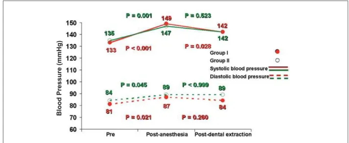
In relation to blood pressure, variations are shown in Figure 1. No significant difference was observed between the groups in the three study phases: pre, post-anesthesia, and post-dental extraction.
Results in Table 3 show the occurrence of ST-segment depression (1.0 mm) in three patients in whom epinephrine and the primary endpoint of this study was the occurrence of
the following variables detecting myocardial ischemia during or after dental treatment: 1) ST-T segment alterations, as assessed by Holter monitoring; 2) left ventricular segmental hypocontractility, as assessed by Doppler-echocardiography and 3) elevation of biochemical markers. The secondary endpoint was the detection of occurrence of: 1) chest pain during the dental procedure; 2) arrhythmias, as assessed by Holter monitoring and 3) mitral regurgitation, as assessed by Doppler-echocardiography.
Methods
From May, 2004 to May, 2005, 54 patients diagnosed with chronic coronary artery disease confirmed by coronary angiography and undergoing treatment in the Coronary Angioplasty Section of the Instituto Dante Pazzanese de Cardiologia of the State of São Paulo were included in the study. This study was approved by the Research Ethics Committee of that Institution and written informed consent was obtained from all patients.
Inclusion criteria were: patients of both genders with indication for dental extraction and no restriction as to the age range; patients with chronic coronary artery disease confirmed by previous coronary angiography, and with stable angina on exertion. Patients with unstable angina; acute myocardial infarction (occurring < 3 months); imminent indication of cardiac surgery or angioplasty; heart diseases associated with coronary disease; heart failure; recent stroke (< 3 months); VHYHUH K\SHUWHQVLRQ 63 PP+J DQGRU '3 mmHg) and uncontrolled diabetes mellitus were excluded.
Prior to the dental intervention, an electrocardiogram (ECG) was performed, and the patients had their blood drawn for general biochemical tests (blood count; platelet count; coagulation tests; BUN, creatinine, sodium, chloride, potassium, and fasting plasma glucose levels) and determinations of biochemical markers of myonecrosis (CKMB activity, CKMB mass, and troponin T); a Holter monitor was installed to obtain an ECG during the dental procedure, and the first blood pressure reading was taken.
After the pre-dental intervention tests were obtained, patients were referred to the Doppler-echocardiography laboratory, where the pre-anesthetic echocardiogram for the analysis of left ventricular measurements, left ventricular function and mitral flow was performed.
Also in the Doppler-echocardiography laboratory, the patients underwent local anesthesia with 2% mepivacaine anesthetic salt combined with 1:100,000 epinephrine, or 3% mepivacaine without vasoconstrictor, administered according to the envelopes previously drawn.
After two-minute latency for the mepivacaine anesthetic salt, heart rate was measured and post-anesthetic echocardiogram was performed for the analysis of the same measurements.
The patients were then referred to the dental office for dental extraction and second blood pressure measurement.
7DEOH&OLQLFDOGDWDRIWKHSDWLHQWVGLYLGHGDFFRUGLQJWRWKHW\SHRIDQHVWKHWLFV*URXS,ZLWKYDVRFRQVWULFWRU
and Group II, control group (without vasoconstrictor)
Variables Group I - with
vasoconstrictor n=27
Group II - without
vasoconstrictor n=27 p
Age, mean (SD) (years) 58 (7.98) 55.3 (8.57) 0.236
Variation (min. and max. ages) 46 a 71 43 a 73
(OGHUO\!\HDUV 1 1
0DOHJHQGHUQ 16 (59.3) 18 (66.7)
Previous cardiovascular events 0.362
ST-segment elevation myocardial infarction (n/%) 13 (48.1) 14 (51.8)
Non-ST segment elevation myocardial infarction (n/%) 5 (18.5) 7 (25.9)
Clinical presentation
Coronary insufficiency (n/%) 27 (100) 27 (100)
-Arterial hypertension (n/%) 26 (96.3) 26 (96.3) 1.000
Diabetes mellitus (n/%) 7 (26) 9 (33.3) 0.551
Dyslipidemia (n/%) 14 (51.8) 9 (33.3) 0.268
Types of coronary intervention
Stent implantation
Anterior descending artery (n/%) 20 (74.1) 15 (55.5) 0.154
Circumflex artery (n/%) 5 (18.5) 4 (14.8) 1.000
Right coronary artery (n/%) 5 (18.5) 10 (37) 0.129
Marginal artery (n/%) 1 (3.7) 1 (3.7) 1.000
Balloon catheter angioplasty
Circumflex artery (n/%) 1 (3.7) - (-) 1.000
Anterior descending artery (n/%) - (-) 2 (7.4) 0.491
n - number; % - percentage; SD - standard deviation.
7DEOH7HFKQLFDOFKDUDFWHULVWLFVDQGFOLQLFDOV\PSWRPVRIWKHFRURQDU\SDWLHQWVLQFRQYHQWLRQDOGHQWDOH[WUDFWLRQV
Variables Group I (with vasoconstrictor) n=27
Group II (without vasoconstrictor)
n=27 p
Conventional dental extraction 1.000
One tooth extracted (n/%) 15 (55.5) 15 (55.5)
Two teeth extracted (n/%) 8 (29.6) 7 (25.9)
Three or more teeth extracted (n/%) 4 (14.8) 5 (18.5)
Mean and standard deviation 1.67 (0.96) 1.81 (1.21)
1DQHVWKHWLFFDUWULGJHV 0.161
Q 24 (88.9) 20 (74.1)
> 2 (n/%) 3 (11.1) 7 (25.9)
Mean and standard deviation 1.56 (0.87) 1.89 (0.79)
Symptoms (periprocedural)
Chest pain (n/%) - (-) - (-)
-Pain at dental luxation (n/%)l 1 (3.7) 9 (33.3%) 0.005
Palpitation (n/%) - (-) - (-)
-Diaphoresis (n/%) - (-) - (-)
was used, all during anesthetic injection. However, no ST-segment alteration was observed when anesthetics without vasoconstrictor were used in the two phases of the dental treatment: anesthesia and dental extraction.
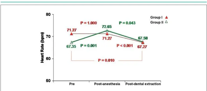
Figures 2 and 3 show intragroup variations of mean heart rates and ejection fraction during dental treatment in the 54 coronary patients of groups I and II.
Pre and post-dental intervention levels of biochemical markers of myonecrosis in the two groups of patients are shown in Table 4.
Discussion
The high prevalence of cardiovascular diseases in the population, particularly ischemic heart diseases, shows that dental surgeons will treat this type of patient with increasing frequency8.
The use of local anesthetics with vasoconstrictors in coronary patients remains very controversial in the literature. In 1955, the New York Heart Association recommended and stipulated that the maximum dose of
epinephrine should be 0.2mg in local anesthesia when used in cardiac patients9.
This recommendation was approved by the American Dental Association and American Heart Association (1964)10, which specify that vasoconstrictors are not
contraindicated in patients with heart diseases, provided that a safe anesthetic technique is used with a minimum amount of anesthetics and previous aspiration, although the use of vasoconstrictors should be avoided in high risk cardiovascular disease patients.
Malamed11 and Bennet12 and, more recently, Budentz13
in 2000, have recommended lower doses of 0.04mg of vasoconstrictor, which corresponds to approximately one 1:50,000 epinephrine cartridge; two 1:100,000 epinephrine cartridges, or four 1:200,000 epinephrine cartridges in each dental treatment session for patients with severe cardiovascular diseases. However, these authors do not specify criteria to categorize severe heart diseases.
In the present study, only three patients (12.0%) in group I had mild ST-segment ischemic depression (1.0 mm), which was observed only in the initial period of anesthetic action
7DEOH67VHJPHQWDOWHUDWLRQLQGLFDWLQJWKHRFFXUUHQFHRIP\RFDUGLDOLVFKHPLDGHSUHVVLRQ!PPGXULQJ
anesthesia or dental extraction, compared with the pre-anesthetic phase
Group I (with vasoconstrictor)
Q Group II (without vasoconstrictor)Q
Variable AnesthesiaQ Dental extractionQ AnesthesiaQ Dental extractionQ Total
ST-segment depression
Yes 3 (12) - (-) - (-) - (-) 3
No 22 (88) 25 (100) 24 (100) 24 (100) 49
Total 25 25 24 24
n - number; % - percentage.
Fig. 1 -Mean systolic and diastolic blood pressure variations in the three phases of dental treatment in all 54 patients of groups I and II.
B
lo
o
d
Pr
e
s
s
u
re
(m
m
H
g
7DEOH6HUXPOHYHOVRIELRFKHPLFDOPDUNHUVRIP\RQHFURVLVGHWHUPLQHGSUHDQGSRVWGHQWDOLQWHUYHQWLRQLQWKHSDWLHQWVVWXGLHG
Group I (with vasoconstrictor) n=27
Group II (without vasoconstrictor) n=27
Biochemical marker 1RUPDOQ AbnormalQ 1RWPHDVXUHGQ 1RUPDOQ AbnormalQ 1RWPHDVXUHGQ
CKMB mass 18(66.7) 2(7.4) 7(25.9) 10(37.0) - (-) 17(62.9)
CKMB activity 24(88.9) - (-) 3(11.1) 24(88.9) - (-) 3(11.1)
Troponin T 21(77.8) - (-) 6(22.2) 10(37.0) - (-) 17(62.9)
QQXPEHUSHUFHQWDJHQRUPDOUDQJH&.0%PDVV QJPO&.0%DFWLYLW\ 8OO7URSRQLQ7 QJPO
Fig. 2 -Heart rate variations in the three phases of dental treatment in all 54 patients of groups I and II.
(Table 3). In group II, no patients presented ST-segment alterations. No significant difference was observed between the two groups in relation to this ST-segment shift (p = 0.235).
We should point out that in the three cases with ST-segment depression the simultaneous occurrence of any other alteration considered as detectors of myocardial ischemia (left ventricular hypocontractility and elevation of myonecrosis markers) was not observed.
Vanderheyden et al14 from the Veterans Administration
Medical Center, Los Angeles, also conducted a study in which they assessed the effects of dental treatment with local anesthesia with 1:100,000 epinephrine in 20 coronary patients and did not demonstrate the occurrence RIP\RFDUGLDOLVFKHPLD67VHJPHQWGHSUHVVLRQPP during the different phases of the procedure.
On the other hand, Doppler-echocardiography has shown to be sensitive to detect myocardial ischemia using the analysis of left ventricular segmental contractility during physical or pharmacological stress. Depending on the size and location of the myocardial ischemia, some degree of mitral regurgitation may also occur under these circumstances. As demonstrated, in this study we included these two echocardiographic variables in the analysis of occurrence of myocardial ischemia during dental extractions. However, no significant alterations of these data were observed between the groups in any of the study phases.
Also in relation to the primary endpoints of our investigation, we point out the determination of biochemical markers, whose alterations may be a manifestation of myocardial ischemia.
Three markers were analyzed: CKMB activity, CKMB mass, and troponin T.
In this study (Table 4), only two patients of group I presented mild CKMB mass elevation versus no cases in group II (p = 0.540). However, none of the two cases presented other manifestations of myocardial ischemia, considering the other data analyzed in the study.
Mean systolic and diastolic blood pressures obtained before anesthesia, during anesthesia, and after dental extraction are shown in Figure 1. As can be observed in this figure, the systolic blood pressure increased somewhat DIWHUORFDODQHVWKHWLFDGPLQLVWUDWLRQSLQERWK groups, and decreased after dental extraction, reaching values close to those of baseline. The diastolic blood pressure also increased in the post-anesthetic phase when compared to the pre-anesthetic phase (p > 0.05), and did not suffer significant changes between study phases 2 and 3 (p > 0.05).
As for the other variables, Chernow et al’s observations15
show that heart rate drops immediately after the use of anesthetics without vasoconstrictor, but remains increased by two to ten beats per minute with the use of vasoconstrictors. Another study added that if higher doses
of vasoconstrictor are used, the heart rate elevation will be even greater16. This finding was corroborated in the present
study, in which the heart rate decreased (Figure 2) in group I in the pre-dental extraction phase when compared with the pre and post-anesthesia phases (67.27 vs 71.27, p < 0.001); however, the ejection fraction (Figure 3) had a significant increase in the post-dental extraction period in relation to the pre-anesthesia period (57.92 vs 56.72, p = 0.009). This observation is of the utmost importance since the presence of ischemia causes a decrease in ejection fraction, and not its elevation.
In group II, an increase in heart rate (Figure 2) was observed when comparing the post-anesthesia with the pre-anesthesia period (72.65 vs 67.35, p = 0.001), and a significant decrease occurred in the post-dental extraction period (72.65 vs 67.58, p = 0.043); no significant changes were observed in ejection fraction (Figure 3) during the three study phases (57.35 vs 57.08vs 57.19, p = 0.988).
The routine use of local anesthetics with vasoconstrictors in dental offices requires care and careful assessment on the part of dental surgeons, since there are absolute contraindications to the use of vasoconstrictors, particularly in high risk cardiac patients. On the other hand, the results of this study, like those of other studies in the literature, do not systematically show the presence of myocardial ischemia in the assessments during dental extractions, thus corroborating that the benefit of the use of these anesthetics is greater than the risk of some cardiac complication.
Therefore, contact between the dental surgeon and the patient’s cardiologist is suggested for a perfect knowledge of the patient’s heart disease and medications used to ensure that the patient is controlled from a cardiac point of view, and can be allowed to undergo dental treatment.
We should also point out the importance of pre-procedural blood pressure measurement; of measures to reduce patient’s stress during dental extractions; and of monitoring laboratory tests to detect any clinical alteration.
As a limitation of the present study we point out that the observations of this investigation refer to a selected group of patients with chronic coronary diseases of moderate clinical and anatomical complexity. Therefore, they should not be extrapolated to high risk coronary disease patients, such as those with unstable angina or complex arrhythmias; those with three-vessel coronary artery disease, and those with severe left ventricular dysfunction.
In conclusion, dental extractions performed under the use of anesthesia with 1:100,000 epinephrine do not imply additional ischemic risks, provided that they are performed with good anesthetic technique and maintenance of the pharmacological treatment prescribed by the cardiologist.
Potential Conflict of Interest
References
1. Conrado VCLS, Andrade ACP, Angelis GAMC, Timerman L. Efeitos cardiovasculares da anestesia local com vasoconstritor durante exodontia convencional em coronariopatas [resumo]. In: 27º Congresso da Sociedade de Cardiologia do Estado de São Paulo. Campos do Jordão, SP, de 25 a 27 de maio de 2006. Rev Soc Cardiol Estado de São Paulo. 2006;16 (2 Supl B):140.
2. Magalhães HM. Tratamento odontológico no cardiopata. São Paulo: Sarvier; 1993. p. 1.
3. Franken RA, Franken M. Avaliação de risco cardiovascular para procedimentos odontológicos. Rev Soc Cardiol Estado de São Paulo. 2000; 10: 406-13.
4. Andrade ED, Ranali J, Volpato MC. Pacientes que requerem cuidados especiais. In: Andrade ED. Terapêutica medicamentosa em odontologia. São Paulo: Artes Médicas; 1999. p. 93-140.
5. Malamed SF. Handbook of medical emergencies in the dental office. 3rd ed. St Louis: Mosby-Year Book; 1987.
6. Perusse R, Goulet JP, Turcotte JY. Contraindications to vasoconstrictors in dentistry: Part I. Cardiovascular diseases. Oral Surg Oral Med Oral Pathol. 1992; 74: 679-86.
7. Perusse R, Goulet JP, Turcotte JY. Contraindications to vasoconstrictors in dentistry: Part II. Hyperthyroidism, diabetes, sulfite sensitivity, cortico-dependent asthma, and pheochromocytoma. Oral Surg Oral Med Oral Pathol. 1992; 74: 687-91.
8. Jowett NI, Cabot LB. Patients with cardiac disease: considerations for the dental practitioner. Br Dent J. 2000; 189: 297-302.
9. Use of epinephrine in connection with procaine in dental procedures. Report of the Special Committee of the New York Heart Association. J Am Dent Assoc. 1955; 157: 854.
10. Akutsu A, Chiba T, Takahashi H, Shimoda M, Suematsu T. American Dental Association and American Heart Association. Management of dental problems in patients with cardiovascular disease. J Am Dent Assoc. 1964; 68: 333-42.
11. Malamed SF. Handbook of local anesthesia. 2nd ed. St Louis: Mosby-Year Book; 1986.
12. Bennett CR. Monheim’s local anesthesia and pain control in dental practice. 7th ed. St Louis: Mosby-Year Book; 1984.
13. Budentz AW. Local anesthetics and medically complex patients. J Calif Dent Assoc. 2000; 28: 611-9.
14. Vanderheyden PJ, Williams RA, Sims TN. Assessment of ST segment depression in patients with cardiac disease after local anesthesia. J Am Dent Assoc. 1989; 119: 407-12.
15. Chernow B, Balestrieri F, Ferguson CD, Terezhalmy GT, Fletcher JR, Lake CR. Local dental anesthesia with epinephrine: minimal effects on the sympathetic nervous system or on hemodynamics variables. Arch Intern Med. 1983; 143: 2141-3.