ABSTRACT
http://dx.doi.org/10.1590/1678-7757201302200
Biocompatibility and setting time of CPM-MTA
and white Portland cement clinker with or without
calcium sulfate
1, Marcia Magro KATO2, Gerson Francisco de ASSIS3 ,
Norberti BERNARDINELI1, Ivaldo Gomes de MORAES1!", Ronald ORDINOLA-ZAPATA2,
Alexandre Silva BRAMANTE2
1- DDS, PhD, Full Professor, Discipline of Endodontics, Department of Operative Dentistry, Endodontics and Dental Materials, Bauru School of Dentistry, University of São Paulo, Bauru, SP Brazil.
2- DDS, PhD in Endodontics, Department of Operative Dentistry, Endodontics and Dental Materials, Bauru School of Dentistry, University of São Paulo, Bauru, SP Brazil.
3- DDS, PhD, Full Professor, Discipline of Histology, Department of Biological Sciences, Bauru School of Dentistry, University of São Paulo, Bauru, SP, Brazil. 4- DDS, PhD, Associate Professor, Discipline of Histology, Department of Biological Sciences, Bauru School of Dentistry, University of São Paulo, Bauru, SP, Brazil.
#$ Clovis Monteiro Bramante - Departamento de Dentística, Endodontia e Materiais Odontológicos - Faculdade de Odontologia de Bauru - Universidade de São Paulo - Al. Octávio Pinheiro Brisola, 9-75 - 17012-901 - Bauru - SP - Brasil - Phone: 55-14-32358344 - e-mail: [email protected]
%&$ '()*+)4 ;$<=)*+(4#$<=)*+(
O
bjective: To evaluate the biocompatibility and the setting time of Portland cement clinker with or without 2% or 5% calcium sulfate and MTA-CPM. Material and Methods:Twenty-four mice (
with Portland cement clinker with or without 2% or 5% calcium sulfate and MTA. After 15, 30 and 60 days of implantation, the animals were killed and specimens were prepared for microscopic analysis. For evaluation of the setting time, each material was analyzed ! "#$& Number C266-08 guideline. Data were analyzed by ANOVA and Tukey’s test for setting time ')*+ /479; < 44 4 (p>0.05) among the materials in the subcutaneous tissues. For the setting time, clinker 4 ?@EG@ s), followed by clinker with 2% calcium sulfate (9.22 s/25.33 s), clinker with 5% calcium sulfate (10.06 s/42.46 s) and MTA (15.01 s/42.46 s). Conclusions: All the tested materials 4 setting times of the white Portland cement clinker.
Key words: Dental material. Calcium sulfate.
INTRODUCTION
Mineral Trioxide Aggregate (MTA) has been used for surgical and nonsurgical treatment such * ! 4 ! K 6,17,20,21,24,26. Previous researches showed adequate sealing ability, antimicrobial activity and biocompatibility for this material3,5,14-16,24,26. MTA is basically Portland cement with bismuth oxide added for radiopacity12,15. According to Camilleri7 (2007) the main components of Portland cement are: lime (CaO) 60-66%, silica
culture tests22,29, and in human and animal direct pulp capping procedures17,20,21,23. These studies showed similar results for Portland cement and MTA.
In the cement production process, gypsum is added to the Portland cement clinker in amounts of 3-6% to delay the setting time. The exclusion 4 4 R production showed decrease of the setting time of
the cement8#
helpful in clinical procedures because MTA exhibits a longer setting time2,4,5,19. The Portland cement clinker has advantageous properties, such as an adequate setting time, alkaline pH and calcium release28. The aim of this study was to evaluate the subcutaneous tissue response of white Portland cement clinker with 2% and 5% calcium sulfate. The setting time of each material was also evaluated.
MATERIAL AND METHODS
The materials used in this study were: White Portland cement clinker without calcium sulfate-WPCC (Grupo Votorantim, Itaboca, MG, Brazil), WPCC with 2% or 5% calcium sulfate (Carlo Erba Reagents, Italia) and MTA-CPM (EGEO SRL, Buenos Aires, Argentina). White Portland cement clinker was sieved under laboratory conditions to a particle size of 0.062 mm (Bronzinox, São Paulo, SP, Brazil). The materials were mixed using a spatula and glass plate and the powder/liquid ratio used for all tested materials was in the 3:1 ratio.
Biocompatibility test
Twenty-four adult male Wistar rats ( norvegicus albinus) weighing between 200-250
g were used for this experiment after approval by the institutional Ethics Committee for Animal Research. The animals were anesthetized with an intramuscular dose of 25 mg/kg ketamine chloride and 10 mg/kg xylazine chloride, and then four incisions were made through the dorsal skin using a #5 scalpel blade. A blunt dissecting instrument was used to create a 20 mm deep pocket in the subcutaneous tissue to receive the implants. Polyethylene tubes (1 mm diameter and 10 mm ] the tested materials. Each animal received four implants with the different tested materials. The wounds were closed and sutures were removed after 7 days.
Eight animals were used for each evaluated period (15, 30 and 60 days). The animals were killed by an intracardiac anesthetic injection. Subcutaneous tissues with the implanted tubes were removed and immediately placed in 10% formalin. After histological procedures, 5 μm-thick serial sections were prepared and stained with hematoxylin and eosin.
The subcutaneous response of the tissue in direct contact with the cements (capsule) was evaluated using descriptive parameters and by 4^ using an optical microscope (Leitz, Aristoplan, Germany). The counting procedure was done at a 4___`$ 4 { 4 4 ] network of test lines crossed to form 25 volumetric points. For this procedure, 3 slices with 4 semi-serial sections were selected for the analyses. In each ! 4 morphometric procedure. In each point (Pi), the 4^ ! cell density was determined by dividing the total number of points analyzed (P) by the number of points that include the histological structure.
$^ by semi-quantitative analysis at 400X and scored ;_;^?}; ~G^? }G;G*G ^? };G inflammatory cells (severe reaction). Kruskal-Wallis and Dunn tests were used to determine the 444^ of the test materials (p<0.05).
Setting time test
Setting time of the materials was measured under controlled temperature and humidity (37±1°C and 95±5% relative humidity), according to the "#$&G*_@ $ materials were mixed and inserted in metallic ring molds (10 mm diameter and 2 mm thick). Three specimens of each cement were made. After 180 s, each specimen was indented using a 113.5 g Gilmore needle for determining initial setting time. Final setting time was obtained using a 456.5 g Gilmore needle. Data were subjected to statistical analysis using ANOVA and Tukey test for multiple comparisons (p<0.05).
15 d 30 d 60 d
CPM-MTA 2 1 0
Clinker 2 0 0
Clinker+2%
CaSO4
1 1 0
Clinker+5%
CaSO4
2 1 0
Table 1-
RESULTS
Microscopic analysis
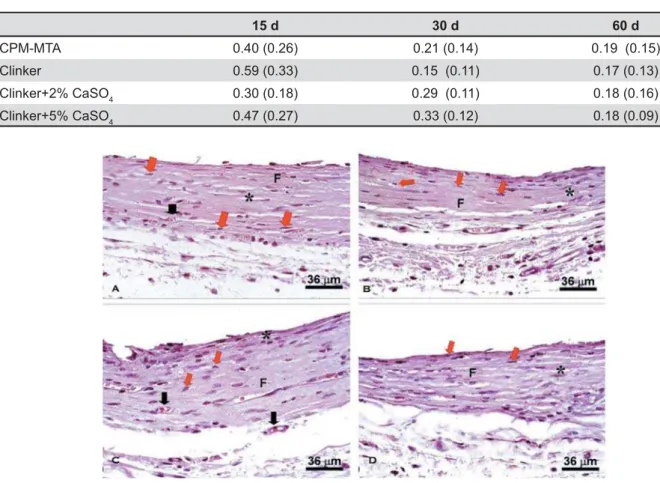
Table 1 shows the mean scores attributed to ^ 4 Table 2 shows the results of morphometric analysis adjacent to the implanted material surface at 15, 30 and 60 days.
15 days
Tissue response for all cements was very similar in this period. Among the main characteristics, it 4 the polyethylene tube. The capsule was thick, with $ 4! synthetic activity. This area also presented small 4 ^ 44 materials (p>0.05).
30 days
Tissue response was similar to the 15 days period. However, more organized capsules were
4 !4 ^ ! were thicker, blood vessels less numerous and there was fewer empty spaces than in the 15-day-period, indicating a better degree of tissue organization. $ 44 among the materials (p>0.05).
60 days
The tissue showed a thick fiber structure positioned parallel to the cement surface. There 4 ! ] 4 structures, decreasing in quantity. There were few 4^ $ 44 among the materials (p>0.05). Representative images of the tested materials are shown in Figure 1.
In a general way, blood vessels decreased when compared to shorter periods, demonstrating a better organization and tissue maturation with " ! 4 ^ ?! and multinucleated giant cells) circumscribed the
15 d 30 d 60 d
CPM-MTA 0.40 (0.26) 0.21 (0.14) 0.19 (0.15)
Clinker 0.59 (0.33) 0.15 (0.11) 0.17 (0.13)
Clinker+2% CaSO4 0.30 (0.18) 0.29 (0.11) 0.18 (0.16)
Clinker+5% CaSO4 0.47 (0.27) 0.33 (0.12) 0.18 (0.09)
Table 2-
adjacent to the implanted pure clinker, 2% calcium sulfate clinker; 5% calcium sulfate clinker; CPM (MTA-Egeo)
material in the 15 days period. The number of these cells decreased in the 30 and 60 days periods.
Setting time
$ 4 are shown in Table 3. Clinker without calcium 4 times (6.18 s/21.48 s), followed by clinker with 2% calcium sulfate (9.22 s/25.33 s), clinker with 5% calcium sulfate (10.06 s/42.46 s) and MTA (15.01 s/42.46 s). Regarding the initial setting time, MTA ?~__ than the other materials. Only the addition of 5% 4 ?~__ ) setting time, the clinker with 5% calcium sulfate " 44 (p<0.05) was observed in the comparison of pure clinker and MTA.
DISCUSSION
This study evaluated the setting time and the subcutaneous tissue response to the white Portland cement clinker with or without 2% and 5% calcium sulfate. Clinically, some situations such 4 4! require a faster setting time to avoid the dissolution 4 ^ these situations, the use of MTA without calcium sulfate (Portland cement clinker) could be more advantageous. The results showed that all tested materials presented a similar behavior with small differences regarding to the evaluated periods. In the literature review were found no biocompatibility
in vivo studies about the white Portland cement clinker with or without calcium sulfate in different proportions. The biocompatibility of MTA and R and is basically attributed to calcium hydroxide formation after hydration10,12,17.
The biocompatibility of MTA is related to its capacity of releasing calcium ions and, consequently, to the alkaline pH produced by the material.
When exposed to a humid medium, MTA releases calcium silicate and calcium oxide. The calcium oxide released produces calcium hydroxide after ^ ! calcium ions. Calcium ions are important for their participation in the activation of calcium-dependent adenosine triphosphatase. Calcium carbonate from the reaction of calcium ion with carbon K 4 and favors the mineralization. A rich extracellular )4 crystals strongly supports the role of calcite crystals 4 of hard tissue. Although this calcium ion release promoted by MTA contributes to turning the medium inhospitable for bacterial growth, its high concentration after hydration might contribute to
4 1,15,28.
Regarding the setting time, previous studies have shown an initial setting time of 40 min for MTA ProRoot and 12 min for MTA-Angelus13,18. MTA
;_*_13,18,26
R were 70 min and 170 min18 respectively. It was observed that the Portland cement clinker without calcium sulfate exhibited the smallest initial setting time (6.18 min) followed by Portland cement clinker with 2% calcium sulfate (9.22 min), and Portland cement clinker with 5% calcium sulfate (10.06 min). The results of setting time of white Portland cement clinker are similar to the results reported by Camilleri, et al.10 (2005)who reported initial and 4 @
! ^ 4 white Portland cement clinker with or without 2% or 5% calcium sulfate and MTA Egeo
were
similar when they were implanted in subcutaneous tissue for 15, 30 and 60 days. White Portland cement clinker without calcium sulfate presented the shortest initial setting time. The 2% and 5% calcium sulfate clinkers showed intermediary setting times while MTA showed the longest. Calcium sulfate delayed the white Portland cement clinker setting time.REFERENCES
1- Aguilar FG, Garcia LFR, Pires-de-Souza FCP. Biocompatibility of new calcium aluminate cement (EndoBinder). J Endod. 2012;38:367-71.
2- Al Anezi AZ, Zhu Q, Wang YH, Safavi KE, Jiang J. Effect of selected accelerants on setting time and biocompatibility of mineral trioxide aggregate (MTA). Oral Surg Oral Med Oral Pathol Oral Radiol Endod. 2011;111:122-7.
3- Al Hiyasat AS, Al Sa’Eed OR, Darmani H. Quality of cellular * "# 2012;20:82-8.
*#!<!#R 4R9 MTA to improve handling characteristics and decrease setting time. J Endod. 2007;33:1231-4.
Material Initial setting time
Final setting time
MTA 15’01’’ 27’36’’
Clinker 6’18’’ 21’48’’
Clinker+2% CaSO4
9’22’’ 25’33’’
Clinker+5%
CaSO4
10’06’’ 42’46’’
5- Bortoluzzi EA, Broon NJ, Bramante CM, Garcia RB, Moraes IG, Bernardineli N. Sealing ability of MTA and radiopaque Portland 4 * Endod. 2006;32:897-900.
6- Broon NJ, Bramante CM, Assis GF, Bortoluzzi EA, Bernardineli N, Moraes IG, et al. Healing of root perforations treated with Mineral Trioxide Aggregate (MTA) and Portland cement. J Appl Oral Sci. 2006;14:305-11.
7- Camilleri J. Hydration mechanisms of mineral trioxide aggregate. Int Endod J. 2007;40:462-70.
8- Camilleri J. Characterization of hydration products of mineral trioxide aggregate. Int Endod J. 2008;41:408-17.
9- Camilleri J, Montesin FE, Brady K, Sweeney R, Curtis RV, Pitt Ford TR. The constitution of mineral trioxide aggregate. Dent Mater. 2005;21:297-303.
10- Camilleri J, Montesin FE, Di Silvio L, Pitt Ford TR. The chemical constitution and biocompatibility of accelerated Portland cement for endodontic use. Int Endod J. 2005;38:834-42.
11- Camilleri J, Montesin FE, Juszczyk AS, Papaioannou S, Curtis RV, Donald FM, et al. The constitution, physical properties and 4 / & 2008;24:341-50.
12- Camilleri J, Pitt Ford TR. Mineral trioxide aggregate: a review of the constituents and biological properties of the material. Int Endod J. 2006;39:747-54.
13- Chng HK, Islam I, Yap AU, Tong YW, Koh ET. Properties of a * G__};*@
14- Cunha AS, Rached FJ Jr, Alfredo E, León JE, Perez DE. Biocompatibility of sealers used in apical surgery: a histological study in rat subcutaneous tissue. Braz Dent J. 2011;22:299-305. 15- Estrela C, Bammann LL, Estrela CR, Silva RS, Pécora JD. Antimicrobial and chemical study of MTA, Portland cement, calcium hydroxide paste, Sealapex and Dycal. Braz Dent J. 2000;11:3-9. 16- Gonçalves JL, Viapiana R, Miranda CE, Borges AH, Cruz Filho AM. Evaluation of physico-chemical properties of Portland cements and MTA. Braz Oral Res. 2010;24:277-83.
17- Holland R, Souza V, Murata SS, Nery MJ, Bernabé PF, Otoboni Filho JA, et al. Healing process of dog dental pulp after pulpotomy and pulp covering with mineral trioxide aggregate or Portland cement. Braz Dent J. 2001;12:109-13.
18- Islam I, Chng HK, Yap AU. Comparison of the physical and mechanical properties of MTA and Portland cement. J Endod. 2006;32:193-7.
19- Kogan P, He J, Glickman GN, Watanabe I. The effects of various additives on setting properties of MTA. J Endod. 2006;32:569-72. 20- Menezes R, Bramante CM, Letra A, Carvalho VG, Garcia RB. Histologic evaluation of pulpotomies in dog using two types of mineral trioxide aggregate and regular and white Portland cements as wound dressings. Oral Surg Oral Med Oral Pathol Oral Radiol Endod. 2004;98:376-9.
21- Menezes R, Bramante CM, Garcia RB, Letra L, Carvalho VG, Carneiro E, et al. Microscopic analysis of dog dental pulp after pulpotomy and pulp protection with mineral trioxide aggregate and white Portland cement. J Appl Oral Sci. 2004;12:104-7. 22- Min KS, Kim HI, Park HJ, Pi SH, Hong CU, Kim EC. Human pulp cells response to Portland cement in vitro. J Endod. 2007;33:163-6.
23- Nair PN, Duncan HF, Pitt Ford TR, Luder HU. Histological, ultrastructural and quantitative investigations on the response of healthy human pulps to experimental capping with mineral trioxide aggregate: a randomized controlled trial. Int Endod J. 2008;41:128-50.
24- Parirokh M, Asgary S, Eghbal MJ, Stowe S, Eslami B, Eskandarizade A et al. A comparative study of white and grey mineral trioxide aggregate as pulp capping agents in dog's teeth. Dent Traumatol. 2005;21:150-4.
25- Saidon J, He J, Zhu Q, Safavi K, Spångberg LS. Cell and tissue reactions to mineral trioxide aggregate and Portland cement. Oral Surg Oral Med Oral Pathol Oral Radiol Endod. 2003;95:483-9. 26- Torabinejad M, Chivian N. Clinical applications of mineral trioxide aggregate. J Endod. 1999;25:197-205.
27- Torabinejad M, Ford TR, Abedi HR, Kariyawasam SP, Tang HM. $ * and mandible of guinea pigs. J Endod. 1998;24:468-71. 28- Vivan RR, Zapata RO, Zeferino MA, Bramante CM, Bernardineli N, Garcia RB, et al. Evaluation of the physical and chemical properties of two commercial and three experimental root-end #& R9 2010;110:250-6.