Involvement of beta absolute power in motor areas after
hand immobilization: An EEG study
Dionis MachadoI, Jadna Helena dos Santos FrançaII, Silmar TeixeiraIII, Victor Hugo do Vale BastosIII, Maurício CagyIV, Alberto Souza de Sá FilhoV, Sérgio MachadoV,VI, Bruna VelasquesI,VII,VIII, Pedro RibeiroI,VII,VIII
DOI: 10.5935/MedicalExpress.2016.05.03
I Universidade Federal do Rio de Janeiro (UFRJ), Instituto de Psiquiatria (IPUB), Laboratório de Mapeamento Cerebral e Integração Sensório Motora, Rio de Janeiro,
RJ, Brasil
II Universidade Federal do Piauí (UFPI), Parnaíba, PI, Brasil
III Universidade Federal do Piauí (UFPI), Laboratório de Mapeamento Cerebral e Funcionalidade (LAMCEF), Parnaíba, PI, Brasil IV Universidade Federal do Rio de Janeiro (UFRJ), Programa de Bioengenharia (COPPE), Rio de Janeiro, RJ, Brasil
V Universidade Federal do Rio de Janeiro (UFRJ), Instituto de Psiquiatria (IPUB), Laboratório de Pânico e Respiração (LABPR), Rio de Janeiro, RJ, Brasil
VI Universidade Salgado de Oliveira (UNIVERSO), Programa de Pós-Graduação em Ciências da Atividade Física (PGCAF), Laboratório de Neurociência da Atividade
Física (LABNAF), Niterói, RJ, Brasil
VII Universidade Federal do Rio de Janeiro (UFRJ), Escola de Educação Física, Rio de Janeiro, RJ, Brasil VIII Instituto de Neurociências Aplicadas (INA), Rio de Janeiro, RJ, Brasil
OBJECTIVE: The purpose of this study was to analyze changes in beta band absolute power in cortical areas, before and after a condition of hand immobilization for 48 hours.
METHOD: Fifteen healthy volunteers, aged between 20 and 30, were submitted to EEG assessment before and after immobilization, while performing a motor task triggered by a visual stimulus.
RESULTS: Statistical analysis revealed that hand immobilization caused changes in cortical areas. Signiicant increases in beta band absolute power were found after hand immobilization at electrodes Fp2, C3 and P4. In contrast, at electrode C4 a decrease in beta band absolute power occurred after hand immobilization.
CONCLUSION: Predominant hand immobilization, even for 48 hours, is suicient to cause cortical changes that afect movement planning. Such changes may represent a cortical strategy to supply cortical changes in contralateral hemisphere due to immobilization. Further studies are necessary to understand cortical changes due to hand immobilization and movement planning, especially considering how much time of immobilization is necessary to promote such changes.
KEYWORDS: Beta band, Hand immobilization, Neural plasticity, Electroencephalography.
Machado D, França JHS, Teixeira S, Bastos VHV, Cagy M, Sá-Filho AS, Machado S, Velasques B, Ribeiro P. Involvement of beta absolute power in motor areas after hand immobilization: An EEG study. MedicalExpress (São Paulo, online). 2016;3(5):M160503
Received for Publication on April 4, 2016; First review on May 11, 2016; Accepted for publication on September 20, 2016; Online on 17 October, 2016
E-mail: [email protected]
■
INTRODUCTIONElectroencephalography (EEG) has been frequently used to analyze neural activity during motor tasks in order to understand how the human brain controls movements and thus shed light on mechanisms associated with motor learning.1-5 Neuroimaging represents a helpful tool for neurological rehabilitation, i.e., it is able to analyze how
a specific task triggers brain activation differently in
subjects should not have mental or physical illnesses (as evaluated through a previous anamnesis) and should not use any psychoactive or psychotropic substances during the duration of the study.Due to hand laterality, the Edinburgh inventory24,25 was used to identify the right- vs left-handed laterality predominance of the participants. Left-handed individuals were excluded from the experiment. The subjects were instructed not to use tobacco, coffee or alcoholic drinks 10 hours before the test because these
substances may influence cortical activation recorded
by QEEG brain mapping.26-28 The study was approved by the ethics committee of Veiga de Almeida University and complied with the ethical standards of the Declaration of Helsinki.
Tasks and Procedures
A room with acoustic and electrical isolation wa s u s e d . T h e l i g h t s we re d i m m e d d u r i n g t h e electroencephalography (EEG) signal acquisition and the subjects were sitting in a chair with armrests in order to minimize muscle artifact during EEG signal acquisition.In front of the subjects, on a table, there was a 15-inch monitor that was placed facing the subjects and turned on only when the subjects performed the task (i.e., flexion and extension of the index finger). Initially, the EEG signal acquisition lasted for 2 minutes (at rest) with the monitor turned off and facing the subjects. Then, a sensor to measure acceleration (accelerometer) was placed on the right index finger. A visual stimulus appeared on the monitor and the subjects performed the task (i.e., flexion and extension of the index finger). The accelerometer was connected to the EEG through an additional channel (i.e., channel 21) and thereby provided a signal to EEG recording when the subjects performed the movement.
The research team developed a task where
participants performed index finger flexion and extension
when visual feedback was generated by a random image (i.e., a yellow ball) on the monitor. The complete task involved 6 blocks of 15 trials. In order to avoid muscle fatigue, a 3-minute break between each block was given to the subjects. Thus, the task had 1 minute in each block with a 3-minute interval between blocks, adding up to 24 minutes for the whole task. After completing the task, the monitor was turned off and the subjects were submitted again to EEG during 2 minutes (at rest). After EEG recording, a plaster cast was applied on the subjects’ right hand and kept on for 48 hours. The plaster cast was
applied with hand and fingers in flexion in order to prevent any hand or finger movement. After this period, subjects
returned to the laboratory to remove the plaster cast. Five minutes after cast removal, they were submitted to the same task procedures that had been performed before immobilization.
information about brain function. Cognitive impairment is generally followed by an increase in theta and delta power, whereas alpha and beta power usually decrease in such conditions.8-10
The Beta band (14-30 Hz)11 is associated to cortical activity involved in mental processes required for motor and somesthetic processes12,13 and must be regarded as a remarkable feature of the primate nervous system, namely the somatomotor network.14,15 Traditionally, this neural activity is related to motor functions and their preparation and execution.16 Neural oscillations in the beta band occur predominately in primary somatosensory, motor and premotor cortical areas.11 Some studies proposed that beta oscillations supply a mechanism to bind sensory to motor cortical areas during movement.1,17,18 In spite of accumulated knowledge about beta oscillations in motor cortex activity, less is known about its behavior in situations of movement deprivation. It is well established that oscillatory cortical activity in the beta band is suppressed during dynamic movements. This frequency band has been extensively studied for upper limb movements.6 In this study, we investigate the absolute power which represents the power of a signal at a particular frequency band. It
reflects the amount of energy presented, i.e., the band
activation in a specific pair of electrodes.19,20
Beta band was chosen because its activity seems related to the maintenance of the current sensorimotor state. Voluntary, imagery and even passive movements21 may decrease beta band activity; on the other hand, an increase occurs after movement (beta rebound) and during steady contractions.22 A link-up mechanism between sensory and motor cortical areas has been associated to beta oscillations.11 This type of neural activity has a strong relation to motor functions, including the preparation and execution of movement, in which beta band activity is attenuated.16
Thus, this study aimed to analyze changes in beta band absolute power in cortical areas before and after a condition of hand immobilization for 48 hours. Our hypothesis is that changes in this band absolute power, may occur in sensory and motor areas. An associated article on gamma absolute power in the same procedural setup is published simultaneously with this report.23
■
METHODSSample
Data acquisition - Electroencephalography
The International 10/20 system for electrodes was used with 20-channel Braintech-3000 EEG system (EMSA-Medical Instruments, Brazil). The 20 electrodes were arranged in a nylon cap (ElectroCap Inc., Fairfax, VA, USA), yielding mono-pole derivations to linked earlobes. Different sizes of the nylon cap were used according to the subject’s cranial perimeter. In addition to those, two 9-mm-diameter electrodes were attached above and on the external corner of the right eye, in a bipolar electrode montage, to monitor artifacts on eye-movements (EOG). Impedance of EEG and EOG electrodes was kept under
5-10 KΩ. Acquired data had total amplitude of less than 100 µV. The EEG signal was amplified with a gain of 22,000 times analogically filtered between 0.3 Hz (high-pass) and
100 Hz (low-pass), and sampled at 240 Hz. A Delphi 5.0
Data Acquisition software was employed to filter the raw data with a 60 Hz notch filter.
Data processing
A visual inspection and independent component analysis (ICA) was applied to identify and remove any remaining artifacts, i.e., eye blinks and ocular movements.29 ICA was applied to the EEG recordings in order to interpret the source of underlying electrocortical signals in the contaminated artifact of electrical potentials on the scalp. Data from individual electrodes exhibiting loss of contact
with the scalp or high impedance levels (>10 kΩ) were
discarded, and data from single-trial epochs exhibiting excessive movement artifacts (± 100 µV) were also deleted. ICA is an information maximization algorithm that blinds EEG signals related to the artifacts. It was applied to identify and remove any artifacts after the initial visual inspection.29-31 Independent components resembling eye-blink or muscle artifacts were removed and the remaining components were then projected back onto the electrode data by multiplying it by the inverse matrix of the spatial
filter coefficients derived from ICA, using established procedures. The ICA-filtered data were then reinspected for
residual artifacts using the same rejection criteria described above. Then, a classic estimator was applied for the power spectral density, or directly from the square modulus of the Fourier Transform performed by MATLAB (Matworks, Inc.). Quantitative EEG parameters were reduced to 4s periods (the selected epoch started 2s before and ended 2s after visual stimulus).
Selected derivations and frequency band
In this study, we chose the derivations Fp1, Fp2, F3, F4, F7, F8 and Fz located in frontal region. In the sensorimotor region, the derivations C3, Cz and C4 were selected. In the parietal region, we chose the derivations P3, Pz and P4, and in the occipital regions, the O1, Oz and O2 derivations were also used.32-34
Statistical analysis
The statistical design allowed the investigation about the cortical functioning before and after 48 hours of HI. Data were normalized into values of absolute power using a natural logarithmic (LogN) in order to approximate values to a normal distribution35,36 and normality and
homoscedasticity data were verified by the Levene and
Shapiro-Wilk tests (p > 0.05). Thus, a two-way repeated measures ANOVA was employed for beta band with factor moment (before vs after visual stimuli) and factor
conditions (before vs after 48-hour HI). The significance levels were set at p ≤ 0.05. The analyses were conducted
using the SPSS for Windows version 18.0 (SPSS Inc., Chicago, Il, USA).
■
RESULTSOur findings show that an increase in beta band
absolute power after immobilization was found in the contralateral motor cortex, indicating less participation of this area in movement planning. The results of the two-way repeated measures ANOVA indicated a main effect for condition (i.e., before immobilization vs after
immobilization) at Fp2 derivation (F(1.3823) = 4.147; p = 0.042). An increase in beta band absolute power was seen at Fp2 derivation between before (mean 1.499 ± 0.024) and after HI (mean 1.570 ± 0.025), suggesting that
immobilization influenced beta oscillations (Figure 1). In
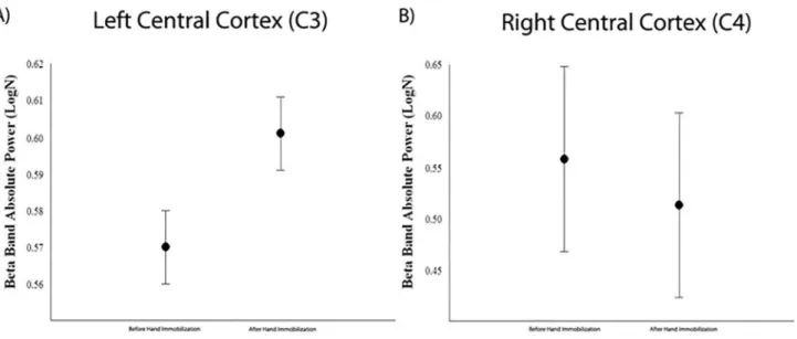
the central area, the two-way repeated measures ANOVA found a main effect for condition at C3 (F(1.3563) = 5.005; p = 0.025) and C4 (F(1.3657) = 11.858; p = 0.001). At C3 derivation, there was an increase in beta band absolute power between before (mean 0.570 ± 0.010) and after HI (mean 0.601 ± 0.010). On the other hand, at C4 derivation there was a decrease in beta band absolute power between before (mean 0.558 ± 0.09) and after HI (mean 0.513 ± 0.09). These results showed that C3 and C4 derivations were differently affected by HI (Figure 2).
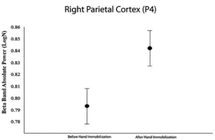
At P4 derivation the analysis implemented by
two-way repeated measures ANOVA demonstrated a significant
difference in beta band absolute power (F(1.3290) = 5.114; p = 0.024) (Figure 3). An increase occurred between before (mean 0.793 ± 0.015) and after HI (mean 0.842 ± 0.015).
There were no significant effects on any of other analyzed
derivations, presumably because neural activity at those locations is not involved in the requirements of the task.
■
DISCUSSIONFigure 1 - Mean and SE indicate main efect for condition (before immobilization versus
after immobilization) observed in right fronto polar cortex (Fp2) (p = 0.042).
Figure 2 - A) Main efect for condition (before immobilization versus after immobilization) observed in the left central cortex (C3) by mean and SE (p=0.025); B) In the right central
cortex (C4) mean and SE point main efect for condition (p < 0.001).
involved the analysis of changes in beta band absolute power that occur in cerebral cortex 2 sec before and 2 sec after the onset of visual stimuli linked to a motor task in two conditions: before and after immobilization of the right hand in right-handed subjects. Our hypothesis was that hand immobilization could lead to changes in beta absolute power in sensory and motor areas. We observed that Fp2, C3, C4 and P4 derivations were sensitive to changes in beta band absolute power after a condition of hand immobilization. There were no changes between conditions in the other derivations.
The frontopolar cortex is reported in the studies because it was activated in tasks that involve planning or problem solving.37 Moreover, this cortical region may
reflect a specific human feature and is thought to be
related to alternative courses of action.38 We observed that Fp2 derivation was sensitive to changes in beta band
absolute power after a condition of hand immobilization. We employed this derivation, located in the ipsilateral frontopolar cortex, because the task was performed by the right hand. Decrease in the beta band is frequently associated to movement planning and execution.37 Thus, we understand that this increase in beta band absolute power at the Fp2 derivation, should be understood as a smaller participation of this area in the condition after HI. Immobilization seems to limit the functioning of this cortical area during movement planning. This should be correlated to neuroplasticity, a feature that central nervous system exhibits while reorganizing and changing
its functions to adapt to external and internal influences.39
In this case, a maladaptive plasticity39 occurred, which may indicate that even a short period of immobilization (48h) is enough to hinder the performance of motor tasks, although easy to perform.
It is worth noting that the frontopolar cortex has a considerable role in executive function and its neural substrates. Byunk et al.40 examined changes in psychological mood states after a single bout of cycloergometer at mild intensity using non-invasive functional near-infrared spectroscopy while performing a color-word matching Stroop task. They found acute effects on executive function. The single bout of aerobic exercise led to improved Stroop performance, possibly correlated with increased arousal levels. In the same way, cortical activations regarding Stroop interference on the left dorsolateral prefrontal cortex and frontopolar area were evoked. Such activations corresponded to improved cognitive performance and increased arousal levels.40 Moreover, executive motor
deficits were associated with a decrease in cortical
may denote less activation of this area after a condition of movement deprivation (immobilization). In contrast, C4 exhibited smaller absolute power values in beta after immobilization and, by analogy, this region was more active in order to allow the execution of the task.
Immobilization is used as a therapeutic resource to enhance functional recovery in patients with motor
deficits due to damage to the motor cortex. This concept
is the basis of constraint-induced therapy, which means immobilization of the healthy arm and the forced use of the affected limb.48-51 Our results showing a decrease in beta band absolute power after HI at the C4 derivation may corroborate this concept. HI, even for as little as 48 hours could promote cortical changes at C4 derivation, increasing its excitability to compensate for cortical changes occurred in the contralateral hemisphere due to immobilization of the right hand. Thus, the decrease in beta band absolute power at C4 derivation after HI may indicate a cortical compensatory strategy translated as reduced activation of this area during movement planning.52
Studies involving neuroimaging consider the areas related to the parietal lobes as sites of multisensory integration53-55 and some authors highlight that the posterior parietal cortex subserves higher-level cognitive functions associated to action, i.e., intentions or early movement plans.56 Structural and functional reorganization of the sensorimotor cortex may result in changes in the motor function. Previous investigations revealed that sensorimotor restriction caused by chronic weightless bearing and reduction in limb movement may decrease sensorimotor function. Using a hindlimb unloading model, involving microscopy Trinel et al.57 showed that morphological changes due to sensorimotor restriction cause functional reorganization of the motor cortex, leading to impaired motor function. The study demonstrated dendritic spine remodeling in a period of 14 days. Our
findings of increased beta band absolute power at P4 may confirm changes in the sensorimotor cortex due to
sensorimotor restriction.
The study by Sainburg42 showed that, when subjects executed a task, new information was created and the contralateral cortex activity increased while the ipsilateral activity decreased. As seen in the other derivations in this study, the immobilization produced cortical changes at P4 that correspond to the right somatosensory cortex (ipsilateral) with a lower activation of this area after HI. This represents an adjustment for the sensorimotor integration
and may reflect a mechanism of functional inhibition,
perhaps to supply its cortical function.57 Due to transcallosal
inhibition the intensity of the influences of neurons in the
left hemisphere on cells in the right hemisphere may be
changed significantly after the immobilization.58
The presentation of visual stimuli in our task may be accepted as the moment of movement planning. Moreover,
Figure 3 - Main efect for condition (before versus after immobilization) in the right
parietal cortex (P4) derivation by mean and SE (p = 0.024).
By contrast, our results showed increased absolute beta power in Fp2 derivation, indicating a lower activation of the cortex frontopolar suggesting that damage to executive functioning and activity of this region may occur after a short of HI period.
Studies suggest that there is an inter-hemispheric “rivalry” observed in conditions of imbalance caused by injury or deprivation of brain functions.2,42,43 A hemisphere starts to inhibit the other via the corpus callosum and for this reason unilateral hand movements are associated with ipsilateral cerebral deactivation, including decreased
blood flow.44
We are assuming that because the task involved only the right hand, the right frontopolar cortex (Fp2) showed less activation. Such activity seems to occur due to the presence of increased beta band absolute power after HI, because beta band is normally decreased while planning or executing a motor task.6 At Fp1 derivation beta band absolute power did not show any changes between conditions (before vs. after immobilization). In other words, the cortical area corresponding to Fp2 derivation showed less activation, while the area corresponding to Fp1 maintained its activation both before and after 48 hours of immobilization.
Using intracortical microstimulation combined with behavioral testing, a study unraveled the effects of limb immobilization on movement representations in the rat primary motor cortex (M1). Changes in M1 were bilateral
and specific for the forelimb area, but they were stronger
the analysis of EEG was performed 2 sec before and 2 sec after the execution of motion. Then, the increase in beta band absolute power found in ipsilateral cortical areas associated to movement planning may be interpreted as less participation of these areas, indicating that the immobilization generated cortical changes so that the ipsilateral hemisphere readjusted its functions after HI.33 Furthermore, the ipsilateral motor cortex showed a decrease in beta band absolute power after immobilization that could be interpreted as a compensatory strategy to supply cortical changes in contralateral motor cortex due to HI.
Several studies showed that immobilization, even for short periods, result in changes in skeletal muscle properties.45,59-61 Changes in cerebral plasticity precede notable effects of immobilization, such as muscle strength loss and atrophy.62-65 Through our findings, we see an effect of a short, 48-hour immobilization on cortical activation and its comprehension is useful to understand impact damage and treatment possibilities.
Further studies are necessary to understand cortical changes due to HI and movement planning, especially considering how much time of immobilization is necessary to promote such changes. The impossibility to exactly identify when (how many hours after immobilization) cortical changes appear may be understood as a limitation of the study and could be further explored in future studies as well as the involvement of more complex tasks and the use of control groups.
■
CONCLUSIONCorroborating our hypothesis, an increase in beta band absolute power after immobilization was found in the contralateral motor cortex, indicating less participation of this area in movement planning. This may be relevant to therapeutic strategies that seek to activate regions responsible for the execution of a movement, such as transcranial magnetic stimulation and constraint-induced movement therapy.
■
CONFLICT OF INTERESTThe authors declare no conflict of interest regarding
this project.
■
AUTHOR PARTICIPATIONBastos VHV, Cagy M, de Sá Filho AS, Velasques B
reviewed the literature and the final version of the article.
Machado D, França JHS, Teixeira S, Ribeiro P, Machado S developed the project, contributed in work orientation,
discussed the data, and reviewed the final draft of the article.
ENVOLVIMENTO DA POTÊNCIA ABSOLUTA DA BANDA BETA APÓS IMOBILIZAÇÃO DA MÃO: ESTUDO ELETROENCEFALOGRÁFICO
OBJETIVO: O objetivo deste estudo foi analisar mudanças na potencia absoluta da banda beta em áreas corticais, antes e depois de uma condição de imobilização da mão por 48 horas.
MÉTODO: Quinze voluntários saudáveis, com idades entre 20 e 30 anos, foram submetidos à avaliação EEG antes e depois da imobilização, durante a execução de uma tarefa motora desencadeada por um estímulo visual.
RESULTADOS: A análise estatística revelou que a imobilização da mão causou mudanças em áreas corticais.
Um aumento significativo na potencia absoluta da banda beta
foi encontrado após imobilização da mão nos eletrodos Fp2 (F (1,3823) = 4,147; p = 0,042), C3 (F (1,3563) = 5,005; p = 0,025) e P4 (F (1,3290) = 5,114; p = 0,024). No C4 eletrodo (F (1,3657) = 11,858; p = 0,001) uma diminuição da potencia absoluta da banda beta ocorreu após imobilização da mão.
CONCLUSÃO: A imobilização da mão predominante,
mesmo para 48 horas, é suficiente para causar alterações
corticais que afetam o planejamento movimento. Tais mudanças podem representar uma estratégia cortical para
fornecer alterações corticais em hemisfério contralateral
devido à imobilização. Mais estudos são necessários para entender as mudanças corticais devido a imobilização da mão e planejamento do movimento, especialmente considerando quanto tempo de imobilização é necessário para promover essas mudanças.
PALAVRAS-CHAVE: Banda Beta, imobilização,
plasticidade neural, eletroencefalografia
■
REFERENCES1. Wheaton L, Fridman E, Bohlhalter S, Vorbach S, Hallett M. Left parietal activation related to planning, executing and suppressing praxis hand movements. Clin Neurophysiol. 2009;120(5):980-6. http://dx.doi. org/10.1016/j.clinph.2009.02.161
2. Fortuna M, Teixeira S, Machado S, Velasques B, Bittencourt J, Peressutti C, et al. Cortical Reorganization after Hand Immobilization: The beta qEEG Spectral Coherence Evidences. Plos One. 2013;8(11):e79912. http://dx.doi.org/10.1371/journal.pone.0079912
3. Gould IC, Nobre AC, Wyart V, Rushworth MFS. Effects of decision vari-ables and intraparietal stimulation on sensorimotor oscillatory activity in the human brain. J Neurosci. 2013;32(40):13805-18. http://dx.doi. org/10.1523/JNEUROSCI.2200-12.2012
4. Paek AY, Agashe HA, Contreras-Vidal JL. Decoding repetitive finger
movements with brain activity acquired via non-invasive electro-encephalography. Front Neuroeng. 2014, 7(1):1-18. http://dx.doi. org/10.3389/fneng.2014.00003
5. Cannon EN, Yoo KH, Vanderwert RE, Ferrari PF, Woodward AL, Fox
NA. Action experience, more than observation, influences Mu rhythm
desynchronization. PlosOne. 2014;9(3):e92002. http://dx.doi. org/10.1371/journal.pone.0092002
7. Makin TR, Cramer AO, Scholz J, Hahamy A, Henderson Slater D, Tracey I, et al. Deprivation-related and use-dependent plasticity go hand in hand. eLife. 2013;2:e01273. http://dx.doi.org/10.7554/eLife.01273 8. Roh JH, Park MH, Ko D, Park KW, Lee DH, Han C, et al. Region and
fre-quency specific changes of spectral power in Alzheimer’s disease and
mild cognitive impairment. Clin. Neurophysiol. 2011;122(11):2169-76. http://dx.doi.org/10.1016/j.clinph.2011.03.023.
9. Rodriguez G, Arnaldi D, Picco A. Brain functional network in Alzheimer’s disease: diagnostic markers for diagnosis and moni-toring. Int J Alzheimer’s Dis. 2011;2011:481903. http://dx.doi. org/10.4061/2011/481903.
10. Machado DC, Lima GC, Santos RS, Ramos AJ, Menezes de Sousa CC, Moreira Dos Santos RP, et al. Comparative analysis electroencephalo-graphic of alpha, beta and gamma bands of a healthy individual and one with hemiparesis J Phys Ther Sci. 2014;26(6):801-4. http://dx.doi. org/10.1589/jpts.26.801.
11. Brovelli A, Ding M, Ledberg A, Chen Y, Nakamura R, Bressler SL. Beta oscillations in a large-scale sensorimotor cortical network:
direc-tional influences revealed by Granger causality. Proc Natl Acad Sci.
2004;101(26):9849-54. DOI: 10.1073/pnas.0308538101
12. Silva JG, Knackfuss IG, Portella CE, Bastos VH, Machado D de C, Basile L, et al. EEG spectral coherence at patients submitted to tendon transfer surgery: study pre- and post-surgery. Arq Neuropsiquiatr. 2006,64:473-7. http://dx.doi.org/10.1590/S0004-282X2006000300023 13. Kimura T, Fujiwara T, Nishimura N, Ohira M, Yanagihashi R, Oshita S.
Changes in the inter-cortical correlation of electroencephalograph in motor learning process. J Phys Ther Sci. 1999;11(2):87-94. http://doi. org/10.1589/jpts.11.87
14. Kim J, Lee B, Lee HS, Shin KH, Kim MJ, Son E. Differences in brains wa-ves of normal persons and stroke patients during action observation and motor imagery. J Phys Ther Sci. 2014;26(2):215-8. http://dx.doi. org/10.1589/jpts.26.215
15. Van Ede F, Maris E. Somatosensory demands modulate muscu-lar beta oscillations, independent of motor demands. J Neurosci. 2013;33(26):10849-57. http://dx.doi.org/10.1523/JNEUROS-CI.5629-12.2013.
16. Pfurtscheller G, Lopes da Silva FH. Event-related EEG/MEG syn-chronization and desynsyn-chronization: basic principles. Clin Neuro-physiol. 1999;110(11):1842-57. http://dx.doi.org/10.1016/S1388-2457(99)00141-8
17. Baker SN. Oscillatory interactions between sensorimotor cortex and the periphery. Curr Opin Neurobiol. 2007;17(6):649-55. http://dx.doi. org/10.1016/i.conb.2008.01.007
18. Gilbertson T, Lalo E, Doyle L, Di Lazzaro V, Cioni B, Brown P. Existing motor state is favored at the expense of new movement during 13-35 Hz oscillatory synchrony in the human corticospinal system. J Neurosci. 2005;25:7771-9. http://dx.doi.org/10.1523/JNEUROS-CI.1762-05.2005
19. Singh Y, Singh J, Sharma A. FFT transformed quantitative EEG analysis of short term memory load. Ann Neurosci. 2015;22(3):176-9. http:// dx.doi.org/10.5214/ans.0972.7531.220308
20. Zhang Z, Parhi KK. Low-Complexity Seizure Prediction From iEEG/ sEEG Using Spectral Power and Ratios of Spectral Power. IEEE Trans. Biomed. Circuits Syst. 2015;10(3):693-706. http://dx.doi. org/10.1109/TBCAS.2015.2477264
21. Alegre M, Labarga A, Gurtubay IG, Iriarte J, Malanda A, Artieda J. Beta eletroencephalograph changes during passive movements:sensory afferences contribute to beta event-related desynchronization in hu-mans. Neurosci Lett. 2002;331(1):29-32. http://dx.doi.org/10.1016/ S0304-3940(02)00825-X
22. Engel AK, Fries P. Beta-band oscillations signaling the status quo? Curr Opin Neurobiol. 2010;20(2):156-65. http://dx.doi.org/10.1016/j. conb.2010.02.015
23. Machado D, França JHS, Teixeira S, Bastos VHV, Santos RPM, Cagy M, et al. Gamma absolute power reveals activation on motor areas after hand immobilization. MedicalExpress. 2016;3(5):M160504. http:// dx.doi.org/10.5935/MedicalExpress.2016.05.04
24. Oldfield RC. The assessment and analysis of handedness: the Edin
-burgh inventory. Neuropsychologia. 1971;9(1):97-113. http://dx.doi. org/10.1016/0028-3932(71)90067-4
25. Catanzariti JF, Guyot MA, Agnani O, Demaille S, Kolanowski E, Donze C. Eye-hand laterality and right thoracic idiopathic scoliosis. Eur Spine J. 2014 Jun;23(6):1232-6. http://dx.doi.org/10.1007/s00586-014-3269-z
26. Dixit A, Goyal A, Thawani R, Vaney N. Effect of caffeine on infor-mation processing: evidence from stroop task. Indian J Psychol Med. 2012;34(3):218-22. http://dx.doi.org/10.4103/0253-7176.106013.
27. Knott V, Bisserbe JC, Shah D, Thompson A, Bowers H, Blais C, et al.
The moderating influence of nicotine and smoking on resting-state
mood and EEG changes in remitted depressed patients during trypto-phan depletion. Biol Psychol. 2013 Dec;94(3):545-55. http://dx.doi. org/10.1016/j.biopsycho.2013.09.008.
28. Martinovic J, Jones A, Christiansen P, Rose AK, Hogarth L, Field M. Electrophysiological responses to alcohol cues are not asso-ciated with pavlovian-to-instrumental transfer in social drinkers. PLoS One. 2014;9(4):e94605. http://dx.doi.org/10.1371/journal. pone.0094605
29. Daly I, Nicolaou N, Nasuto SJ, Warwick K. Automated arti-fact removal from the electroencephalogram: a comparative study. Clin EEG Neurosci. 2013;44(4):291-306. http://dx.doi. org/10.1177/1550059413476485.
30. Gross J. Analytical methods and experimental approaches for electrophysiological studies of brain oscillations. J Neurosci Meth-ods. 2014 May 15;228:57-66. http://dx.doi.org/10.1016/j.jneu-meth.2014.03.007.
31. Stewart AX, Nuthmann A, Sanguinetti G. Single-trial classification
of EEG in a visual object task using ICA and machine learning. J Neurosci Methods. 2014;228:1-14. http://dx.doi.org/10.1016/j. jneumeth.2014.02.014
32. Fabbri S, Strnad L, Caramazza A, Lingnau A. Overlapping representa-tions for grip type and reach direction. NeuroImage 2014;94:138-46. http://dx.doi.org/10.1016/j.neuroimage.2014.03.017
33. Manaia F, Teixeira S, Velasques B, Bittencourt J, Salles JI, Arias-Carrión O, et al. Does immobilization of dependent hand promote adaptative changes in cerebral cortex? An analysis through qEEG asymmetry. Neurosci Lett. 2013 Mar 22;538:20-5. http://dx.doi.org/10.1016/j. neulet.2012.12.030.
34. Machado DCD, Santos RPM, Silva AP, Santana SB, Alves GVS, Cagy M,
et al. Análise eletroencefalográfica na hemiparesia à esquerda: um
estudo de caso. Rev Bras Neurol. 2013;49(4):129-36.
35. Jiang Z, Zheng L. Inter- and intra-hemispheric EEG coherence in patients with mild cognitive impairment at rest and during working memory task. J Zhejiang Univ Sci B. 2006;7:357-64. http://dx.doi. org/10.1631/jzus.2006.B0357
36. Van Albada SJ, Robinson PA. Transformation of arbitrary distribu-tions to the normal distribution with application to EEG test–retest reliability. J Neurosci Methods. 2007;161(2):205-11. DOI: 10.1016/j. jneumeth.2006.11.004
37. Dreher JC, Koechlin E, Tierney M, Grafman J. Damage to the fronto-polar cortex is associated with impaired multitasking. PLoS One. 2008;16;3(9):e3227. http://dx.doi.org/10.1371/journal. pone.0003227.
38. Kovach CK, Daw ND, Rudrauf D, Tranel D, O’Doherty JP, Adolphs R. Anterior prefrontal cortex contributes to action selection through tracking of recent reward trends. J Neurosci. 2012;20;32(25):8434-42. http://dx.doi.org/10.1523/JNEUROSCI.5468-11.2012
39. Lundbye-Jensen, Nielsen JB. Central nervous adaptations following 1 wk of wrist and hand immobilization. J Appl Physiol. 2008;105(1):139-51. http://dx.doi.org/10.1152/japplphysiol.00687.2007
41. Netto TM, Greca DV, Ferracini R, Pereira DB, Bizzo, B, Doring, T, et al. Correlation between frontal cortical thickness and executive
func-tions performance in patients with human immunodeficiency virus
infection. Radiol Bras. 2011;44(1):7-12. http://dx.doi.org/10.1590/ S0100-39842011000100006.
42. Sainburg RL. Evidence for a dynamic-dominance hypothesis of handedness. Exp Brain Res. 2002 Jan;142(2):241-58. http://dx.doi. org/10.1007/s00221-001-0913-8.
43. Brol AM, Bortoloto F, Magagnin NMS. Tratamento de restrição e indução do Movimento na reabilitação funcional de Pacientes pós
acidente vascular encefálico: Uma revisão bibliográfica. Fisioter. Mov.
2009;22(4):497-509.
44. Liepert J, Dettmers C, Terborg C, Weiller C. Inhibition of ipsilateral motor cortex during phasic generation of low force. Clin Neuro-physiol. 2001;112(1):114-21. http://dx.doi.org/10.1016/S1388-2457(00)00503-4
45. Viaro R, Budri M, Parmiani P, Franchi G. Adaptive changes in the motor cortex during and after longterm forelimb immobilization in adult rats. J Physiol. 2014;592(10):2137-52. http://dx.doi.org/10.1113/ jphysiol.2013.268821.
46. Yi W, Qiu S, Qi H, Zhang L, Wan B, Ming D. EEG feature comparison and
classification of simple and compound limb motor imagery. J Neuroeng
Rehabil. 2013;10:106. http://dx.doi.org/10.1186/1743-0003-10-106 47. Berends HI, Wolkorte R, Ijzerman MJ, van Putten MJ. Differential cor-tical activation during observation and observation-and-imagination. Exp Brain Res. 2013;229(3):337-45. http://dx.doi.org/10.1007/ s00221-013-3571-8.
48. Liepert J, Miltner WH, Bauder H, Sommer M, Dettmers C, Taub E, et al. Motor cortex plasticity during constraint-induced movement therapy in stroke patients. Neurosci Lett. 1998;250(1):5-8. http://dx.doi. org/10.1016/S0304-3940(98)00386-3
49 Fleet A, Page SJ, MacKay-Lyons M, Boe SG. Modified constraint-induced
movement therapy for upper extremity recovery post stroke: what is the evidence? Top Stroke Rehabil. 2014 Jul-Aug;21(4):319-31. http:// dx.doi.org/10.1310/tsr2104-319.
50. Chen JC, Shaw FZ: Progress in sensorimotor rehabilitative physical the-rapy programs for stroke patients. World J Clin Cases. 2014;2(8):316-26. http://dx.doi.org/10.12998/wjcc.v2.i8.316.
51. Ragaie AHM, Zamzam ML, Fathalla MM, El-Badawy MA, El Nahhas N,
El-Nabil LM, et al. Efficacy of modified constraint induced movement
therapy in acute stroke. Eur J Phys Rehabil Med. 2015;51(4):371-9. 52. Kobayashi M, Hutchinson S, Theoret H, Schlaug G, Pascual-Leone A..
Repetitive TMS of the motor cortex improves ipsilateral sequential
simple finger movements. Neurology. 200413;62(1):91-8
53. Calvert GA. Crossmodal processing in the human brain: insights from functional neuroimaging studies. Cerebral Cortex. 2001;11:1110-23. http://dx.doi.org/10.1093/cercor/11.12.1110
54. Calvert GA, Hansen PC, Iversen SD, Brammer MJ. Detection of audio--visual integration sites in humans by application of electrophysio-logical criteria to the BOLD effect. Neuroimage. 2001;14(2):427-38. http://dx.doi.org/10.1006/nimg.2001.0812.
55. Molholm S, Sehatpour P, Mehta AD, Shpaner M, Gomez-Ramirez M, Ortigue S, et al. Audio-visual multisensory integration in superior pa-rietal lobule revealed by human intracranial recordings. J Neurophysiol. 2006;96(2):721-9. http://dx.doi.org/10.1152/jn.00285.2006 56. Andersen RA, Buneo CA. Intentional maps in posterior parietal cortex.
Rev. Neurosci. 2002;25:189-220. http://dx.doi.org/10.1146/annurev. neuro.25.112701.142922
57. Trinel D, Picquet F, Bastide B, Canu MH. Dendritic spine remodeling induced by hindlimb unloading in adult rat sensorimotor cortex. Behav Brain Res, 2013. 15:1-7. http://dx.doi.org/10.1016/j.bbr.2013.04.015 58. Bogdanov AV, Galashina AG. Correlated activity of sensorimotor cortex neurons in the left and right hemispheres of the rabbit brain immo-bilization catatonia. Neurosci Behav Physiol. 2010 Sep;40(7):801-6. http://dx.doi.org/10.1007/s11055-010-9329-x.
59. Thom JM, Thompson MW, Ruell PA Bryant GJ, Fonda JS, Harmer AR, et al. Effect of 10-day cast immobilization on sarcoplasmic reticulum calcium regulation in humans. Acta Physiol Scand. 2001;172(2):141-7. http://dx.doi.org/10.1046/j.1365-201X.2001.00853.x
60. Clark BC, Taylor JL, Hoffman RL, Dearth DJ, Thomas JS. Cast immobili-zation increases long-interval intracortical inhibition. Muscle Nerve. 2010;42(3):363-72. http://dx.doi.org/10.1002/mus.21694. 61. Bolzoni F, Bruttini C, Esposti R, Cavallari P. Hand immobilization affects
arm and shoulder postural control. Exp Brain Res. 2012;220(1):63-70. http://dx.doi.org/10.1007/s00221-012-3115-7.
62. Santos-Junior FFU, Alves JSM, Machado AAN, Nogueira AA, Carlos PS, Ferraz ASM, et al. Morphometric alterations in respiratory muscle of rats submitted to paw immobilization. Rev Bras Med Esp. 2010;16(3):215-8. http://dx.doi.org/10.1590/S1517-86922010000300012. 63. Kannus P. Immobilization or early mobilization after an acute
soft--tissue injury? Phys Sportsmed. 2000;28(3):55-63. http://dx.doi. org/10.3810/psm.2000.03.775.
64. Kannus P, Parkkari J, Järvinen TL, järvinen TA, Järvinen M. Basic science and clinical studies coincide: active treatment approach is needed after sports injury. Scand J Med Sci Sports. 2003;13(3):150-4.