THE VALUE OF TRANSVULVAR ULTRASONOGRAPHY IN THE
ASSESSMENT OF RELEVANT ANATOMICAL PARAMETERS
IN THE MANAGEMENT OF FEMALE STRESS URINARY
INCONTINENCE*
Frederico Teixeira Brandt1
, Carla Daisy Costa Albuquerque2
, Felipe Rinald Barbosa Lorenzato2 , Daniela Saraiva Guerra Lopes3
, Adriana Scavuzzi Carneiro da Cunha4
, Rosângela Falcão da Costa5
OBJECTIVE: The aim of this study was to describe the role of transvulvar ultrasonography in the assess-ment of anatomical changes following fascia lata sling or tension-free vaginal tape procedures. MATERIALS AND METHODS: Forty women in the age range between 30 and 60 years had fascia lata sling (20 patients) or tension-free vaginal tape (20 patients) placement for treating stress urinary incontinence. Transvulvar ul-trasonography was utilized, both pre- and post-operatively, as the main investigational tool in these cases for evaluating the urethrovesical junction vertical and horizontal distances, pubo-urethral distance and proxi-mal urethra length. RESULTS: The urethrovesical junction vertical distance did not vary significantly after the fascia lata sling surgery (p ≥≥≥≥≥ 0.10). Both the pubo-urethral distance and the proximal urethra length became shorter (p < 0.003), and the urethrovesical junction horizontal distance was reduced only at rest (p = 0.03). The tension-free vaginal tape procedure resulted in a reduction of the urethrovesical junction ver-tical distance (p = 0.0005) and of the proximal urethra length (p = 0.02). CONCLUSION: Transvulvar ul-trasonography was of utmost importance in the demonstration that both the fascia lata sling and tension-free vaginal tape surgical procedures elongate the proximal urethra, even though the fascia lata sling tech-nique does it more effectively. The fascia lata sling techtech-nique is more focused on shortening the pubo-ure-thral distance, and the tension-free vaginal tape, on the correction of the vertical displacement of the urethrovesical junction.
Keywords: Transvulvar ultrasound; Perineal ultrasound; Urethrovesical junction; Stress urinary incontinence; Tension-free vaginal tape; Sling procedure.
A importância da ultra-sonografia transvulvar na avaliação de parâmetros anatômicos relevantes no trata-mento de mulheres com incontinência urinária de esforço.
OBJETIVO: Descrever a importância da ultra-sonografia transvulvar na avaliação das diferenças anatômicas induzidas pelas cirurgias de sling de fáscia lata e tension-free vaginal tape. MATERIAIS E MÉTODOS: Qua-renta mulheres com incontinência urinária de esforço, com idades entre 30 e 60 anos, foram tratadas por sling de fáscia lata (20 pacientes) ou tension-free vaginal tape (20 pacientes). A ultra-sonografia transvulvar da junção uretrovesical e da uretra proximal foi a principal ferramenta de investigação pré- e pós-operatória. Os parâmetros estudados foram: distância vertical e distância horizontal da junção uretrovesical, distância pubouretral e comprimento da uretra proximal. RESULTADOS: A distância vertical da junção uretrovesical não variou significativamente após a sling de fáscia lata (p≥≥≥≥≥ 0,10). A distância pubouretral e a uretra pro-ximal tornaram-se menores (p < 0,003) e a distância horizontal da junção uretrovesical tornou-se menor só no repouso (p = 0,03) após a sling de fáscia lata. A tension-free vaginal tape diminuiu o deslocamento vertical da junção uretrovesical (p = 0,0005) e o comprimento da uretra proximal (p = 0,02). CONCLUSÃO: A ultra-sonografia transvulvar foi fundamental para documentar que as cirurgias de sling de fáscia lata e ten-sion-free vaginal tape alongam a uretra proximal, sendo a sling de fáscia lata de forma mais eficaz. A sling de fáscia lata enfoca a diminuição da distância pubouretral e a tension-free vaginal tape, o deslocamento vertical da junção uretrovesical.
Unitermos: Ultra-sonografia transvulvar; Ultra-sonografia perineal; Junção uretrovesical; Incontinência uri-nária de esforço; Tension-free vaginal tape; Cirurgia de sling.
Abstract
Resumo
* Study developed at Universidade Federal de Pernambuco (UFPE), Recife, PE, Brazil. 1. Private Docent, Professor of Urology, Universidade Federal de Pernambuco (UFPE), Recife, PE, Brazil.
2. Associate Professor Doctors, Universidade Federal de Pernambuco (UFPE), Recife, PE, Bra-zil.
3. Student of Medicine supported by Scientific Initiation Scholarship, Universidade Federal de Pernambuco (UFPE), Recife, PE, Brazil.
4. Master in Maternal-Infantile Health, Gynecologist/Obstetrician at Hospital das Clínicas da Universidade Federal de Pernambuco (UFPE), Recife, PE, Brazil.
5. Fellow Master Degree in Surgery, Universidade Federal de Pernambuco (UFPE), Sonogra-phist at Clínica Lucilo Ávila, Recife, PE, Brazil.
Mailing address: Prof. Dr. Frederico Teixeira Brandt. Avenida Dezessete de Agosto, 2475, ap. 2801, Bairro Monteiro. Recife, PE, Brazil, 52061-540. E-mail: [email protected]
INTRODUCTION
Urinary incontinence affects million of women, with high worldwide prevalence, achieving 28% in some European coun-tries, and up to 37% among North Ameri-can women, according to a recent survey conducted in the United States of America(1). The magnitude of this problem
can be measured by the amount of about 15 billion dollars annually spent in USA for diagnosis and treatment of this condition, besides the high impact on the women’s quality of life, leading to low self-esteem, constraint and social isolation. Only 25% of patients affected by some degree of uri-nary incontinence and about half of those with significant incontinence seek medical assistance(1–6).
Several surgical methods such as sling techniques have been adopted to achieve the four fundamental objectives of the treatment for stress urinary incontinence (SUI) with minimal complications. The theoretical principles are the following: positioning of the vesicourethral segment in its normal topography; adequately sus-tained continence under increased intra-abdominal pressure; absence of injury to the urethrovesical segment; and absence of urinary obstruction by urethral compres-sion(7–10).
In spite of having been developed in 1907, only in the last decades the sling technique started being more frequently utilized in the management of female SUI employing an autologous, heterologous or synthetic material placed in a retropubic position and through the vagina, involving the proximal urethra to support the poste-rior urethral wall. The rate of success of this technique ranges between 70% and 100%, with widespread acceptance as the first option for treating I-type and II-type SUI where, at strain, the urethrovesical junction (UVJ) descends lower than the pubis(7–10).
Historically, there was a marked con-cern with the clinical results from the sur-gical treatment for SUI, without consider-ing the objective analysis of both the pre-and postoperative UVJ pre-and proximal ure-thra (PU) positioning(8,10).
In the current decade, some studies started showing more concern about ana-tomical aspects of the UVJ and PU,
par-ticularly in relation their vertical position-ing. So, the concept of primary measure-ment of parameters such as urethrovesical junction vertical distance (UVJVD), urethrovesical junction horizontal distance (UVJHD), pubourethral distance (PUD), and PU, was associated with the evaluation of the vertical UVJ mobility in order to identify more accurately the urethral topog-raphy in a system of orthogonal coordi-nates, comparing these pre- and postopera-tive sonographic measurements for correc-tion of SUI by means of several surgical techniques(8,10).
Although the sling surgical technique in the management SUI is globally wide-spread, producing satisfactory results, it is highly expensive as heterologous material, synthetic or not, are utilized, hindering or even preventing its application(11,12).
The utilization of the homologous fas-cia lata has substantially reduced the cost of this surgery, so this technique is avail-able for a higher number of patients, espe-cially in less privileged regions(12).
The present study is aimed at describ-ing the anatomical changes resultdescrib-ing from fascia lata sling (FLS) and tension-free vaginal tape (TVT) procedures in the man-agement of female SUI, by means of post-voiding transvaginal ultrasound. Topo-graphic parameters such as UVJVD, UVJHD, PUD, and PU are evaluated by pre- and postoperative transvaginal ultra-sound.
MATERIALS AND METHODS
In the period between August 2004 and January 2006, the authors developed the present transverse and prospective study in the Unidade de Pesquisa em Incontinên-cia Urinária (UPIU) (Division of Research in Urinary Incontinence) of Hospital das Clínicas da Universidade Federal de Per-nambuco (HC-UFPE). The research proto-col was reviewed and approved by the UFPE Committee for Ethics in Research previously to the enrollment of the first volunteer.
After being given an explanation on aspects of the study, the patients who had accepted to participate were invited to sign a term of free and informed consent. Forty randomly selected women were included in
the present study; 20 of them were submit-ted to FLS, and 20 to TVT.
Inclusion criteria were: age range be-tween 30 and 60 years, symptoms or signs of SUI, vertical UVJ displacement > 9 mm (determined by transvulvar ultrasound), absence of other diseases associated with urinary incontinence, absence of previous history of surgery involving the bladder, urethra or vagina. Exclusion criteria were: onset of acute disease hindering the proce-dure, patient’s giving up on the surgery, diagnosis of pregnancy during the investi-gational period.
All of the 40 patients answered a struc-tured questionnaire and underwent a thor-ough urogynecological examination. Addi-tionally, they were evaluated by a single sonographist with more-than-five-year ex-perience in transvulvar ultrasonography, so minimizing inter- and intraobserver biases, for sonographic documentation in the pre-operative period, and 30 days after the sur-gery by means of clinical follow-up.
The measurement by transvulvar ultra-sound was performed, with the patient in the dorsolithotomy position, with legs bent, immediately after voiding and having a barely empty bladder (< 50 ml of urine). The transducer previously covered by a condom and lubricated with water-soluble gel was placed touching the vulva, in a localization that allowed the sonographist to identify the urethra, the bladder, the vesical cervix and the pubic symphysis – structures pre-senting characteristic echotextures.
The equipment used for the examina-tions was an Aloka SSD500, connected to a 7 MHz convex vaginal transducer and an electronic selector for real image measure-ment, equipped with computer and photo-graphic camera with instantaneous resolu-tion capability.
monitor screen, the first one with the pa-tients at rest, and the second during the Valsalva maneuver, aiming at determining the amplitude of the displacement of the anatomical structures of interest. The dis-placement resulting from the difference between the measurements was evaluated as follows: above the inferior border of the pubic symphysis, it was standardized with a positive (+) value; below the inferior border of the pubic symphysis, with a nega-tive (–) value; above the inferior border of the pubic symphysis at rest, and below the inferior border of the pubic symphysis dur-ing Valsalva maneuver, the result from summation of these values.
The following parameters were ana-lyzed:
– UVJVD: length of a longitudinal line between the inferior border of the pubic symphysis and a straight transverse line originating from the UVJ;
– UVJHD: length of the straight trans-verse line originating from the UVJ to the longitudinal line originating from the pu-bic symphysis;
– PUD: length of a horizontal line from the inferior border of the pubic symphysis to the urethra;
– PU: distance between the UVJ and the pubourethral distance middle point.
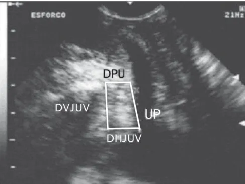
The criteria utilized for the diagnosis of SUI (UVJ hypermobility) have been sug-gested by Brandt et al., who describe the UVJ vertical displacement as the most ac-curate parameter for this purpose(8,13)
(Fig-ure 1).
The TVT surgery was performed ac-cording to the technique primarily de-scribed by Ulmsten(14). The fascia lata sling
procedure involved the removal of an au-tologous segment of aponeurosis from the internal face of the thigh, measuring on average 2 cm × 6 cm. The insertion of this aponeurotic sling was performed by means of an abdominal Pfannenstiel incision.
One month after the surgery, the patients were submitted to a new transvulvar ultra-sound for a reevaluation of the UVJ and PU measurements. These data were compared with those obtained when the patients were enrolled in the present study.
The data were analyzed with the Epi-Info 2002 software version 1.0. The chi-squared (χ²) test was utilized for analysis
of the contingency tables, the Fisher exact test for mean differences and variances ra-tios, with a statistical significance level (p
value) = 5%.
RESULTS
The patients’ age ranged between 30 and 60 years (mean age = 50.3 ± 9.6 years). Mean variation of pre- and postoperative UVJ vertical distances measurements in patients submitted to correction of stress urinary incontinence by fascia lata
pubo-vaginal sling or TVT techniques are de-scribed on Table 1.
Mean variation of sonographic mea-surements of urethrovesical junction hori-zontal distance are described on Table 2.
Mean ultrasound findings regarding measurements of PUD are described on Table 3.
Among the findings of the authors in the present study, the most significant trans-vulvar sonographic measurements were those related to the PU measurements de-scribed on Table 4.
UP
DVJUV
DPU
DHJUV
Figure 1. Transvulvar ultrasound of urethrovesical junction at stain. (UVJVD, urethrovesical junction ver-tical distance; UVJHD, urethrovesical junction horizontal distance; PUD, pubourethral distance; PU, proximal urethra).
UVJVD (mm)
Mean SD SS
Mean SD SS
Pre
16.3 5.7
16.0 4.6
Post
17.4 4.8
16.6 3.2
Pre
2.7 11.5
2.1 15.3
Post
12.6 5.7
8.4 9.6
Pre
13.6 11.4
16.7 7.7
Post
4.8 5.8
10.7 7.3 At rest At strain Displacement
FLS
p = 0.58 p = 0.10 p = 0.12 TVT
p = 0.26 p = 0.28 p = 0.0005
Table 1 Urethrovesical junction vertical distance (UVJVD), in the pre- and postoperative periods in pa-tients submitted to correction of stress urinary incontinence by fascia lata pubovaginal sling (FLS) or ten-sion-free vaginal tape (TVT) techniques.
DISCUSSION
The existence of more than a hundred surgical techniques for the management of SUI may mean that the physiopathology of this disease, and consequently, the logics of its surgical correction are still to be com-pletely known(8,10,15).
It seems that the main issues involved are related to the urethral resistance factor with urinary loss pressure, and the UVJ and PU positioning. On the one hand, the urodynamic testing is considered as a valu-able tool, on the other hand, the utilization of pre- and postoperative ultrasound have increasingly been considered as a relevant investigative tool in the evaluation of the UVJ and PU(8,10,15).
Among the different types of surgery for management of SUI, the former sling-type surgeries were updated with basis on the postulates defined on the TVT technique, with frequent reports on advantages in re-lation to the PU positioning as compared with other similar procedures. However, the majority of authors utilize the urody-namic testing as a supplementary investi-gational method, without taking relevant sonographic parameters into consider-ation(8,10).
As regards the supplementary pre- and postoperative investigational method, al-though there is some divergence about the ideal diagnostic method for SUI, there is a reasonable agreement that the majority of these patients present isolated or associated anatomical alterations in the UVJ and PU. Additionally, it is important to note that approximately 95% of women complaining of SUI present UVJ and PU hypermobil-ity(8,10,16–18).
Independently from the urodynamics, there is a remarkable progress in the under-standing of factors that may affect the re-sults of a UVJ and PU sonographic study, such as the vesical volume and the neces-sary strain to cause UVJ and PU mobility, so increasing the ultrasound interest and re-liability(13,19).
These considerations have led the au-thors of the present study to adopt trans-vulvar ultrasound for a specific evaluation of the UVJ and PU functional topography. On the other hand, the urodynamic testing evaluates only some preoperative
param-UVJHD (mm)
Mean SD SS
Mean SD SS
Pre
13.4 3.0
11.4 6.3
Post
10.3 4.4
11.0 5.5
Pre
14.0 9.4
16.4 7.6
Post
9.2 6.1
15.4 8.9
Pre
10.3 0.6
7.2 5.2
Post
–1.1 7.6
5.6 4.0 At rest At strain Displacement
FLS
p = 0.03 p = 0.11 p = 0.60 TVT
p = 0.33 p = 0.28 p = 0.18
Table 2 Urethrovesical junction horizontal distance (UVJHD), in the pre- and postoperative periods in patients submitted to correction of stress urinary incontinence by fascia lata pubovaginal sling (FLS) or tension-free vaginal tape (TVT) techniques.
SD, standard deviation; SS, statistical significance.
PUD (mm)
Mean SD SS
Mean SD SS
Pre
11.6 3.0
13.4 5.0
Post
10.9 1.7
10.9 2.8
Pre
17.8 4.6
18.3 7.5
Post
10.7 2.8
14.4 6.9
Pre
6.2 4.2
5.7 5.0
Post
–0.2 2.7
4.8 4.3 At rest At strain Displacement
FLS
p = 0.38 p = 0.001 p = 0.001 TVT
p = 0.24 p = 0.26 p = 0.43
Table 3 Pubourethral distance (PUD), in the pre- and postoperative periods in patients submitted to correction of stress urinary incontinence by fascia lata pubovaginal sling (FLS) or tension-free vaginal tape (TVT) techniques.
SD, standard deviation; SS, statistical significance.
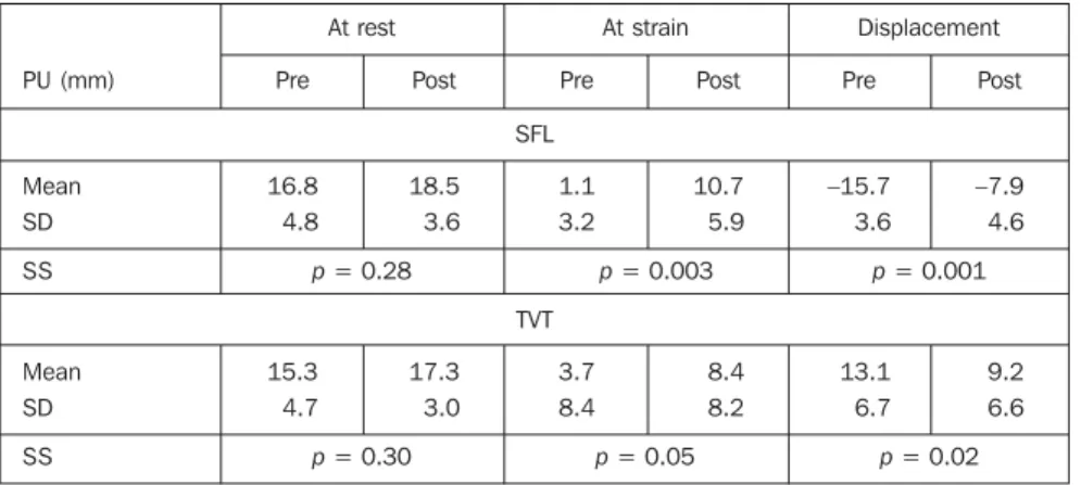
PU (mm)
Mean SD SS
Mean SD SS
Pre
16.8 4.8
15.3 4.7
Post
18.5 3.6
17.3 3.0
Pre
1.1 3.2
3.7 8.4
Post
10.7 5.9
8.4 8.2
Pre
–15.7 3.6
13.1 6.7
Post
–7.9 4.6
9.2 6.6 At rest At strain Displacement
SFL
p = 0.28 p = 0.003 p = 0.001 TVT
p = 0.30 p = 0.05 p = 0.02
Table 4 Proximal urethral length (PU), in the pre- and postoperative periods in patients submitted to correction of stress urinary incontinence by fascia lata pubovaginal sling (FLS) or tension-free vaginal tape (TVT) techniques.
eters, in an indirect attempt to observe the UVJ mobility, and fails to objectively record the UVJ and PU anatomical posi-tioning both from the static and dynamic point of view(8,10,18,20).
The fascia of the rectum-abdominal muscle is quite utilized, but the superior-ity of the fascia lata sling surgery is recog-nized, particularly for the lower tissue in-jury as a result of the possibility of obten-tion of larger tissue segments, the lower risk for complications on the donor site, shorter hospital stay, and, as a result, an earlier return of the patients to their daily activities(12).
It was important to observe by means of transvulvar ultrasound, that the UVJVD to the inferior border of the pubic symphysis considered as one of the main anatomical parameters related to the urinary inconti-nence, has significantly changed only after the TVT surgery, specifically in relation to the UVJ displacement, as shown on Table 1. That is to say, ultrasound can demon-strate that TVT technique significantly hin-ders the UVJ downwards displacement, al-lowing a certain degree of voiding obstruc-tion. Likewise, the FLS method also cor-rected the SUI in most of patients submit-ted to this surgery and even so has resulsubmit-ted in a not-so-remarkable suspension of the UVJ.
On the other hand, from the dynamic, physiopathological point o view, the UVJHD was not affected by any of these techniques. It is important to note that the FLS technique resulted in a more signifi-cant change in the resting angle of the UVJ, but this static anatomical change did not remained significant under the dynamic abdominal pressure (Table 2).
The results of the present study also demonstrated that the FLS surgery for the management of SUI resulted in reduced PUD at strain, and decreased movement of the PU, differently from the TVT technique results (Table 3).
Transvulvar ultrasonography has al-lowed the authors to observe that maybe among their findings the most important one was the elongation of the proximal urethra, both at strain and displacement after correction of SUI by either of the tech-niques described in the present study (Table 4). So, this is the importance of restoring
the proximal urethral pressure by means of an increase in area resulting from the PU elongation to achieve an effective urinary continence.
In summary, transvulvar ultrasound, taking the inferior border of the pubic sym-physis, demonstrate that the pubovaginal sling surgery could simultaneously reduce the UVJHD at rest, and extend the PU length at strain, allowing these structures to return to a more anatomical position, and restoring the urinary continence by reduc-ing the bladder neck mobility.
Thirty days after the surgeries, the au-thors could observe that both the FLS and the TVT methods were effective in 85% of the patients, These results are compliant with data reported in the literature(9).
It is important to highlight that a cure rate cannot be estimated in a short term, neither is this hypothesis supported by the present study, considering the consensus that a mean ten-year follow-up is necessary to confirm a cure. In fact, the present study is aimed at reinforcing the thesis that SUI is directly related to a passive urinary con-tinence established principally by the ana-tomical positioning of the UVJ and PU. Therefore, the surgical correction should be focused on the functional recovery of these structures to restore the passive urinary continence.
The relevant role played by the postop-erative ultrasound should be highlighted as an objective follow-up parameter, where a 5 mm cut-off for UVJVD is the mean mea-surement for young, continent patients(20)
suggesting a satisfactory surgical correc-tion(21), that is to say, leading to an
effec-tive urination control and possibly playing a role as a predictive factor for SUI recur-rence, a matter already approached by other authors(22), and that is the objective of a
new ongoing investigation by the authors of the present study.
Therefore it can be assumed that both the lateral vastus fascia lata sling and the TVT techniques are equivalent in relation to the immediate clinical results and elon-gation of the proximal urethra. Besides the few postoperative anatomical differences above described requiring further studies with larger samples, it is necessary to con-sider that the fascia lata sling technique implies lower operative costs as compared
with those of the synthetic materials uti-lized in TVT, and shorter hospital stay in relation to the techniques which utilized abdominal fasciae.
CONCLUSION
Transvulvar ultrasound plays a signifi-cant role in the pre- and postoperative as-sessment of women with SUI. This method can objectively record the measurements of the essential parameters in the surgical res-toration of the normal voiding, besides al-lowing the comparison among several re-sults from different surgical techniques.
With the aid of transvulvar ultrasonog-raphy, the authors could conclude that the TVT surgery has elongated the PU at strain and displacement, as well as reduced the UVJVD; and the FLS procedure reduced the distance between the UVJ and the pu-bis both by the decrease in the UVJHD at rest and in the PUD at strain and displace-ment. Furthermore, the FLS technique re-sulted in a higher extension of the PU length at strain and displacement.
REFERENCES
1. Waetjen LE, Subak LL Shen H, et al. Stress uri-nary incontinence surgery in the United States. Obstet Gynecol 2003;101:671–676.
2. Hampel C, Artibani W, Espuna Pons M, et al. Understanding the burden of stress urinary incon-tinence in Europe: a qualitative review of the lit-erature. Eur Urol 2004;46:15–27.
3. Contreras O. Stress urinary incontinence in the gynecological practice. Int J Gynaecol Obstet 2004;86:6–16.
4. Minassian VA, Drutz HP, Al-Badr A. Urinary in-continence as a worldwide problem. Int J Gynae-col Obstet 2003;82:327–338.
5. Birnbaum HG, Leong SA, Oster EF, Kinchen KS, Sun P. Cost of stress urinary incontinence: a claims data analysis. Pharmacoeconomics 2004; 22:95–105.
6. Kinchen KS, Orsini S, Crown W, Swindle R. A retrospective claims analysis of the direct costs of stress urinary incontinence. Int Urogynecol J Pelvic Floor Dysfunct 2003;14:403–411. 7. Bullock TL, Ghoniem G, Klutke CG, Staskin DR.
Advances in female stress urinary incontinence: mid-urethral slings. BJU Int 2006;98 (Suppl 1): 32–40.
8. Brandt FT, Santos Junior MW, Albuquerque CDC, Lorenzato FRB, Viana LA, Cunha ASC. Modificações da junção uretrovesical e uretra proximal após a cirurgia de alça sem tensão em mulheres de 45 a 72 anos Rev Bras Saúde Mater Infant 2005;5:185–191.
analysis from The Netherlands TVT database. Am J Obstet Gynecol 2006;195:439–444. 10. Ribeiro CBL, Brandt FTB, Albuquerque CDC,
Arraes F, Pinho Neto JS, Ávila M. Modificações da uretra proximal e da junção uretrovesical de-correntes da cirurgia do tipo Kelly-Kennedy. Acta Cir Bras 2001;17:21–23.
11. Wilson L, Brown JS, Shin GP, Luc KO, Subak LL. Annual direct cost of urinary incontinence. Obstet Gynecol 2001;98:398–406.
12. Norton P, Brubaker L. Urinary incontinence in women. Lancet 2006;367:57–67.
13. Brandt FT, Albuquerque CDC, Arraes AF, Albu-querque GF, Barbosa CD, Araújo CM. Influên-cia do volume vesical na avaliação ultra-sonográ-fica da junção uretrovesical e uretra proximal. Radiol Bras 2005;38:33–36.
14. Ulmsten U. An introduction to tension-free
vagi-nal tape (TVT) – a new surgical procedure for treatment of female urinary incontinence. Int Urogynecol J Pelvic Floor Dysfunct 2001;12 (Suppl 2):S3–4.
15. Howden NS, Zyczynski HM, Moalli PA, Sagan ER, Meyn LA, Weber AM. Comparison of autolo-gous rectus fascia and cadaveric fascia in pubo-vaginal sling continence outcomes. Am J Obstet Gynecol 2006;194:1444–1449.
16. McGuire EJ. Urinary incontinence: a diverse con-dition. J Urol 2005;173:1453–1454.
17. Dietz HP. Why pelvic floor surgeons should uti-lize ultrasound imaging. Ultrasound Obstet Gyne-col 2006;28:629–634.
18. Dalpiaz O, Curti P. Role of perineal ultrasound in the evaluation of urinary stress incontinence and pelvic organ prolapse: a systematic review. Neu-rourol Urodyn 2006;25:301–306.
19. Brandt FT, Nóbrega LV, Albuquerque CDC, et al. Aferição simultânea da pressão abdominal na avaliação ultra-sonográfica de mulheres com in-continência urinária de esforço. Radiol Bras 2006;39:91–95.
20. Brandt FT, Albuquerque CDC, Lorenzato FR, Amaral FJ. Perineal assessment of urethrovesical junction mobility in young continent females. Int Urogynecol J 2000;11:18–22.
21. Vierhout ME, Hol M. Vaginal ultrasound studies before and after successful colposuspension and in continent controls. Acta Obstet Gynecol Scand 1998;77:101–104.