Introduction
The various hormonal, physiological and anatomical changes that occur during pregnancy can result in changes in the biomechanics of women, capable of altering their balance 1, postural control2-4 and their performance in carrying out functional activities5,6 such as sit-to-stand.
The ability to stand up from a sitting position imposes chal-lenges on postural and motor control. According to Nicholls7, women in the third trimester report that the most dificult activities in this period are: sit-to-stand, picking up objects on the loor,
seated work, driving, and getting out of the car. Limiting fac-tors for these activities include postural instability, lumbopelvic pain, and fatigability7.
The sit-to-stand (STS) movement is operationally deined
as a successful transfer of the body’s center of mass (CM) from a sitting position where there is a large support base (thighs and feet) to a stable standing position8,9, followed by obtaining orthostatic balance when body oscillation must be controlled (stabilization phase)10–15. Therefore, it requires good coordination between the central nervous system (CNS) and the neuromuscular system8,9.
Performing the action of going from a sitting to a standing
position is lexible and varies according to adaptation to the task
requirements, the individual and the environment. However, studies that attempt to analyze this movement from the perspec-tive of the daily routine of the population of pregnant women are still scarce. In this sense, the objective of this study was
to investigate the inluence of a virtual reality-based exercise
protocol on STS movement kinematic variables of women in their second and third gestational trimesters. We adopted the
position that virtual reality-based exercises would inluence
STS movement kinematic variables of pregnant women as the hypothesis.
Material and methods
This study is a randomized controlled clinical trial developed at the Laboratory of Intervention and Movement Analysis (LIAM) of the Physiotherapy Department of the Federal University of Rio Grande do Norte - UFRN, between April, 2014 and May, 2015.
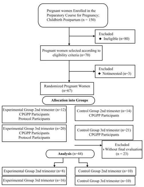
The study population consisted of women in their second (14th to 26th week) and third (between 27 and 37 weeks) gestational trimester, living in the city of Natal-RN and participating in the Preparatory Course for Gestation, Childbirth and Postpartum (CPGPP), promoted by the Physiotherapy Department of UFRN. The total population enrolled in the CPGPP was 150 women during the study period through a non-probabilistic sampling process. The study sample consisted of 44 women allocated into 4 groups: Control Group - 2nd Trimester (CG2T), Experimental Group - 2nd Trimester (EG2T), Control Group - 3rd Trimester (CG3T) and Experimental Group - 3rd Trimester (EG3T).
The volunteers from Control Group (CG) participated in
edu-cational lectures (CPGPP) and those in the Experimental Group (EG) were additionally submitted to the exercise protocol using Wii-Rehabilitation, according to the low chart below (Figure 1).
Original article (short paper)
Inluence of a virtual reality-based exercise
protocol on the sit-to-stand activity kinematic
variables in pregnant women: a randomized
controlled trial
Silvia Oliveira Ribeiro Vanessa Patricia Soares de Sousa Elizabel de Souza Ramalho Viana
Universidade Federal do Rio Grande do Norte, Natal, RN, Brazil
Abstract — Aims: Changes resulting from the gestational period may lead to changes in the biomechanics of women, which can alter the performance of functional activities such as sit-to-stand. Thus, the objective of this study was
to investigate the inluence of a virtual reality-based exercise protocol on the kinematic variables of the sit-to-stand
movement in women in their second and third gestational trimesters. Methods: The sample consisted of 44 women selected according to the eligibility criteria, allocated into 4 groups: control group, 2nd trimester (CG2T); experimental group, 2nd trimester (EG2T); control group, 3rd trimester (CG3T); and experimental group, 3rd trimester (EG3T). All the volunteers answered the identiication and evaluation form and were sent to the kinematic evaluation through the Qualisys Motion Capture System®. An intervention with game therapy was performed in 12 sessions of 30 minutes each, three times a week. Results: No statistically signiicant differences were found intra- (P> 0.54) and inter-groups
(P> 0.059) for kinematic variables. However, there was a tendency for improvement in the analyzed variables after the
proposed protocol. Conclusions: The data obtained suggest that the use of the Nintendo Wii Fit Plus® was not able to
inluence sit-to-stand kinematic variables in the analyzed women.
The inclusion criteria were: (1) no clinical or obstetric changes (low-risk pregnancy); (2) being between 18 and 37 years of age;
(3) being in the second or third gestational trimesters conirmed
by ultrasonography, and between 14 to 21 weeks or 27 to 32 weeks at the initial evaluation; (4) being nulliparous; (5) at-tending prenatal care and having medical clearance to practice physical activity; (6) not using any medication/substances that affect balance; (7) self-report of no history of changes in bal-ance before pregnancy; (8) absence of previous surgeries in the spine, pelvis, hip and knee; (9) no history of epilepsy or photosensitivity; and (10) no musculoskeletal, cardiorespiratory or neurological disorders that would impede accomplishing the evaluation and treatment protocols.
Produceres
The project was submitted to the Research Ethics Committee for Human Subjects of UFRN, approved under the opinion nº 719.939,
and registered in the Brazilian Registry of Clinical Trials under the Registry number RBR-4j35g5. All the volunteers signed the Free and Informed Consent Form (ICF) in accordance with the provisions of Resolution 466/12 of the National Health Council.
All participants were initially evaluated regarding sociode-mographic, clinical, anthropometric and obstetric information. The volunteers were subsequently sent to kinematic evaluation (cinemetry), through a photogrammetry system based on video - Qualisys Motion Capture System® (QUALISYS MEDICAL AB, 411 13 Gothenburg, Sweden), which enables evaluating kinematic parameters with angular, spatial and temporal data. The system consists of 8 cameras (Qualisys Oqus 300) and is based on three-dimensional (3D) reconstruction of passive
(relective) markers, positioned on speciic bony prominences
that delineate body segments.
To enable marker tracking and 3D data transformation, it is necessary for the system to recognize each camera’s positioning and orientation, as well as the volume where the data will be captured. In this sense, system calibration process was performed
Pregnant women Enrolled in the Preparatory Course for Pregnancy; Childbirth Postpartum (n = 150)
Pregnant women selected according to eligibility criteria (n=70)
Excluded
Notinerested (n=3)
Randomized Pregnant Women (n=67)
Experimental Group 2nd trimester (n=12) CPGPP Participants
Protocol Participants
Experimental Group 3rd trimester (n=20) CPGPP Participants
Protocol Participants
Control Group 2nd trimester (n=14) CPGPP Participants
Control Group 3rd trimester (n=21) CPGPP Participants
Excluded Without final evaluation
(n = 23)
Experimental Group 2nd trimester (n=8) Control Group 2nd trimester (n=10)
Control Group 3rd trimester (n=10) Experimental Group 3rd trimester (n=16)
Analysis (n=44)
Excluded
Ineligible (n=80)
Allocation into Groups
through an L-shaped reference metal structure, positioned in the
center of the walkway. This structure is composed of relective markers located on the two axes (two on the shortest axis - X axis, and three on the longest axis - Y axis), enabling deinition of the reference coordinates, represented by the X (mid-lateral), Y (anteroposterior) and Z axes (near-distal).
Spherical passive markers with 19 and 15 mm diameter
were used in this study. Error prediction and maximum residual
parameters were determined at 15 mm and 5 mm, respectively. 2D data capture was performed by the Qualisys Track Manager 2.6 - QTMacquisition software, at a frequency of 120 Hertz
(Hz). Next, the data generated in the QTM was exported to Visual 3D processing software (VISUAL3D Standard, 4.75.33 – CMotion, Rockville, MD, USA). This software is responsible for building the individual’s biomechanical model, which allows for analyzing the spatial-temporal variables (velocity and total duration of movement) and angular variables range of motion
of the trunk, hip, knee and ankle/foot complex)16.
In order to delineate the segments and build the biomechanical
model for analysis, relexive markers were used in the following
anatomical marks: C7; Trunk (acromion, bilaterally); Jugular incisures; Pelvis (highest point of the iliac crest, bilaterally); Thigh (greater trochanter, bilaterally; lateral epicondyle and medial femoral, bilaterally); Leg (lateral and medial malleolus,
bilaterally); and foot (irst and ifth metatarsal heads and distal
end of the calcaneus, bilaterally) 17. Trace markers were placed in the following locations: Pelvis (square-shaped plaque ixed at
the right and left postero-superior iliac spine (PSIS) level using a Velcro fastened elastic band); Thigh (Rectangular shaped plate
placed in the middle of the thigh ixed around the limb by velcro,
bilaterally); Leg (Rectangular shaped plate placed in the middle
of the thigh, ixed around the limb by velcro, bilaterally); Foot
(in the aforementioned bony prominences - lateral malleolus,
ifth metatarsal head and distal calcaneal extremity).
Thus, the marks deined the trunk, pelvis, thigh, leg and
foot segments. The static position (reference position) analysis was performed with the volunteer standing, keeping their arms crossed on their chest and their feet apart for 3 seconds.
The STS movement was performed with the volunteer sit-ting on two blocks of wood without a backrest. The knees were
lexed at 90 degrees and the feet were 10 centimeters apart at the
heels 18. The pregnant woman was instructed to “keep her arms crossed on her chest, stand up securely without support and at her comfortable speed, without changing the positioning of the feet”. The volunteers performed the movement twice in order
to conirm assimilation, correct positioning and safety. Five
data collections were subsequently carried out for recording.
Reduction of Kinetic analysis data
After data capturing and processing on the Qualisys Track Manager (QTM, version 2.6) with the named markers and their
deined trajectories, study movement cycles were selected in
order to allow frame interpolation in each of these cycles.
Next, data processed in the QTM were exported to the Visual 3D software. For this, static marker position and anthropometric
data (height and weight) of the participant were used in the
collection. For data reduction and analysis, three of the ive
performed tests were selected17. The tests chosen for analysis were the most homogeneous, meaning those that had values close to the average/mean generated from the 5 tests.
Each model segment was deined through an association
of anatomical marks, and arranged sequentially: the markers attached to the medial portion of the iliac crest, greater tro-chanter of the femur and the cluster positioned at the sacrum
base deine the pelvis segment; the anatomical marks of the
major trochanter, lateral and medial epicondyle of the femur, associated with the cluster positioned on the lateral side and the
middle third of the thigh deine the thigh segment; while the leg segment is deined by the lateral and medial epicondyle of
the femur, lateral and medial malleoli of the ankle, along with the cluster positioned on the side and middle third of the leg.
The ankle-foot complex is deined by the markers located on
the lateral and medial malleoli, calcaneus and on the 1st and 5th metatarsal heads17.
To eliminate the noises caused by marker movement, a low
pass ilter (Low Pass Butterworth) with the cutoff frequency set at 6 HZ to the marker trajectories was applied19.
Each angular displacement was obtained by associating the segments with a coordinate system that uses a sequence of the
Cardan angles, deined as the coordinate system orientation of
a segment in relation to a reference coordinate system 20. In order to characterize the beginning of the sitting to standing motion, the onset moment of trunk movement was recorded
considering the irst anterior displacement of the CV7 marker.
In order to characterize the end of the movement, we considered the moment in which horizontal displacement of marker CV7
(axis y; sagittal plane) remained stable, forming a plateau (from
the moment that the horizontal values of marker CV7 remained the same, after three frames of movement) and the individual reached the upright position17.
The following events were deined to determine the be -ginning and end of the studied movement and the phases of this movement17:
- Initial Movement (IM): Moment at the beginning of trunk move-ment observed by the irst anterior displacemove-ment of CV7 marker.
- Maximal ankle dorsilexion (Max AD): Moment the ankle reached maximum dorsilexion.
- End of extension (EXT): End of hip extension, accompanied
by the end of trunk and knee extension.
- Final Movement (FM): the moment that CV7 marker remained stable and the individual reached an erect position.
Three phases for movement were deined for this study using previously identiied events, namely:
- Flexion phase (P1): from the Initial Movement (IM) until maximal dorsilexion of the ankle (Max AD).
- Stabilization phase (P3): from EXT until the moment that
CV7 marker remained stable and the individual reached an erect position (FM).
Intervention Protocol
The intervention with the virtual reality program was developed in twelve (12) sessions lasting thirty (30) minutes, not including rest time which was about 2 minutes rest after each game. The frequency was three (3) times per week for a period of four (4) weeks. All participants were instructed not to do the Nintendo Wii Fit Plus® balance exercises at home.
Individual sessions were conducted with a Wii balance board® (WBB), recently validated as a strength platform (20) and as an instrument for equilibrium analysis in the orthostatic position (2).
In the virtual training environment there was a television connected to a Wii console, equipped with sensors responsible for obtaining the data sent by the balance board and controls via wireless transfer. The balance board was placed directly on the
loor at a distance of 2.4 meters from the TV; a physiotherapist was constantly positioned next to the volunteer, being respon -sible for providing guidance and monitoring the participant throughout the duration of the intervention.
The Wii Fit Plus® packagegames used for the balance
train-ing were: Balance bubble, Tightrope, Ski jump, Penguin slidee and Soccer heading. All participants engaged in ive games in the order described above, with designated time of 4 minutes
for each set, corresponding to approximately to 2 cycles. The
volunteers had one familiarization session with the games.
Statistical Analysis
Initially, descriptive statistics from sociodemographic, clinical, anthropometric and obstetric variables were conducted with the objective of characterizing the sample through measures of central tendency, dispersion, absolute and relative values.
In order to verify the interaction between the groups, 4x2
repeated measures ANOVA test was used adopting time factors
and analysis groups for the cinemetry variables. Next, the mean
of the obtained values from the three tests selected for each individual was obtained for kinematic variable data analysis.
A signiicance level of P <0.05 was used.
Results
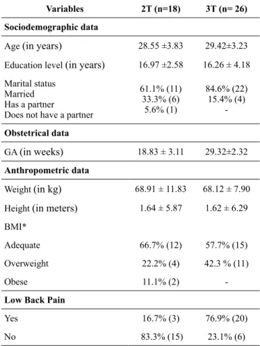
Sample characterization regarding sociodemographic, obstetric and anthropometric variables at the time of the initial evalu-ationis shown in Table 1.
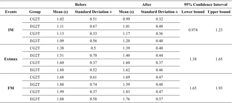
Table 2 shows the instant of occurrence (in seconds) of the events in the sit-to-stand (STS) movement in the four groups
evaluated from the initial and inal evaluations.
When considering the STS movement events, no sta-tistically significant differences were found for intra or intergroup conditions (IM– intragroup [F= 0.06; P= 0.79];
Table 1. Sociodemographic, obstetrical and anthropometric charac-teristics of pregnant women participating in the study at the initial evaluation
Variables 2T (n=18) 3T (n= 26)
Sociodemographic data
Age (in years) 28.55 ±3.83 29.42±3.23
Education level (in years) 16.97 ±2.58 16.26 ± 4.18
Marital status Married Has a partner
Does not have a partner
61.1% (11) 33.3% (6)
5.6% (1)
84.6% (22) 15.4% (4)
-Obstetrical data
GA (in weeks) 18.83 ± 3.11 29.32±2.32
Anthropometric data
Weight (in kg) 68.91 ± 11.83 68.12 ± 7.90
Height (in meters) 1.64 ± 5.87 1.62 ± 6.29
BMI*
Adequate 66.7% (12) 57.7% (15)
Overweight 22.2% (4) 42.3 % (11)
Obese 11.1% (2)
-Low Back Pain
Yes 16.7% (3) 76.9% (20)
No 83.3% (15) 23.1% (6)
intergroup [F=0.30; P=0.82]; EXTmax – intragroup [F=1.52; P= 0.69]; intergroup [F= 0.11; P= 0.94]; FM - in-tragroup [F=2.20; P= 0.14]; intergroup [F=0.41; P= 0.74]). Table 3 shows the time (in seconds) of the STS movement phases and the phase time percentage in relation to the total time of the
movement in the initial and inal evaluation.
When considering the STS movement phases and total
duration, no statistically signiicant differences were found for
intra and intergroup conditions (P1 - intragroup [F= 0.140; P= 0.71]; intergroup [F=2.69; P=0.059]; P2 - intragroup [F=1.96; P= 0.16]; intergroup [F=0.127; P= 0.94]; P3 - intragroup [F=0.329; P=0.57]; intergroup [F=0.743; P=0.53]; Total Time - intragroup [F=0.365; P= 0.54]. Intergroup [F=0.303; P= 0.82]).
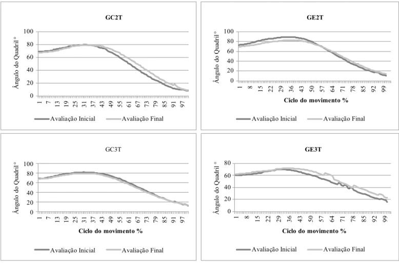
Regarding the comparison of the angular displacement of
the hip joint between the IM and Extmax in the STS movement, no statistically signiicant difference was observed in intra and
intergroup conditions (ROM hip) - intragroup [F=1.09; P=0.30]; intergroup [F=0.37; P=0.77]).
Figure 2 shows the mean angular displacement of the hip joint in the 4 study groups: CG2T, EG2T, CG3T and EG3T.
Hip lexion is observed in the STS movement, followed by the extension of this joint. Regarding comparison of the angular displacement of the hip joint ROM, no statistically signiicant
Table 2. Comparison of the occurrence instant of STS events considering the two moments of evaluation between the analyzed groups
Before After 95% Conidence Interval
Events Group Mean (s) Standard Deviation ± Mean (s) Standard Deviation ± Lower bound Upper bound
IM
CG2T 1.02 0.51 0.99 0.32
0.974 1.23
EG2T 1.11 0.67 1.01 0.48
CG3T 1.13 0.33 1.17 0.36
EG3T 1.09 0.56 1.20 0.40
Extmax
CG2T 1.38 0.5 1.39 0.40
1.38 1.65
EG2T 1.51 0.70 1.40 0.44
CG3T 1.60 0.37 1.60 0.37
EG3T 1.68 0.52 1.62 0.46
FM
CG2T 1.68 0.61 1.69 0.47
1.65 1.93
EG2T 1.88 0.74 1.59 0.48
CG3T 1.99 0.37 1.83 0.47
EG3T 1.88 0.58 1.76 0.57
NOTE: Values are expressed as mean ± Standard Deviation. 4x2 ANOVA for repeated measures test was used. LEGEND: IM - initial movement; Extmax - Maximum hip extension; FM - Final movement; CG2T - Control Group 2nd trimester; EG2T - Experimental Group 2nd trimester; CG3T - Control Group 3rd trimester; EG3T - Experimental Group 3rd trimester.
Table 3. Comparison of the phase duration and the total time of the movement in seconds considering the two evaluation moments between the analyzed groups
Before After 95% Conidence Interval
Events Group Mean (s) Deviation ±Standard Phase
time % Mean (s)
Standard
Deviation ± time %Phase
Lower
bound Upper bound
P1
CG2T 0.64 0.25 27.35 0.68 0.34 29.95
0.557 0.702
EG2T 0.55 0.25 21.48 0.62 0.38 25.94
CG3T 0.76 0.20 28.89 0.55 0.28 21.23
EG3T 0.52 0.29 19.40 0.68 0.27 24.72
P2
CG2T 0.47 0.22 20.08 0.45 0.18 19.82
0.398 0.506
EG2T 0.46 0.19 17.96 0.40 0.18 16.73
CG3T 0.44 0.19 16.73 0.35 0.17 13.51
EG3T 0.53 0.27 19.77 0.48 0.22 17.45
P3
CG2T 0.19 0.09 8.11 0.19 0.13 8.37
0.264 0.330
EG2T 0.19 0.08 7.42 0.34 0.43 14.22
CG3T 0.39 0.18 14.82 0.37 0.22 14.28
EG3T 0.33 0.29 12.31 0.31 0.20 11.27
Total Time
CG2T 2.34 0.96 100 2.27 0.55 100
2.32 2.73
EG2T 2.56 0.84 100 2.39 0.74 100
CG3T 2.63 0.70 100 2.59 0.86 100
EG3T 2.68 0.54 100 2.75 0.60 100
Discussion
The STS movement requires that adequate torque is developed at each joint 21. The dificulty of moving from sitting to stand -ing on both feet requires the use of several strategies in order
to facilitate its execution. When older individuals perform this movement, for example, there is an increase in postural stability
by the use of the upper limbs as support. This adaptation allows the mass center of the support surface to be approached by
in-creased torso lexion or time spent in performing the movement. These strategies result in increased energy expenditure in this
population as a result of the higher recruitment of motor units 22. The dificulty in performing the movement may also be
related to physiological factors, the initial position of the body segments and environmental factors. Physiological factors in-clude decreased proprioceptive acuity, muscle strength, postural
balance deicit, and joint pain, limiting the range of motion 22. Factors such as seat height, depth and inclination/slope of
the seat can also make STS more dificult. Increased seat height, for example, decreases the muscular force required to perform
the movement. This increase may result in new biomechanical requirements (such as the need to move the body center of mass at a greater distance) or an altered strategy (such as the “stabili-zation strategy”). This is due to the biomechanical requirements imposed by the foot, torso, or arm position 10,15. However, Lou,
Chou, Chou, Lin, Chen 23 observed that increasing the time to perform the STS movement has become more signiicant for
seats with lower heights 23.
In our study we chose to use two wooden blocks (Balance Master® accessories) without backrest, keeping the knees bent at 90 degrees and the feet 10 centimeters apart at the heels 18.
Studies that analyze STS tend to restrict the use of the arms for performing the movement. Individuals are advised to adopt the orthostatic position by keeping their hands in certain posi-tions, such as: cradles at the level of the chest, arranged at the side of the body, or resting on the knees 12,15. The presence of arm support is an important environmental strategy for the older adult population and also for pregnant women. Therefore we adopted the crossed arms on the chest position during kinematic analysis of the movement.
For Carr and Shepherd 12, the restricted arms position during STS seemed to cause a different pattern of angular displacement of the ankle, with a much larger mean standard deviation than that of free arms 12,15. The results of the study by Takeda, Katsuhira, Takano 24 found that when an armrest is used, the existing reaction force facilitates a forward and upward center of gravity displace-ment. In addition, a symmetrical dispersion of the load on the legs occurs, thus minimizing the load placed on the feet. Therefore, this suggests that the use of an arm support would decrease the muscular load when performing the sit-to-stand activity 24.
0 20 40 60 80 100 1 7 1 3 1 9 2 5 3 1 3 7 4 3 4 9 5 5 6 1 6 7 7 3 7 9 8 5 9 1 9 7 Â ng ul o do Q ua dr il ° GC2T
Avaliação Inicial Avaliação Final
0 20 40 60 80 100
1 8 15 22 29 36 43 50 57 64 71 78 85 92 99
 ng ul o do Q ua dr il °
Ciclo do movimento % GE2T
Avaliação Inicial Avaliação Final
0 20 40 60 80 100
1 7 13 19 25 31 37 43 49 55 61 67 73 79 85 91 97
 ng ul o do Q ua dr il °
Ciclo do movimento %
GC3T
Avaliação Inicial Avaliação Final
0 20 40 60 80 1 8 1 5 2 2 2 9 3 6 4 3 5 0 5 7 6 4 7 1 7 8 8 5 9 2 9 9 Â ng ul o do Q ua dr il °
Ciclo do movimento % GE3T
Avaliação Inicial Avaliação Final
The indings of Carr and Shepherd12 and Takeda, Katsuhira, Takano24 corroborate those by Lou, Chou, Chou, Lin, Chen23, who stated that it is useful to instruct pregnant women to adopt relatively higher-seating chairs and to use the armrests to help ensure postural stability and avoid putting pressure on the abdomen23. This adaptation would also help to decrease the joint torque of the hips and knees when there is loss of contact with the seat22.
The movement under analysis includes lexion of the torso and extension of the back, hips and knees, made dificult by painful symptoms in the lower back and/or extremities23. As pregnancy progresses, maximum torso lexion is reduced and the
body mass is increased as a result of the impregnated uterus. In
addition, hip joint peak and lexion speed decrease23,25. Therefore, it is expected that with gestation advancement, the activity of raising/lifting oneself from a chair becomes more dificult to
perform. Consequently, the kinematic and kinetic variables would be altered as strategies to complete the task26.
In our results, we did not obtain signiicant differences for
the STS kinematic movement variables when compared to the
analysis groups. Likewise, the indings by Takeda Katsuhira,
Takano24 did not ind any differences for STS movement between the gestational trimesters24. This suggests that pregnant women seem to be aware of postural instability during functional activi-ties and the fear of falls seems to make them more cautious. This fact could cause the propulsion phase to be minimized,
and as a consequence, the extension phase duration25. However, no signiicant differences were observed between the analyzed
groups (P=0.94) for this variable.
Savelberg, Fastenau, Willems, Meijer27 investigated the inluence of increased load on the movement of rising from a
chair in healthy young women. The volunteers performed the movement using a vest with an additional 0%, 15%, 30% and 45% of their body mass. The authors observed that in the 45% condition, the movement lasted on average 22% longer than in the 0% 27. Thus, we expected that in the third trimester of gestation, pregnant women would present a longer total STS movement duration. However, the total duration of STS
move-ment between groups did not differ signiicantly (P=0.82). It is
necessary, however, to take into account that the mass distribu-tion added to the volunteers’ bodies of the study by Savelberg Fastenau, Willems, Meijer 27 may have been different from the physiological distribution of weight gain that happens in pregnant women.
Mazzà, Benvenuti, Bimbi, Stanhope 10 suggests that the higher degree of dificulty of a given task associated with the functional status of the individual inluences the compensa -tory strategies that will be used when raising oneself from a chair 10. Such claims can justify the absence of difference for this variable when analyzing the groups of pregnant women. The need to control the upright balance at the end of the climb is paramount, and participants may have voluntarily limited the propulsion momentum to maintain upright posture
under varying conditions. In this context, the obtained result
suggests that the pregnant women sought to perform a more weighted movement26.
Conclusion
To our knowledge, this study is the irst to be conducted using virtual reality to observe its inluence on the sit-to-stand move -ment kinematics in pregnant women. Although the hypothesis of
this study (that the virtual reality-based exercise protocol would inluence the kinematic variables of pregnant women during the sit-to-stand) was not conirmed, it is important to emphasize the
absence of studies that analyze the effect implementing virtual reality games on the biomechanics of the sit-to-stand movement in pregnant women.
The non-conirmation of the hypothesis of this study can be justiied by: 1) the responses produced by the volunteers seem to
have been more sensitive to the adaptation of sensory systems and the integration between them than to the biomechanical altera-tions to which the woman’s body is subjected; and 2) changes in the biomechanics of pregnant women suggest adaptations in order to minimize the limitation effects, thus increasing stability during movement.
The contradictions between the studies regarding the analysis instruments of the sit-to-stand movement kinematics, along with
the scarcity of research that describe the inluence of physiologi -cal, hormonal and anatomical changes on the postural control of pregnant women evidence the need for further studies analyzing postural control pattern in pregnancy during everyday activities.
One of the limitations of the study was the small sample number and the non-accomplishment of follow-up. In
addi-tion, the intervention time may have been insuficient so that
the answers promoted by the training with virtual reality could be acquired and automated, enabling the transfer of this motor
task from the games to execution of the movement during the
evaluation. However, the results demonstrated in this study are
important and justify further studies analyzing the inluence of
implementing virtual reality on cinemetry variables (occurrence of events and movement phase duration) in pregnant women.
References
1. Ersal T, McCrory JL, Sienko KH. Theoretical and experimental indicators of falls during pregnancy as assessed by postural pertur-bations. Gait Posture [Internet]. Elsevier B.V. 2014;39(1):218–23. 2. Jang J, Hsiao KT, Hsiao-Wecksler ET. Balance (perceived
and actual) and preferred stance width during pregnancy. Clin Biomech (Bristol, Avon) [Internet]. 2008 May [cited 2013 Oct 21];23(4):468–76.
3. McCrory JL, Chambers AJ, Daftary A, Redfern MS. Dynamic postural stability in pregnant fallers and non-fallers. BJOG [Internet]. 2010;117(8):954–62.
4. Gazaneo MM, Oliveira LF. Alteração postural durante gestação. Rev bras ativ fís saúde. 1998;3(2):13–21.
5. Sunaga Y, Anan M, Shinkoda K. Biomechanics of rising from a chair and walking in pregnant women. Appl Ergon [Internet]. Elsevier Ltd; 2013;44(5):792–8.
7. Nicholls JA.Performance of physical tasks in pregnancy. Ergonomics. 1992; 35(3): 301-11.
8. Ramsey VK, Miszko TA, Horvat M. Muscle activation and force production in Parkinson’s patients during sit to stand transfers. Clin Biomech (Bristol, Avon) [Internet]. 2004;19(4):377–84. 9. Mathiyakom W, McNitt-Gray JL, Requejo P, Costa K. Modifying
center of mass trajectory during sit-to-stand tasks redistributes the mechanical demand across the lower extremity joints. Clin Biomech (Bristol, Avon) [Internet]. 2005;20(1):105–11. 10. Mazza C, Benvenuti F, Bimbi C, Stanhope SJ. Association
Between Subject Functional Status, Seat Height, and Movement Strategy in Sit-to-Stand Performance. JAm Geriatr Soc. 2004;52:1750–4.
11. Dehail P, Bestaven E, Muller F, Mallet A, Robert B, Bourdel-Marchasson I, et al. Kinematic and electromyographic analysis of rising from a chair during a “Sit-to-Walk” task in elderly sub-jects: role of strength. Clin Biomech (Bristol, Avon) [Internet]. 2007;22(10):1096–103.
12. Carr JH, Shepherd R. Reabilitação Neurológica: Otimizando o Desempenho Motor. 1ºedição. São Paulo: Editora: Manole, 1998, pag:74.
13. Mourey F, Grishin A, d’Athis P, Pozzo T, Stapley P. Standing up from a chair as a dynamic equilibrium task: a comparison between young and elderly subjects. J Gerontol A Biol Sci Med Sci [Internet]. 2000;55(9):425-31.
14. Riley P, Schenkman ML, Mann RW, Andrew W. Mechanics of a consttrained chair-rise. J Biomechanics. 1991;24(I): 77-85. 15. Janssen, Win GM, Hans BJ Bussmann HJS. Research Report
Determinants of the Sit-to-Stand Movement : A Review. J Am Phys Ther Assoc Fysiotherapeut. 2002;82:866–79.
16. Visual. Visual3D Online Documentation C-Motion.
17. Lana C. Caracterização Cinemática da Atividade de Passar de Sentado para Andar em Indivíduos com Doença de Parkinson. Belo Horizonte. Dissertação [Mestrado]- Universidade de Belo Horizonte; 2010.
18. Brech GC, Alonso AC, Luna NMS, Greve JM. Correlation of postural balance and knee muscle strength in the sit-to-stand test among women with and without postmenopausal osteoporosis. Osteoporos Int [Internet]. 2013;24(7):2007–13.
19. Robertson DG, Dowling JJ. Design and responses of butterworth and critically dampted digital ilters. J Electromyogr Kinesiol. 2003;13(6):69-73
20. Cole GK, Nigg BM, Ronsky JL, Yeadon MR. Application of the joint coordinate system to three-dimensional joint attitude and movement representation: a standardization proposal. J Biomech Eng.1993;115(4):344-9.
21. Schultz AB, Alexander N B, Ashton-Miller JA. Analyses of rising from a chair. J Biomech. 1992;25(12):1383–91
22. Goulart F, Chaves CM, Chagas MLD, Carvalho A, Saiki KR. O movimento de passar de sentado para de pé em idosos : implicações para o treinamento funcional. Acta fisiátrica. 2003;10(3):138-143
23. Lou S, Chou Y, Chou P, Lin C, Chen U. Sit-to-stand at different periods of pregnancy. Clin Biomech. 2001;16:194–8.
24. Takeda K, Katsuhira J, Takano A. Effects of handrail use dur-ing sit-to-stand in the third trimester. Int J Ind Ergon [Internet]. Elsevier Ltd; 2009;39(6):988–94.
25. Gilleard W, Crosbie J, Smith R. A longitudinal study of the effect of pregnancy on rising to stand from a chair. J Biomech [Internet]. 2008;41(4):779–87.
26. Gilleard W, Lai DT, Levinger P BR. Detecting trunk motion changes due to pregnancy using pattern recognition techniques. Eng Med Biol Soc. 2008;
27. Savelberg HH, Fastenau A, Willems PJ, Meijer K. The load/ capacity ratio affects the sit-to-stand movement strategy. Clin Biomech (Bristol, Avon). 2007;22(7):805-12.
Corresponding author
Silvia Oliveira Ribeiro
Postal Address: Department of Physical Therapy, Federal University of Rio Grande do Norte, Avenue Senador Salgado Filho, 3000, Natal, Rio Grande do Norte, Brazil. Email: [email protected]
Manuscript received on May 1, 2017
Manuscript accepted on May 7, 2017