2009; 59: 2: 187-193 SCIENTIFIC ARTICLE
RESUMO
Helayel PE, Conceição DB, Nascimento BS, Kohler A, Boos GL, Oli-veira Filho GR — Curva de Aprendizado da Sonoanatomia do Plexo Braquial na Região Axilar.
JUSTIFICATIVA E OBJETIVOS: A proficiência em bloqueios guia-dos por ultrassom exige quatro habilidades: reconhecimento da sonoanatomia, capacidade de geração de imagens, alinhamento da agulha ao feixe ultrassonográfico e reconhecimento da dispersão do anestésico local. O objetivo deste estudo foi construir e avali-ar curvas de aprendizado da geração de imagens e identificação ultrassonográfica das estruturas neurovasculares axilares.
MÉTODO: Sete médicos em especialização em Anestesiologia re-ceberam noções teóricas e práticas sobre princípios básicos da ultrassonografia e sonoanatomia axilar visando identificar ramos terminais do plexo braquial e vasos axilares. Cada um dos ME re-alizou seis exames. Foram avaliados a acurácia e o tempo trans-corrido para identificação das estruturas. Em cada exame foram calculadas taxas de sucesso. Regressão linear simples avaliou o tempo para identificação de cada estrutura em relação ao número do exame.
RESULTADOS: Os vasos axilares foram identificados em 100% dos exames. O nervo mediano foi identificado em 83% dos exa-mes entre o primeiro e quinto. O nervo radial foi identificado em 100% dos exames. O nervo ulnar foi encontrado em 67% dos ca-sos no primeiro exame e em 83% dos caca-sos do segundo ao quin-to procedimenquin-to. O nervo músculo-cutâneo foi localizado em 50% dos casos no primeiro exame, em 83% no quarto e no quinto
exa-mes. Todas as estruturas foram corretamente detectadas no sex-to exame. O tempo médio para identificação das estruturas dimi-nuiu significativamente entre o primeiro e sexto exames (r = – 0,37).
CONCLUSÕES: A memorização sonoanatômica axilar e a aquisi-ção de habilidade manual de exame ultrassonográfico foram obri-gatórias para progressão do aprendizado e taxas de sucesso crescentes se associaram com redução significativa do tempo para identificação das estruturas.
Unitermos: ANESTESIA, Regional: EQUIPAMENTOS, Ultrassom: TÉC-NICAS ANESTÉSICAS, Regional
SUMMARY
Helayel PE, Conceição DB, Nascimento BS, Kohler A, Boos GL, Oli-veira Filho GR — Learning Curve for the Ultrasound Anatomy of the Brachial Plexus in the Axillary Region.
BACKGROUND AND OBJECTIVES: Proficiency in ultrasound-guided blocks demands four skills: recognition of the ultrasound anatomy, capacity to generate images, aligning the needle with the ultrasound beam, and recognizing the dispersion of the local anesthetic. The objective of this study was to construct and evaluate learning curves for image generation and ultrasound identification of the neurovascular structures in the axilla.
METHODS: Seven Anesthesiology residents received theoretical and practical notions on the basic principles of ultrasound and the ultrasound anatomy of the axillary region with the objective to identify the terminal branches of the brachial plexus and axillary vessels. Each resident performed six exams. The accuracy and the time to identify the structures were evaluated. The success rate of each exam was calculated. Simple linear regression evaluated the time necessary to identify each structure in relation to the number of the exam.
RESULTS: The axillary vessels were identified in 100% of the exams. The median nerve was identified in 83% of the cases from the first to the fifth exams. The radial nerve was identified in 100% of the exams. The ulnar nerve was identified in 67% of the cases in the first exam, and in 83% of the cases from the second to the fifth exams. The musculocutaneous nerve was identified in 50% of the cases in the first exam and in 83% of the cases in the fourth and fifth exams. All structures were identified correctly on the sixth exam. The mean time for the correct identification of the structures decreased considerably from the first to the sixth exam (r = - 0.37).
CONCLUSIONS: Learning progression required the memorization of the ultrasound anatomy of the axillary region and acquisition of manual ability, and increasing success rates were associated with a significant reduction in the time to identify the structures.
Key Words: ANESTHESIA, Regional; ANESTHETIC TECHNIQUES, Regional; EQUIPMENT, Ultrasound.
Curva de Aprendizado da Sonoanatomia
do Plexo Braquial na Região Axilar*
Learning Curve for the Ultrasound Anatomy
of the Brachial Plexus in the Axillary Region
Pablo Escovedo Helayel, TSA1, Diogo Bruggeman da Conceição2, Bruno S. Nascimento3, Anderson Kohler3,
Gustavo Luchi Boos2, Getúlio Rodrigues de Oliveira Filho, TSA4
*Recebido (Received from) doHospital Governador Celso Ramos — CET/SBA Integrado de Anestesiologia da Secretaria de Estado da Saúde de Santa Catarina (SES-SC), Núcleo de Ensino e Pesquisa em Anestesia Regional (Nepar), Florianópolis, SC
1.Anestesiologista; Instrutor Co-Responsável do CET/SBA Integrado de Anestesiologia da SES-SC, Coordenador e Pesquisador do Nepar do CET/SBA Integrado de Anestesiologia da SES-SC
2.Anestesiologista; Pesquisador do Nepar do CET/SBA Integrado de Anestesiologia da SES-SC
3.ME do CET/SBA Integrado de Anestesiologia da SES-SC
4.Anestesiologista; Doutor em Anestesiologia; Responsável do CET/SBA In-tegrado de Anestesiologia da SES-SC; Pesquisador do Nepar do CET/SBA Integrado de Anestesiologia da SES-SC
Apresentado (Submitted) em 16 de maio de 2008
Aceito (Accepted) para publicação em 17 de novembro de 2008 Endereço para correspondência (Correspondence to): Dr. Pablo Escovedo Helayel
Av. Governador Irineu Bornhausen, 3440/204 — Agronômica 88025-200 Florianópolis, SC
E-mail: [email protected]
flexão das ondas de ultrassom de um ângulo de 90° em re-lação à estrutura insonada 10. Em consequência ocorre uma
perda de energia sonora que retorna ao transdutor diminu-indo a intensidade de eco e a visualização nervosa. Com pequenos ajustes na inclinação do transdutor é possível aproximar-se dessa inclinação ideal do feixe de ultrassom obtendo-se a visualização nervosa adequada 11. Outros
fa-tores de dificuldade para o reconhecimento adequado dos nervos periféricos na região axilar consistem na grande mobilidade e na variabilidade de posicionamento em rela-ção à artéria axilar 12. Dessa maneira, a pressão imposta
pelo transdutor sobre o braço pode deslocar nervos perifé-ricos vizinhos aproximando-os a ponto de prejudicar a individualização, assim como o posicionamento variável desses nervos pode causar dúvidas em sua identificação pela modificação das relações sonoanatômicas da região. Conforme foi possível demonstrar, o aprendizado das habi-lidades complexas de manuseio do transdutor e do reco-nhecimento (memorização) da sonoanatomia axilar pelo aprendiz demandou um maior número de exames para atin-gir a proficiência. Esses resultados, de acordo com outros estudos 7,13, demonstraram que a velocidade de aquisição
de habilidades manuais para geração de imagens ultrasso-nográficas dependia da habilidade individual do aprendiz. Contudo, apesar dessa variabilidade individual de aprendiza-do e de sua complexidade, observou-se rápida progressão na aquisição de habilidades manuais e de reconhecimen-to sonoanatômico, em reconhecimen-todos voluntários, atingindo um nível de proficiência adequado ao cabo de seis sessões de exame. O nervo músculo-cutâneo foi o que mais se teve dificuldade de identificar nesse estudo. Na região axilar ele encontra-se num septo entre os músculos bíceps e córaco-braquial, apresentando um padrão hiperecoico, com quatro formatos ao longo de seu trajeto 14, o que pode ter dificultado a
iden-tificação durante os exames iniciais. Entretanto, os nervos mediano e ulnar, por se tratarem de estruturas mais super-ficiais, hipoecoicos e sem grandes variações de formato, possivelmente tiveram sua identificação facilitada em com-paração ao nervo músculo-cutâneo. O nervo radial ao nível axilar encontra-se, na maioria das vezes, póstero-medial ou póstero-lateral à artéria axilar 12. Dessa forma, poderia ter
sua identificação prejudicada pelo aumento do ganho de imagem (artefato de imagem) gerada pelo equipamento de ultrassom no aspecto posterior da artéria somado ao brilho excessivo do septo do músculo tríceps 15. Apesar disso, este
nervo foi identificado corretamente em 100% dos exames a partir do segundo exame, possivelmente por apresentar menor variação de posicionamento que os demais ramos terminais do plexo braquial 12 facilitando seu
reconhecimen-to pela memorização de suas relações sonoanatômicas. O padrão anecoico de estruturas vasculares, associado à pulsatilidade da artéria axilar e ao colabamento das veias axilares à compressão pelo transdutor facilitaram a identifi-cação dessas estruturas. Essas características mecânicas e ecotexturais favoráveis apresentadas pelas estruturas
vasculares axilares não são compartilhadas pelos nervos periféricos, tornando sua visualização tecnicamente mais desafiadora. Dessa maneira, as maiores taxas de identifica-ção positiva foram obtidas num menor número de exames e menor tempo para os vasos axilares quando comparados aos ramos terminais do plexo braquial.
Esse estudo demonstrou que a memorização da sonoana-tomia da axila e a aquisição de habilidade manual no exa-me ultrassonográfico foram obrigatórias para progressão do aprendizado e das taxas de sucesso crescentes que se as-sociaram, rapidamente, com redução significativa do tempo para identificação correta das estruturas axilares.
Learning Curve for the Ultrasound
Anatomy of the Brachial Plexus in the
Axillary Region
Pablo Escovedo Helayel, TSA, M.D.; Diogo Bruggeman da Conceição, M.D.; Bruno S. Nascimento, M.D.; Anderson Kohler, M.D.; Gustavo Luchi Boos, M.D.; Getúlio Rodrigues de Oliveira Filho, TSA, M.D.
INTRODUÇÃO
Ultrasound guided nerve blocks represent a new paradigm in regional blocks 1,2. Through the direct visualization of the
nerves and adjacent structures, the ultrasound allows better distribution of the anesthetic around the nerve, increasing the success rate and decreasing the incidence of complications
3,4. The ultrasound has proved to be an invaluable teaching
tool in regional blocks for anesthesiology residents 5.
Howe-ver, to acquire proficiency in ultrasound-guided blocks, one should fulfill four fundamental requirements: recognize the ultrasound anatomy, have the capacity to generate high-qua-lity images, align the needle to the ultrasound beam, and recognize the adequate pattern of dispersion of the local anesthetic 6,7.
The objective of this study was to construct and evaluate the learning curve for image generation and ultrasound identifica-tion of the neurovascular structures in the axillary region.
METHODS
ultrasound principles (image generation, exam technique, and equipment settings) and illustrating the ultrasound anatomy of the brachial plexus and vascular structures of the axilla 8. Immediately before beginning the series of exams all
participants received individual instructions from an anes-thesiologist (with experience of more than 500 ultrasound-guided blocks) on how to handle the device to optimize the images. This was followed by the demonstration of the ultrasound anatomy of the axillary region, emphasizing the characteristics of the following structures: axillary artery and vein, and median, radial, ulnar, and musculocutaneous nerves (Figure 1). Images were generated and stored on the hard drive of an ultrasound machine (Sonoace 8000 SE®,
Medison, South Korea) equipped with a linear, broad-band transducer (5 to 10 MHz). For the study, the residents who were going to be examined were placed in dorsal decubitus with the arm in 90° abduction, the forearm at a 90° flexion and in external rotation so the back of the hand touched the table and the forearm was parallel to the axis of the body. After applying the ultrasound gel on the skin, the transducer was placed on the axillary region to generate a transversal image. In all exams the examiner was holding the transducer with the non-dominant hand and facing the ultrasound equip-ment to prepare the resident for the recommended technique for ultrasound-guided nerve blocks. For three days, each resident examined and had his axillary region examined six times to identify the terminal branches of the axillary plexus and axillary vessels. The accuracy of the identification of each structure and the length of time between the beginning of the exam and the identification of each structure were evaluated. The contact of the transducer with the skin was established as moment zero in order to evaluate the duration of the exam, which was done simultaneously with the evaluation of the resident by the instructor. The end of the exam was characterized by the identification of the last neurovascular structure of the axillary region or when the
resident gave up. Success rates were calculated for each exam. Simple linear regression evaluated the identification time for each structure per exam.
RESULTS
The axillary artery and vein were identified in 100% of the ca-ses in all exams. The median nerve was identified in 83% of the exams from the first to the fifth (Figure 2). The radial nerve was identified in 100% of the cases from the second exam on (Figure 3). The ulnar nerve was identified in 67% of
Figura 1 – Transversal Image of the Axillary Region. Axillary vessels are shown as anechoic (black) round structures. The median, ulnar and radial nerves are located around the axillary artery and show a “fascicular” aspect (internal hypoechoic gray areas in a matrix of hyperechoic [white] connective tissue). Image obtained with a broad-band linear transducer 5 – 10 MHz (Sonoace 8000 SE®, Medison, South Korea). (Pele = skin; biceps = biceps; mediano = median; art axilar = axillary artery; veia axilar = axillary vein; triceps = triceps).
Number of exams Median Nerve
Success Rates %
1o 2o 3o 4o 5o 6o
100
80
60
40
20
0
Figure 2 – Graphic Representation of the Progression of the Success Rate (Percentage) of Median Nerve Identification as a Function of the Number of Exams.
Figure 3 – Graphic Representation of the Progression of the Success Rate (Percentage) of Radial Nerve Identification as a Function of the Number of Exams.
Number of exams Radial Nerve
Success Rates %
1o 2o 3o 4o 5o 6o
100
80
60
40
20
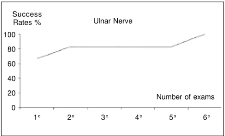
the cases in the first exam and in 83% of the cases from the second to the fifth exam (Figure 4). The musculocutaneous nerve was identified in 50% of the cases in the first exam, in 67% of the second and third exams, and in 83% in the fourth and fifth exams (Figure 5). All structures were identified correctly in 100% of the sixth exam. The mean time to identify the structures decreased significantly between the first and sixth exams (r = - 0.37).
DISCUSSION
The teaching of ultrasound-guided regional blocks should be divided in steps, as follows: recognition of the ultrasound anatomy and image generation in the first step followed by the alignment of the needle in relation to the ultrasound beam, and recognition of the proper pattern of dispersion of the local anesthetic 6,7,9. To succeed in obtaining images of
the brachial plexus, besides the knowledge of the ultrasound anatomy of the region, one also should acquire some abi-lities regarding the generation and interpretation of ultra-sound images. The knowledge of the normal ultraultra-sound anatomy depends on the memorization of the ultrasound characteristics of each nerve and vascular structure and the position pattern of each one in the region being visualized. The acquisition of visual and motor coordination is essential to produce manual movements capable of generating ima-ges with an acceptable recognition pattern. Manual motor coordination is composed by three basic transducer move-ments: sliding, inclination, and rotation. The combination of those three movements allows for the optimization of the characteristics of the image generated. Anisotropy is a form of tissue attenuation caused by irregularities on the surface of a peripheral nerve or its path, changing the incidence and reflection of the ultrasound waves from a 90° angle in relation to the structure being examined10. This causes a reduction
in the energy returning to the transducer, decreasing the intensity of the echo and nerve visualization. Through small adjustments in transducer inclination, it is possible to get closer to the ideal ultrasound beam inclination and obtain adequate nerve visualization 11. Other factors that hinder
proper recognition of peripheral nerves in the axillary region include the great mobility and variability in its position in relation to the axillary artery 12. Thus, the pressure of the
transducer on the arm can dislocate neighboring peripheral nerves, bringing them close together and making their individualization difficult; the variable position of those nerves can also be a source of difficulty by modifying the anatomic relationships in the region.
It was possible to demonstrate that achieving proficiency on complex abilities of transducer handling and recognition (memorization) of the axillary ultrasound anatomy demanded a greater number of exams. According to other studies 7,13,
those results demonstrate that the speed to acquire manual abilities to generate ultrasound images depended on the individual ability of each resident. However, despite this in-dividual variability in learning and its complexity, a fast progression on the acquisition of manual abilities and recog-nition of the ultrasound anatomy was observed in all volun-teers, who reached adequate proficiency level at the end of a six-exam session. In this study, the musculocutaneous nerve was the most difficult to identify. In the axillary region it is located in a septum between the biceps and coracobra-chial muscles, has a hyperechoic pattern, and four different shapes along its trajectory 14, what might have hindered its
identification in the initial exams. However, since the median and ulnar nerves are more superficial, hypoechoic, and do not show wide variability in shape, they were more easily identified. In the axilla, the radial nerve is located, most of the times, posteromedial or posterolateral to the axillary artery 12.
Thus, its identification could be hindered by the increased image gain (artifact) generated by the ultrasound equipment on the posterior aspect of the artery along with the excessive
Figure 4 – Graphic Representation of the Progression of the Success Rate (Percentage) of Ulnar Nerve Identification as a Function of the Number of Exams.
Number of exams Ulnar Nerve
Success Rates %
1o 2o 3o 4o 5o 6o
100
80
60
40
20
0
Figure 5 – Graphic Representation of the Progression of the Success Rate (Percentage) of Musculocutaneous Nerve Identification as a Function of the Number of Exams.
Number of exams Musculocutaneous Nerve
Success Rates %
1o 2o 3o 4o 5o 6o
100
80
60
40
brightness of the muscular septum of the triceps muscle 15.
Despite it all, this nerve was identified correctly in 100% of the cases from the second exam on, probably due to the reduced variation in position than the remainder terminal branches of the brachial plexus 12, facilitating its recognition
by memorizing its anatomic relationships. The anechoic pattern of vascular structures, associated with the pulsation of the axillary artery and collapsibility of axillary veins by the transducer facilitated their identification. Peripheral nerves do not share those favorable mechanical and echo characte-ristics of axillary vascular structures, making their visualization more challenging technically. Thus, the rate of identification of the axillary vessels was higher in a lower number of exams and in shorter time than the terminal branches of the brachial plexus.
This study demonstrated that memorization of the ultrasound anatomy of the axilla and the acquisition of manual ability in ultrasound examination were mandatory for progression of learning and increasing success rates, which were quickly associated with a significant reduction in the time necessary for the correct identification of axillary structures.
REFERÊNCIAS — REFERENCES
01. Helayel PE, Conceição DB, Oliveira Filho GR — Bloqueios nervo-sos guiados por ultrassom. Rev Bras Anestesiol, 2007;57:106-123.
02. Brull R, Perlas A, Chan VW — Ultrasound-guided peripheral nerve blockade. Curr Pain Headache Rep, 2007;11:25-32. 03. Sites BD, Beach ML, Spence BC et al. — Ultrasound guidance
improves the success rate of a perivascular axillary plexus block. Acta Anaesthesiol Scand, 2006;50:678-684.
04. Chan VW, Perlas A, McCartney CJ et al. — Ultrasound guidance improves success rate of axillary brachial plexus block. Can J Anaesth, 2007;54:176-182.
05. Grau T, Bartusseck E, Conradi R et al. — Ultrasound imaging improves learning curves in obstetric epidural anesthesia: a preliminary study. Can J Anaesth, 2003;50:1047-1050.
06. Oliveira Filho GR, Helayel PE, Conceicao DB et al. — Learning curves and mathematical models for interventional ultrasound basic skills. Anesth Analg, 2008;106:568-573.
07. Sites BD, Gallagher JD, Cravero J et al. — The learning curve associated with a simulated ultrasound-guided interventional task by inexperienced anesthesia residents. Reg Anesth Pain Med, 2004;29:544-548.
08. Marhofer P, Greher M, Kapral S — Ultrasound guidance in regi-onal anaesthesia. Br J Anaesth, 2005;94:7-17.
09. Sites BD, Spence BC, Gallagher JD et al. — Characterizing novice behavior associated with learning ultrasound-guided peripheral regional anesthesia. Reg Anesth Pain Med, 2007;32:107-115. 10. Aldrich JE — Basic physics of ultrasound imaging. Crit Care Med,
2007;35:S131-137.
11. Soong J, Schafhalter-Zoppoth I, Gray AT — The importance of transducer angle to ultrasound visibility of the femoral nerve. Reg Anesth Pain Med 2005;30:505.
12. Conceição DB, Helayel PE, Carvalho FAE et al. — Imagens ultrassonográficas do plexo braquial na região axilar. Rev Bras Anestesiol, 2007;57:684-689.
13. Gracias VH, Frankel HL, Gupta R et al. — Defining the learning curve for the Focused Abdominal Sonogram for Trauma (FAST) examination: implications for credentialing. Am Surg, 2001;67: 364-368.
14. Schafhalter-Zoppoth I, Gray AT — The musculocutaneous nerve: ultrasound appearance for peripheral nerve block. Reg Anesth Pain Med, 2005;30:385-390.
15. Sites BD, Brull R, Chan VW et al. — Artifacts and pitfall errors associated with ultrasound-guided regional anesthesia. Part II: a pictorial approach to understanding and avoidance. Reg Anesth Pain Med, 2007;32:419-433.
RESUMEN
Helayel PE, Conceição DB, Nascimento BS, Kohler A, Boos GL, Oli-veira Filho GR — Curva de Aprendizaje de la Sonoanatomía del Plexo Braquial en la Región Axilar.
JUSTIFICATIVA Y OBJETIVOS: El desempeño en bloqueos gui-ados por ultrasonido exige cuatro habilidades: reconocimiento de la Sonoanatomía, capacidad de generación de imágenes, alineación de la aguja al haz ultrasonográfico y reconocimiento de la dispersión del anestésico local. El objetivo de este estudio fue construir y evaluar curvas de aprendizaje de la generación de imágenes e identificación ultrasonográfica de las estructuras neurovasculares axilares.
MÉTODO: Siete médicos en especialización en Anestesiología recibieron nociones teóricas y prácticas sobre principios básicos de la ultrasonografía y sonoanatomía axilar, para identificar ramas terminales del plexo braquial y vasos axilares. Cada ME realizó seis exámenes. Fueron evaluados la exactitud y el tiempo transcurrido para la identificación de las estructuras. Se calcularon las tasas de éxito en cada examen. La regresión lineal simple evaluó el tiempo para la identificación de cada estructura con relación al número del examen.
RESULTADOS: Los vasos axilares fueron identificados en 100% de los exámenes. El nervio mediano fue identificado en un 83% de los exámenes entre el primero y el quinto. El nervio radial fue iden-tificado en 100% de los exámenes. El nervio cubital fue identifica-do en un 67% de los casos en el primer examen y en un 83% de los casos del segundo al quinto procedimiento. El nervio musculocutáneo fue identificado en un 50% de los casos en el primer examen, en un 83% en el cuarto y en el quinto exámenes. Todas las estructuras fueron correctamente identificadas en el sexto examen. El tiempo promedio para la identificación de las estructuras se redujo significativamente entre el primer y el sexto examen (r = - 0,37).