Influence of thermoluminescent dosimeters energy
dependence on the measurement of entrance skin dose
in radiographic procedures*
Influência da dependência energética de dosímetros termoluminescentes na medida da dose na entrada da pele em procedimentos radiográficos
Mércia Liane de Oliveira1, Ana Figueiredo Maia2, Natália Cássia do Espírito Santo Nascimento3, Maria da Conceição de Farias Fragoso4, Renata Sales Galindo5, Clovis Abrahao Hazin6
OBJECTIVE: This study was aimed at evaluating the influence of the energy dependence of thermoluminescent materials on the determination of entrance skin dose in patients submitted to conventional radiographic studies (general radiology, mammography and dental radiology). MATERIALS AND METHODS: Three different thermoluminescent materials were utilized: LiF:Mg,Ti, LiF:Mg,Cu,P and CaSO4:Dy. These materials were exposed to standardized sources of X and gamma radiation and clinical X-ray beams. RESULTS: Calibration and energy dependence curves were obtained. All the materials showed a linear response as a function of the air kerma. As far as energy dependence is concerned, the CaSO4:Dy and LiF:Mg,Ti samples showed the greatest variation on thermoluminescent responses as a function of the effective radiation beam. CONCLUSION: The tested materials showed an appropriate performance for detecting X radiation on standard and clinical X-ray beams. Although CaSO4:Dy and LiF:Mg,Ti samples present a significant energy dependence in the considered energy range, these materials can be utilized for measuring entrance skin doses, provided appropriate correction factors are applied.
Keywords: Thermoluminescent dosimeters; X-ray; Radiation protection; Entrance skin dose.
OBJETIVO: O objetivo do presente trabalho foi avaliar a influência da dependência energética de materiais termoluminescentes na determinação da dose na entrada da pele de pacientes submetidos a exames radio-gráficos (radiologia geral, mamografia e radiologia odontológica). MATERIAIS E MÉTODOS: Três diferentes materiais termoluminescentes foram utilizados: LiF:Mg,Ti, LiF:Mg,Cu,P e CaSO4:Dy. Estes materiais foram expostos a fontes padronizadas de radiação X e gama, e a feixes clínicos de raios X. RESULTADOS: As curvas de calibração e de dependência energética foram obtidas. Todos os materiais apresentaram resposta linear em função do kerma no ar. Com relação à dependência energética, as amostras de CaSO4:Dy e LiF:Mg,Ti mostraram maior variação da resposta termoluminescente em função da energia efetiva do feixe de radia-ção. CONCLUSÃO: Os materiais testados mostraram desempenho adequado para a detecção da radiação X em feixes padronizados e clínicos. Embora as amostras de CaSO4:Dy e LiF:Mg,Ti apresentem dependência energética significativa no intervalo de energia considerado, este materiais podem ser utilizados para medi-ção da dose de entrada na pele se fatores de corremedi-ção apropriados forem utilizados.
Unitermos: Dosímetros termoluminescentes; Raios X; Proteção radiológica; Dose na entrada da pele.
Abstract
Resumo
* Study developed at Centro Regional de Ciências Nucleares do Nordeste – Comissão Nacional de Energia Nuclear (CRCN-NE/CNEN), Recife, PE, Brazil.
1. PhD, Researcher for Comissão Nacional de Energia Nu-clear (CNEN), Centro Regional de Ciências NuNu-cleares do Nordeste – Comissão Nacional de Energia Nuclear (CRCN-NE/CNEN), Recife, PE, Brazil.
2. PhD, Professor at Department of Physics, Universidade Federal de Sergipe (UFS), São Cristóvão, SE, Brazil.
3. Graduate in Biomedicine, Fellow Master degree in Energetic and Nuclear Technologies, Universidade Federal de Pernambuco (UFPE), Recife, PE, Brazil.
4. Graduate in Radiology Technology, Fellow Master degree, Program of Energetic and Nuclear Technologies, Universidade Federal de Pernambuco (UFPE), Recife, PE, Brazil.
5. Chemistry Technician, Technician at Laboratory of Individual Monitoring of Centro Regional de Ciências Nucleares do Nordeste
exposure to artificial radiation sources(1,2). If by one side the technological advances in medicine provide for more accurate di-agnosis, on the other side the dissemination of such technologies leads to an increase in the collective dose, making it essential that medical practices based on ionizing radia-tions be optimized, assuring the benefits of such technologies and reducing associated risks.
An important tool for evaluating the optimization of procedures is the measure-ment of entrance skin dose (ESD) in pa-Oliveira ML, Maia AF, Nascimento NCES, Fragoso MCF, Galindo RS, Hazin CA. Influence of thermoluminescent dosimeters energy dependence on the measurement of entrance skin dose in radiographic procedures. Radiol Bras. 2010;43(2):113– 118.
INTRODUCTION
The use of ionizing radiation in medi-cine represents the main cause of human
– Comissão Nacional de Energia Nuclear (CRCN-NE/CNEN), Recife, PE, Brazil.
6. PhD of Health Physics, Researcher for Comissão Nacional de Energia Nuclear (CNEN), Centro Regional de Ciências Nuclea-res do Nordeste – Comissão Nacional de Energia Nuclear (CRCN-NE/CNEN), Recife, PE, Brazil.
Mailing address: Dra. Mércia Liane de Oliveira. Avenida Pro-fessor Luiz Freire, 200, Cidade Universitária. Recife, PE, Brazil, 50540-740. E-mail: [email protected]
tients submitted to radiographic examina-tions. This value shall be the lowest the more optimized the employed radiographic technique is, without compromising the imaging quality(3–5). ESD represents the dose on the patient’s skin surface added with back scattering radiation.
The ESD can be evaluated by direct methods (measurements with ionization chambers or by using thermoluminescent dosimeters [TLDs]), by indirect methods (by means of the determination of the dose-area product) or also by means of calcula-tions based on the X-ray tube perfor-mance(6).
Thermoluminescent dosimetry presents some advantages: high sensitivity, which allows the use of small-sized dosimeters; response with a low dependence on pho-ton energy and linear response for a wide dose interval; low cost and easy handle; high sensitivity, even for small doses; stable response, even under adverse envi-ronmental conditions; good reproducibil-ity, even for small doses; and simple emis-sion curve, with well defined peaks(7,8). Such dosimetry is based on the fact that materials will emit light when appropri-ately heated, after having been irradiated, with the amount of emitted light being pro-portional to the absorbed radiation energy (in other words, the absorbed dose)(8).
A detector response variation as a func-tion of the incident radiafunc-tion energy de-pends on the process of interaction between the radiation and the detector. In the energy interval of interest for diagnostic radiology (from 20 to70 keV(7)), the interaction be-tween radiation and matter predominantly happens because of the photoelectric ef-fect, whose occurrence probability in-creases with the effective atomic number of the medium(9). This means that TLDs with higher effective atomic numbers will present overestimated responses to radia-tion as compared with the readings done with dosimeters with lower effective atomic numbers. Consequently, the dosim-eter response variation as a function of in-cident radiation energy becomes a decisive factor in the choice of the material to be utilized, as without previous knowledge of such behavior, the ESD values might be unreliable.
Amongst the most frequently utilized thermoluminescent materials is the lithium fluoride activated with magnesium and ti-tanium (LiF:Mg,Ti); such material presents some important characteristics such as the effective atomic number (Zef = 8.2) close
to that of human tissue, not compromising the radiographic images, although present-ing a very complex thermoluminescent emission spectrum(10). Another lithium fluoride based dosimetric material has more recently been developed, using other dopant agents, the lithium fluoride acti-vated with magnesium, copper and phos-phorus (LiF:Mg,Cu,P; Zef = 8.2). This
ma-terial presents some advantageous charac-teristics as compared with LiF:Mg,Ti, among them the 40 times higher sensitiv-ity to gamma radiation. In Brazil, the Ins-tituto de Pesquisas Energéticas e Nucleares (IPEN-CNEN) produces dosimeters of cal-cium sulfide activated with dysprosium (CaSO4:Dy). This material is quite sensitive
to radiation, however, it has a high atomic number (Zef = 14.4) and for this reason it
presents a high dependence on the radiation energy, particularly up to 100 keV(10,11).
The aim of the present study was to evaluate the behavior of three thermolumi-nescent materials widely utilized in dosim-etry of X and gamma radiations (LiF:Mg,Ti; LiF:Mg,Cu,P; and CaSO4:Dy) on different
X radiation beams and the implications in the entrance skin dose estimation in pa-tients submitted to diagnostic radiology procedures (conventional radiology, mam-mography and dental radiology).
MATERIALS AND METHODS
The following dosimetric materials were utilized in the present study: LiF:Mg, Ti (commercially known as TLD-100), marketed by Thermo Scientific, Massachu-setts, USA; LiF:Mg,Cu,P (commercially known as TLD-100H), also marketed by Thermo Scientific, Massachusetts, USA, and CaSO4:Dy, manufactured by IPEN/
CNEN, São Paulo, Brazil. Initially a batch comprising 250 TLDs was gathered; the working batch was selected in such a way that, after five identical thermal treatment, irradiation and readout cycles, the maxi-mum thermoluminescent response
varia-tion was lower than 3%. After this selec-tion, the working batch comprised 24 TLD-100H samples, 36 TLD-100 samples and 39 CaSO4:Dy pellets.
The thermoluminescent materials were characterized according to their main do-simetric characteristics. For this purpose, they were irradiated with three radiation sources:
– Standard cesium 137 source (137Cs)
(JLShepherd & Associates, California, USA, with activity of 390 GBq – 1/1/2009) and energy of 660 keV;
– Standard cobalt 60 source (60Co) (IPEN, São Paulo, Brazil, with activity of 4.47 GBq – 1/1/2009) and mean energy of de 1,250 keV;
– Standard X radiation system, model HF 320 (Pantak Incorporated, Connecticut, USA) operating under the conditions pre-sented on Table 1.
In all exposures to radiation, the samples were individually encapsulated in transparent plastic. This same procedure was utilized in all irradiations. The sensi-tivity factor for each dosimeter was ob-tained after five identical irradiation, read-out and thermal treatment cycles, by calcu-lating the ratio between each dosimeter’s mean response and the mean response of the dosimeter that presented the smallest reading variation after the five measure-ment cycles.
The calibration curves were obtained by simultaneously irradiating all dosimeters with one of the previously mentioned stan-dard radiation sources, varying the air kerma. The samples were irradiated in the air, at the reference distance (1 m). When exposed to the 60Co source, the samples
were covered by a 4 mm-thick acrylic plate in order to assure the condition of elec-tronic equilibrium. When exposed to the
137
Cs source, the electronic equilibrium acrylic plate thickness was 2 mm. Because of the low energy, no electronic equilibrium plates were used when the samples were irradiated by X radiation beams.
to 50% fat and 50% glandular tissue, de-veloped by Oliveira et al.(12), and the fol-lowing equipment:
– Polymat 30/50 Plus general radiology equipment, manufactured by Siemens, Erlangen, Germany;
– M III mammography equipment, manufactured by Lorad Corporation, Con-necticut, USA;
– Intraoral dental radiology equipment, with two tube heads, manufactured by Indústrias Reunidas Rhos Ltda., Rio de Janeiro, Brazil.
In all irradiations with clinical beams, the TLDs were positioned in the center of the radiation field, on the phantom, with a pair of each thermoluminescent material being simultaneously irradiated. The pa-rameters utilized in the irradiations simu-lating chest, abdomen and skull studies are presented on Table 2. In the irradiations made with the mammography equipment, the parameters were the following: semi-automatic exposure control, 28 kVp and 37.6 mAs. On the other hand, in the irra-diations with the dental radiology beams, the following parameters were used: 80 kVp and 1.2 s; with a 22cm-long collima-tor with 6 cm in diameter.
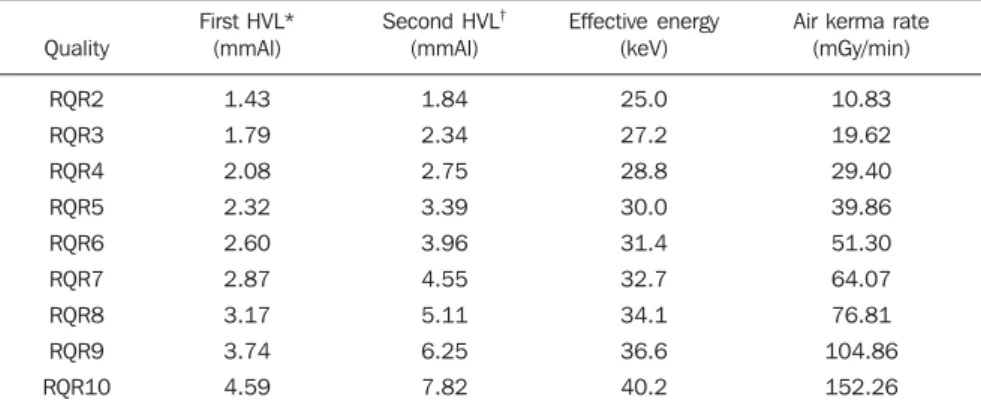
Table 1 Specification of radiodiagnostic qualities established in the Pantak 320kV X radiation system.
Quality RQR2 RQR3 RQR4 RQR5 RQR6 RQR7 RQR8 RQR9 RQR10 First HVL* (mmAl) 1.43 1.79 2.08 2.32 2.60 2.87 3.17 3.74 4.59
Second HVL†
(mmAl) 1.84 2.34 2.75 3.39 3.96 4.55 5.11 6.25 7.82 Effective energy (keV) 25.0 27.2 28.8 30.0 31.4 32.7 34.1 36.6 40.2
Air kerma rate (mGy/min) 10.83 19.62 29.40 39.86 51.30 64.07 76.81 104.86 152.26
* The first half-value layer (HVL) corresponds to the thickness of material required to reduce the radiation beam intensity to one half (50%) of its original value (100%). †The second HVL corresponds to the thickness of material required to reduce the radiation beam intensity from one half (50%) to a quarter (25%) of its original value (100%).
Table 2 Summary of radiographic techniques utilized to simulate chest, abdomen and skull radio-graphic studies, all of them performed anteroposterior projection.
Study Thorax Abdome Skul Distance (cm) 180 150 150 Voltage (kVp) 77 81 81 Product mAs 8 32 20 Current (mA) 200 200 200 Field size (cm2)
36 × 36 51 × 34 20 × 26
The thermoluminescent readout system used was a model 5500 manufactured by Thermo Electron Corporation, Massachu-setts, USA. For the thermal treatment of the samples a PTW-TLDO annealing oven, manufactured by PTW, Freiburg, Germany, was utilized.
With the CaSO4:Dy samples, a
pretreat-ment at 150°C for 20 seconds was per-formed. The readings were integrated from 150°C to 300°C with a heating rate of 10°C/s. After the reading, a thermal treat-ment was performed in the oven during 15 minutes at 300°C. In the case of LiF:Mg,Ti (TLD-100), a pretreatment was performed in the oven, before the reading at 100°C for one hour. The reading was integrated from 50°C to 300°C, with a heating rate of 15°C/s. After the reading, a thermal treat-ment was performed in the oven at 400°C for three hours and at 100°C for 1 hour. With the LiF:Mg,Cu,P (TLD-100H) sam-ples, a pretreatment at 145°C was per-formed for 10 seconds. The reading was integrated from 145°C to 260°C, with heat-ing rate of 10°C/s.
From the reading values of each ther-moluminescent sample, ESD was calcu-lated using the following equation (1):
ESD(mGy) = (L–LBG) ×Si×Cf (1)
where: L is the average of the irradiated dosimeter readings under the same condi-tions (in nC); LBG is the reading of non-ir-radiated dosimeters (background radia-tion); Si is the sensitivity factor for each sample; Cf is the calibration factor (mGy/ nC) obtained from each one of the obtained calibration curves.
RESULTS
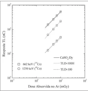
Initially, the calibration curves were obtained (thermoluminescent response ver-sus absorbed dose in air) for the tested dosimetric materials, in the radiation ener-gies described in Materials and Methods. Because of the low air kerma rate of the
60Co source, the calibration curve at this
energy was obtained for absorbed dose in air < 10 mGy. For the other energies (137Cs
and X-radiation qualities) the calibration curves were obtained for values up to 50 mGy. These curves are shown on Figures 1 and 2.
The energy dependence was obtained by irradiating all tested thermoluminescent materials with the same value of absorbed dose in air (50 mGy), under the same geo-metric conditions, and varying only the radiation beam energy. The results are shown on Figure 3.
In order to determine the patients’ ESD, phantoms were exposed to general radiol-ogy, mammography and dental radiology beams, as described in Materials and Meth-ods. The ESD values are presented on Table 3, using three different methods:
– method 1: determination of ESD using the calibration factor Cf for the
60Co
en-ergy;
– method 2: determination of ESD using the calibration factor Cf for the
137Cs
en-ergy;
– method 3: determination of ESD using the calibration factors Cf for the mean energies (determined by HVL values) of the utilized X-radiation beams.
With respect to uncertainties estimation, the factors in equation (1) utilized to deter-mine ESD were considered. The standard deviations only estimate the uncertainties in L and LBG.
Thus the combined uncertainty (k = 1) is in the order of 19%.
DISCUSSION
The excellent calibration curves linear-ity (evaluated by the linear coefficients of the adjustment applied to the experimental points) shows the applicability of the tested materials for dosimetry of X-radiation in the considered air kerma interval. It is
im-portant to note that, in terms of magnitude, such interval corresponds to the values of reference dose for typical adult patients in conventional radiodiagnostic, mammogra-phy and dental radiology studies, accord-ing to the Order (Portaria) 453 of the Bra-zilian Ministry of Health, published in 1998, which is up to 10 mGy for the most common studies(13). However, according to the literature, ESD values between 0.01 and 100 mGy(7)may be found.
With respect to the TLDs energy depen-dence (Figure 3), LiF:Mg,Cu,P (TLD-100H) was the material that presented the lowest response variation in the considered energy interval (less than 10% as compared with the response with 137Cs energy). However,
the LiF:Mg,Ti (TLD-100) and CaSO4:Dy
samples presented a more significant varia-tion; for such materials, the normalized thermoluminescent responses as compared with the response for to 37Cs energy were Figure 3. Energy dependence for: (A) LiF:Mg,Ti and LiF:Mg,Cu,P samples; and (B) CaSO4:Dy pellets. In this figure, the thermoluminescent response is normalized for the energy corresponding to 137Cs.
A B
Figure 1. Thermoluminescent response as a function of absorbed dose in air for the energies of 662 keV (137Cs) and 1250 keV (60Co) for the three
ther-moluminescent materials (CaSO4:Dy, TLD-100 and TLD-100H).
1.4 and 10.9 for TLD-100 and CaSO4:Dy,
respectively. These results are in agreement with those reported in the literature(7,14–16). According to Ministry of Health Order (Portaria) 453(13), the minimum HVL val-ues as a function of peak voltage (kVp) applied to the X-ray tube for general radi-ology equipment should range between 2.1 and 3.5 mmAl for single phase equipment, and 2.3 and 4.9 mmAl for three-phase equipment. For mammography equipment, the HVL value must be between kVp/100 and kVp/100 + 0.1 mm aluminum equiva-lent; and in the case of dental radiology equipment the minimum HVL must range from 1.2 to 2.5 mmAl, as a function of kVp. Such HVL values are in agreement with the HVL values of the X-radiation beams (Table 1) in which the thermolumi-nescent materials were characterized. This means that in the commonly utilized ener-gies in clinical radiodiagnostic beams, be-cause of its effective atomic number, a material may present greater or smaller re-sponse as demonstrated on Figure 3.
Table 3 shows the ESD values for radio-graphic studies utilizing the three thermolu-minescent studied materials calculated by means of equation (1). In the case of the
Of the three tested materials, only LiF:Mg,Cu,P did not present a significant response variation (which was in the same magnitude as the uncertainties) as a func-tion of the effective X-radiafunc-tion beam en-ergy. On the other hand, CaSO4:Dy and
LiF:Mg,Ti presented significant variations in response to X-radiation beams as com-pared with the responses obtained in the
137Cs or 60Co energies. This, however, does
not impair the use of such materials. Be-sides being produced in Brazil, CaSO4:Dy,
is very sensitive, being particularly useful for low dose measurements. On the other hand, LiF:Mg,Ti has an effective atomic number which is very close to that of hu-man tissue, and for this reason it does not cause artifacts on radiographic images, therefore being very useful in measure-ments directly performed by placing the TLD on the patient’s skin.
The three studied thermoluminescent materials can be utilized for patients dosim-etry in clinical beams. However, for the correct determination of ESD in patients submitted to radiographic studies (general radiology, mammography or dental radiol-ogy), the dosimeters must be previously calibrated for the energies corresponding to those utilized in the clinical practice.
Acknowledgments
The authors wish to thank Dr. L.L. Cam-pos and Dr. E.C. Vilela for their assistance in supplying the thermoluminescent mate-rials used in this study, and also the Con-selho Nacional de Desenvolvimento Cien-tífico e Tecnológico (CNPq) for part of the financial support.
REFERENCES
1. Berrington de González A, Darby S. Risk of can-cer from diagnostic X-rays: estimates for the UK and 14 other countries. Lancet. 2004;363:345–51. 2. Covens P, Berus D, Buls N, et al. Personal dose monitoring in hospitals: global assessment, criti-cal applications and future needs. Radiat Prot Dosimetry. 2007;124:250–9.
3. Compagnone G, Pagan L, Bergamini C. Local di-agnostic reference levels in standard X-ray exami-nations. Radiat Prot Dosimetry. 2005;113:54–63. 4. Tung CJ, Tsai HY, Lo SH, et al. Determination of guidance levels of dose for diagnostic radiogra-phy in Taiwan. Med Phys. 2001;28:850–7. 5. Organismo Internacional de Energia Atomica.
Normas básicas internacionales de seguridad para la protección contra la radiación ionizante y para la seguridad de las fuentes de radiación. Colec-ción Seguridad Nº 115. Viena: Organismo Inter-nacional de Energia Atomica; 1997.
Table 3 ESD for general radiology, mammography and dental radiology studies on phantoms. In this table, the mean values and corresponding standard deviations are shown.
Thermoluminescent material
LiF:Mg,Ti
LiF:Mg,Cu,P
CaSO4:Dy
Study Thorax AP Abdome AP Skull AP Breast Periapical 1 Periapical 2 Thorax AP Abdome AP Skull AP Breast Periapical 1 Periapcal 2 Thorax AP Abdome AP Skull AP Breast Periapical 1 Periapical 2 ESD (mGy) Method 1
0.325 ± 0.011 2.178 ± 0.086 1.31 ± 0.10 1.22 ± 0.22 6.08 ± 0.22 5.80 ± 0.17
0.315 ± 0.037 1.638 ± 0.040 0.904 ± 0.037 0.733 ± 0.030 4.02 ± 0.22 4.06 ± 0.10
2.598 ± 0.066 17.22 ± 0.65
9.17 ± 0.30 7.27 ± 0.25 47.8 ± 2.5 47.2 ± 1.4
Method 2
0.305 ± 0.010 2.043 ± 0.074 1.228 ± 0.095 1.14 ± 0.21 5.71 ± 0.19 5.44 ± 0.14
0.323 ± 0.038 1.677 ± 0.033 0.926 ± 0.036 0.751 ± 0.028 4.12 ± 0.22 4.153 ± 0.086
2.477 ± 0.057 16.42 ± 0.59
8.75 ± 0.26 6.93 ± 0.23 45.6 ± 2.3 45.0 ± 1.3
Method 3
0.229 ± 0.012 1.584 ± 0.060 0.952 ± 0.074 0.82 ± 0.15 4.08 ± 0.23 3.89 ± 0.20
0.304 ± 0.036 1.645 ± 0.040 0.908 ± 0.037 0.743 ± 0.027 4.12 ± 0.22 4.16 ± 0.10
0.248 ± 0.034 1.738 ± 0.053 0.926 ± 0.022 0.635 ± 0.017 4.26 ± 0.20 4.20 ± 0.10
ESD, entrance skin dose; AP, anteroposterior.
CaSO4:Dy pellets and LiF:Mg,Ti
(TLD-100) samples, there is a huge discrepancy between the ESD values determined from the calibration curves for the 60Co or 137Cs
energy as compared with the ESD values obtained by knowing the effective beam energy and the behavior of such materials in the energy interval of interest. Only for the LiF:Mg,Cu,P (TLD-100H) samples, the differences between the ESD values deter-mined from any of the calibration curves revealed to be in the same magnitude of uncertainties associated with the measure-ment. In this case, it is important to note that, although LiF:Mg,Ti and LiF:Mg,Cu,P have the same effective atomic number (Zef
= 8.2), LiF:Mg,Cu,P presents an anoma-lous response to radiation, according to Olko et al.(17), which explains the smaller variation in the thermoluminescent re-sponse in the studied energy interval.
CONCLUSIONS
6. Faulkner K, Broadhead DA, Harrison RM. Patient dosimetry measurement methods. Appl Radiat Isot. 1999;50;113–23.
7. Zoetelief J, Julius HW, Christensen P. Recom-mendations for patient dosimetry in diagnostic radiology using TLD. Luxembourg: Office for Official Publications of the European Communi-ties, European Commission; 2000.
8. Becker K. Solid state dosimetry. Ohio: CRC Press; 1973.
9. Knoll GF. Radiation detection and measurement.
2nd ed. New York: John Wiley; 1989. 10. Portal G. Review of the principal materials
avail-able for thermoluminescent dosimetry. Radiat Prot Dosimetry. 1986;17:351–7.
11. Campos LL, Lima MF. Dosimetric properties of CaSO4:Dy teflon pellets produced at IPEN. Radiat Prot Dosimetry. 1986;14:333–5.
12. Oliveira M, Nogueira MS, Guedes E, et al. Aver-age glandular dose and phantom imAver-age quality in mammography. Nucl Inst Meth Phys Res A. 2007;580:574–7.
13. Brasil. Ministério da Saúde. Secretaria de Vigi-lância Sanitária. Diretrizes de proteção radioló-gica em radiodiagnóstico médico e odontológico. Portaria nº 453, de 1º de junho de 1998. Brasília: Diário Oficial da União, 2 de junho de 1998.
14. Campos LL. Termoluminescência de materiais e sua aplicação em dosimetria da radiação. Cerâ-mica. 1998;44:26–8.
15. Davis SD, Ross CK, Mobit PN, et al. The response of LiF thermoluminescence dosemeters to pho-ton beams in the energy range from 30 kV X rays to 60Co gamma rays. Radiat Prot Dosimetry. 2003;106:33–43.
16. Duggan L, Hood C, Warren-Forward H, et al. Variations in dose response with x-ray energy of LiF:Mg,Cu,P thermoluminescence dosimeters: implications for clinical dosimetry. Phys Med Biol. 2004;49:3831–45.