Use of diffusion tensor magnetic resonance imaging
in the assessment of patterns of white matter
involvement in patients with brain tumors: is it useful
in the differential diagnosis?*
Uso do tensor de difusão na avaliação dos padrões de acometimento da substância branca em pacientes com tumores cerebrais: é uma ferramenta útil para o diagnóstico diferencial?
Vanessa Granado Alves Itagiba1, Rafael Borges2, Luiz Celso Hygino da Cruz Jr3, Andre Dietz Furtado4, Romeu Côrtes Domingues5, Emerson Leandro Gasparetto6
OBJECTIVE: The present study was aimed at evaluating the role of diffusion tensor imaging for the assessment of different patterns of white matter involvement in patients with brain tumors and the utility of this technique for differential diagnosis of such neoplasms. MATERIALS AND METHODS: Forty-four patients with brain tumors, including low-grade gliomas, anaplastic astrocytomas, glioblastomas multiforme and metastases, were studied with conventional and diffusion tensor magnetic resonance imaging. White matter tracts around the tumors were characterized as displaced, disrupted, infiltrated and edematous. RESULTS: A significant overlap was observed between patterns of white matter involvement on diffusion tensor images as the displaced pattern was seen in all the types of tumor. Disrupted and infiltrated patterns were found in glioblastomas multiforme and anaplastic astrocytomas. The edematous pattern was found in the cases of metastases. CONCLUSION: Diffusion tensor imaging patterns of white matter involvement assists in the mapping of tracts adjacent to tumors, providing significant data about tumors extent; however, they cannot distinguish low-grade and high-grade gliomas from metastases.
Keywords: Diffusion tensor imaging; White matter; Brain tumors.
OBJETIVO: Propõe-se avaliar o papel do tensor de difusão na avaliação dos diferentes padrões de acometi-mento da substância branca em pacientes com tumores cerebrais e a utilidade desta técnica no diagnóstico diferencial dessas neoplasias. MATERIAIS E MÉTODOS: Quarenta e quatro pacientes com tumores cere-brais, incluindo gliomas de baixo grau, astrocitomas anaplásicos, glioblastomas multiformes e metástases, foram estudados com imagens de ressonância magnética convencional e tensor de difusão. Os tratos de substância branca próximos aos tumores foram caracterizados como deslocados, interrompidos, infiltrados e edematosos. RESULTADOS: Houve significativa sobreposição entre os padrões de acometimento da subs-tância branca pelo tensor de difusão, uma vez que o padrão deslocado foi observado em todos os tipos de tumor. Os padrões interrompido e infiltrado foram encontrados em glioblastomas multiformes e astrocitomas anaplásicos e o padrão edematoso foi observado em metástases. CONCLUSÃO: Os padrões de envolvimento dos tratos de substância branca cerebral avaliados pelo tensor de difusão auxiliam no mapeamento dos tra-tos adjacentes aos tumores e fornecem informações importantes sobre a extensão do tumor, no entanto, eles não possibilitam fazer a distinção entre gliomas de baixo e alto graus e metástases.
Unitermos: Tensor de difusão; Substância branca; Tumores cerebrais.
Abstract
Resumo
* Study developed at CDPI Clínica de Diagnóstico Por Imagem and Clínica Multi-Imagem, Rio de Janeiro, RJ, Brazil.
1. Master Fellow degree, MD, Radiologist at CDPI Clínica de Diagnóstico Por Imagem, Hospital Central do Exército and Uni-versidade Federal do Rio de Janeiro (UFRJ), Rio de Janeiro, RJ, Brazil.
2. MD, Radiology Resident, Universidade Federal do Rio de Janeiro (UFRJ), Rio de Janeiro, RJ, Brazil.
3. Fellow PhD degree, MD, Radiologist at CDPI Clínica de Diag-nóstico Por Imagem, Clínica Multi-Imagem e Universidade Fe-deral do Rio de Janeiro (UFRJ), Rio de Janeiro, RJ, Brazil.
INTRODUCTION
Magnetic resonance imaging (MRI) has been widely used in the evaluation of pa-tients with brain tumors(1,2). However, in Itagiba VGA, Borges R, Cruz Jr LCH, Furtado AD, Domingues RC, Gasparetto EL. Use of diffusion tensor magnetic reso-nance imaging in the assessment of patterns of white matter involvement in patients with brain tumors: is it useful in the differential diagnosis? Radiol Bras. 2010;43(6):362–368.
4. MD, Radiologist, Clinical Fellow in Neuroradiology at Children’s Hospital of Pittsburgh, Pittsburgh, PA, USA.
5. MD, Radiologist, Director, CDPI - Clínica de Diagnóstico Por Imagem and Clínica Multi-Imagem, Rio de Janeiro, RJ, Brazil.
6. PhD, Associate Professor, Universidade Federal do Rio de Janeiro (UFRJ), MD, Radiologist at CDPI - Clínica de Diagnóstico Por Imagem and Clínica Multi-Imagem, Rio de Janeiro, RJ, Bra-zil.
Mailing Address: Dr. Emerson L. Gasparetto. Avenida das Amé-ricas, 4666, sala 325, Barra da Tijuca. Rio de Janeiro, RJ, Bra-zil, 22640-102. E-mail: [email protected]
many cases, conventional MRI findings are not enough for accurately defining a differ-ential diagnosis(1,3,4).
Recently, advanced MRI techniques including spectroscopy, diffusion tensor imaging (DTI), perfusion and blood oxy-gen level dependent (BOLD) have added important information regarding not only the diagnosis of tumors, but also concern-ing treatment options, follow-up and prog-nosis(1,3,5).
Nowadays, the DTI technique is achiev-ing more importance in the study of pa-tients with brain tumors(1,3,6). Diffusion ten-sor imaging can describe the direction of molecular water motion within tissues(1,3,7,8). In structures such as white matter fibers, the molecules diffusion will be more restricted perpendicular than parallel to the micro-structural boundaries, which is called anisotropic diffusion(1,3,7,9). This informa-tion can generate color-coded schematic maps of white matter tracts, where the col-ors brightness is directly proportional to the fractional anisotropy (FA)(1,7). The DTI post-processing (tractography) allows the estimation of the anatomic relationship between the neoplasm and adjacent white matter tracts, allowing the maximization of tumor resection while minimizing the as-sociated surgical morbidity(1,3).
Some authors have suggested a classi-fication of white matter involvement pat-terns in patients with brain tumors accord-ing to DTI findaccord-ings, aimaccord-ing at a better pre-operative assessment of such lesions(1,2,6,10). They considered the following DTI pat-terns of white matter surrounding the tu-mors: edematous, displaced, infiltrated and disrupted(1,6,10). The classification of such different patterns was based on the FA val-ues and on the direction and integrity of the white matter tracts adjacent to the tu-mors(1,2,6). To our knowledge, however, this classification has only be applied in small series of patients.
The present study was aimed at evalu-ating the DTI findings in 44 patients with brain tumors submitted to preoperative MRI. We have attempted to reproduce, in a larger series of brain tumor patients, the different DTI patterns previously re-ported(1,6,10), aiming at evaluating the role of DTI for the assessment of white matter tracts involvement in patients with brain
tumors as well as the utility of this tech-nique for the differential diagnosis of brain tumors.
MATERIALS AND METHODS
Subjects
The cohort study included 44 consecu-tive patients with intracranial neoplasms (24 male and 20 female patients; age range, 3–88 years; mean age, 44 years), who un-derwent MRI for pre-surgical evaluation. The final diagnoses of the tumors were based on the histological evaluation of material obtained either with surgical resec-tion (n = 37) or stereotactic biopsy (n = 7). The series included low-grade gliomas (n = 14), glioblastoma multiforme (n = 12), anaplastic astrocytomas (n = 9), and me-tastases (n = 9). All patients signed a term of free and informed consent and the study was approved by the Committee for Ethics in Research of the institution.
MRI acquisition
The MRI studies were performed in a 1.5 T system (Avanto; Siemens Medical Systems, Erlangen, Germany) using a 8-channel head coil. All patients underwent a conventional MRI protocol, including the following sequences: coronal T2-weighed images (TR: 4410 ms, TE: 98 ms, FOV: 240 mm, matrix: 320 × 320 and 3-mm sec-tion thickness with 30% of interval), axial FLAIR images (TR: 9950 ms, TE: 100 ms, FOV: 220 mm, matrix: 256 × 256 and 5-mm section thickness with 35% of interval), as well as axial and sagittal T1-weighed im-ages (TR: 37 5ms, TE: 11 ms, FOV: 240 mm, matrix: 212 × 256 and 5-mm section thick-ness with 30% of interval) before and after intravenous administration of 0.1 mmol/kg of gadolinium.
DTI acquisition
The DTI was performed using a single-shot echo-planar sequence with accelera-tion factor of two and with the following parameters: TR: 3100 ms, TE: 90 ms, FOV: 250 × 250 mm, matrix: 192 × 192, section thickness: 5 mm, interslice gap: 1.5 mm, bandwidth: 1346 kHz, EPI factor: 128, echo-spacing: 0.83, flip angle: 90º, NEX: 3, diffusion encoding in 12 different direc-tions and b values = zero and 1000 s/mm2.
DTI post-processing and analysis
All images were transferred to a work-station (Leonardo; Siemens Medical Solu-tions, Erlangen, Germany) and post-pro-cessed with the software DTI Task Card (MGH-Martino’s Center; Boston, USA). Values for b, FA, and FA color-coded maps were automatically calculated according to protocols previously described in the litera-ture(7). The FA color-coded map was gen-erated by mapping the major eigenvector x, y, and z components into red, green, and blue color channels, which were weighted by FA. On the color maps, by standard defi-nition, white matter tracts depicted in red have a right/left orientation, green tracts, anterior/posterior orientation, and blue tracts, superior-inferior orientation(1). We defined the FA as the anisotropy index to allow comparison with other studies, con-sidering that most of them have used this index(11–14).
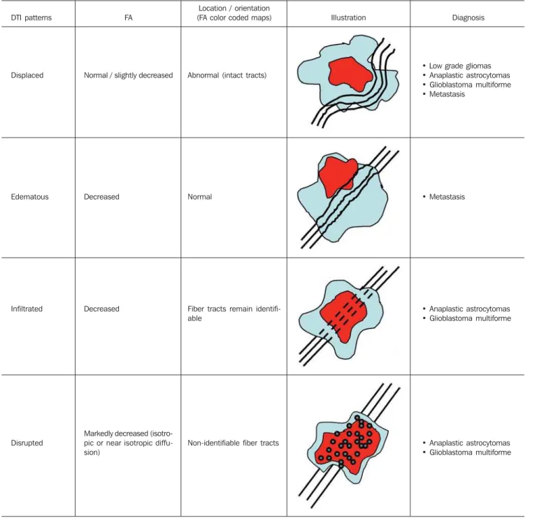
For the images analysis, the white mat-ter tracts adjacent to the tumors were visu-ally compared with the corresponding con-tralateral tracts, and then characterized as follows (Table 1): a) displaced if they main-tained normal or mildly decreased anisot-ropy as related to the corresponding tract on the contralateral hemisphere, but were situated at an abnormal location and/or orientation on the FA color-coded orienta-tion maps; b) edematous if they showed reduced anisotropy, but maintained normal location and orientation on the FA color-coded maps; c) infiltrated if they demon-strated reduced anisotropy, remained iden-tifiable on color-coded orientation maps, but exhibited altered color hues on the di-rectional color maps, not attributable to bulk mass displacement; b) disrupted if color-coded anisotropy was markedly re-duced, such that the tract could not be iden-tified on orientation maps(1,6). Firstly, two experienced neuroradiologists analyzed all the DTI findings before evaluating the struc-tural MR images and consensually reached final decisions regarding the different pat-terns of white matter involvement.
RESULTS
with low-grade gliomas, only the displaced pattern (14/14; 100%) was seen (Figure 1). The cases of anaplastic astrocytoma dem-onstrated the infiltrated (8/9; 88.9%), dis-placed (7/9; 77.8%) and disrupted (7/9; 77.8%) patterns, usually in association (Figure 2). All the patients with glioblas-toma multiforme showed both the dis-placed, disrupted and infiltrated patterns (12/12; 100%) (Figure 3). As regards the
patients with metastasis, all of them dem-onstrated the displaced and edematous pat-terns (9/9; 100%) (Figure 4).
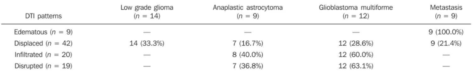
Alternatively, comparing the different DTI patterns with each histological diag-nosis, there is a significant overlap between patterns, since different tumors demon-strated the same DTI patterns of adjacent white matter involvement (Table 2). The displaced pattern (n = 42) was observed in
cases of low-grade gliomas (33.3%), glio-blastoma multiforme (28.6%), metastasis (21.4%) and anaplastic astrocytoma (16.7%). The disrupted pattern (n = 19) was found in patients with glioblastoma multiforme (63.2%) and anaplastic astro-cytoma (36.8%). Finally, the infiltrated pattern (n = 20) was demonstrated in cases of glioblastoma multiforme (60%) and anaplastic astrocytoma (40%).
Table 1 DTI patterns seen in patients with brain tumors.
DTI patterns
Displaced
Edematous
Infiltrated
Disrupted
FA
Normal / slightly decreased
Decreased
Decreased
Markedly decreased (isotro-pic or near isotro(isotro-pic diffu-sion)
Location / orientation (FA color coded maps)
Abnormal (intact tracts)
Normal
Fiber tracts remain identifi-able
Non-identifiable fiber tracts
Illustration Diagnosis
• Low grade gliomas • Anaplastic astrocytomas • Glioblastoma multiforme • Metastasis
• Metastasis
• Anaplastic astrocytomas • Glioblastoma multiforme
• Anaplastic astrocytomas • Glioblastoma multiforme
Figure 1. 32-year-old female patient with a low-grade glioma in the left fronto-temporal region. On A, axial FLAIR shows a well-circumscribed mass with heterogeneous high-signal intensity, located in the left fronto-temporal region. On B, axial, post-contrast T1-weighted image demonstrates unenhanced le-sion. On C, FA map shows the posterior limb of internal capsule medially dis-placed. On D, axial directional color map demonstrates normal anisotropy and abnormal location of the posterior limb of internal capsule (displaced pattern).
Figure 2. 49-year-old female patient with an anaplastic astrocytoma in the left centrum semiovale with extension towards the corpus callosum. On A, axial FLAIR shows a heterogeneous hyperintense mass involving the body of the corpus callosum. On B, axial post-contrast T1-weighted image demonstrates peripheral enhancement of the tumor. On C, FA map shows significant reduc-tion of the FA in the body of the corpus callosum. Displacement of the left corona radiata is also seen. On D, axial directional color map demonstrates complete destruction of corpus callosum fibers (disrupted pattern). The co-rona radiata in the left hemisphere is displaced (displaced pattern) and shows both reduced anisotropy and disorientation of white matter fibers (reflected in asymmetric color hues) (infiltrated pattern).
Table 2 Diffusion tensor MR imaging patterns in 44 patients with brain tumors.
DTI patterns
Edematous (n = 9)
Displaced (n = 42)
Infiltrated (n = 20)
Disrupted (n = 19)
Low grade glioma (n = 14)
—
14 (33.3%)
—
—
Anaplastic astrocytoma (n = 9)
—
7 (16.7%)
8 (40.0%)
7 (36.8%)
Glioblastoma multiforme (n = 12)
—
12 (28.6%)
12 (60.0%)
12 (63.1%)
Metastasis (n = 9)
9 (100.0%)
9 (21.4%)
—
—
DTI, diffusion tensor imaging.
DISCUSSION
The preoperative identification of the extent of malignant cell infiltration within white matter tracts constitutes a significant challenge. Tumor cells can invade and change the white matter fibers structure by widening, displacing, and/or disrupting the fiber bundles(15). The surgical treatment for brain tumors is aimed at achieving the maximum possible resection, while
mini-mizing the neurological deficits resulting from surgical injury to the intact, function-ing brain not affected by the tumor. This requires pre- and intraoperative mapping of the tumor as well as the definition of its relationship with functional structures, so that such structures can be preserved dur-ing surgical resection(10).
In the present study, we evaluated the DTI findings in 44 patients with brain tu-mors who underwent preoperative MRI
pattern was seen in metastases, low and high-grade gliomas; and the edematous pattern was observed only in cases of me-tastases.
Conventional MRI cannot satisfactorily demonstrate relevant anatomical details and does not provide enough information about fiber bundles location and integ-rity(15,16). Recently, advanced MRI tech-niques, including spectroscopy, DTI, per-fusion and BOLD, have added important information regarding not only the diagno-sis of the tumors, but also concerning treat-ment options, follow-up and progno-sis(1,3,5,9). The study of white matter tracts with magnetic resonance DTI allows the noninvasive investigation of neuronal fi-bers, based on the imaging characteristics of anisotropic water diffusion. The anisot-ropy of water diffusion in the brain white
matter is a result of its organization into myelinated axon fibers, where diffusion is faster parallel than perpendicular to the fi-bers tract(1,7,15,17).
Previous studies have assessed the DTI findings in patients with brain tumors, aim-ing at evaluataim-ing different patterns of white matter involvement(1,6,18). The suggested classification considered the FA values, integrity and orientation of white matter tracts on FA color-coded maps. The tracts were rated as displaced in cases of normal or slightly decreased anisotropy, situated at an abnormal location and/or orientation. The infiltrated pattern was associated with decreased anisotropy, without displace-ment of the normal white matter architec-ture, remaining identifiable on color-coded FA maps, but with abnormal color hues. The tracts with reduced anisotropy but
normal location and/or orientation were called edematous. The infiltrated and edematous patterns seem to be associated with infiltration of white matter tracts by tumor and/or edema, and the differentiation between them is not always possible. Both patterns were characterized by substantially decreased FA but, in most of the cases, they differed in their appearance on directional color maps(6). Whereas the edematous pat-tern tracts were normal in location and ori-entation (showed normal color hues on the color maps), the infiltrated pattern tracts exhibited abnormal hues not attributable to bulk mass displacement, which reflects a more severe form of disorganization(6,10,19). Finally, the cases with markedly reduced anisotropy and unidentifiable tracts on di-rectional color maps were rated as dis-rupted. However, the small series included Figure 4. 59-year-old male patient with lung metastasis in the right pariental region. On A, axial FLAIR shows a hyperintense mass in the right fronto-parietal region, surrounded by vasogenic edema. On B, axial, post-contrast T1-weighted image demonstrates peripheral enhancement of the lesion. On C, fractional anisotropy map demonstrates decreased anisotropy in the white matter surrounding the tumor. On D, axial directional color map shows de-creased anisotropy in the area surrounding the metastasis (reflected by dimin-ished color brightness) but with normal location and orientation (reflected by normal hues as compared with the homologous area in the contralateral hemi-sphere) (edematous pattern). In addition, there is a medial deviation of the corona radiata (displaced pattern).
in such studies limit the clinical application of these patterns of involvement of the white matter surrounding brain tumors.
Witwer et al.(1) have studied DTI find-ings in nine patients with brain tumors, applying the previously mentioned DTI classification. They evaluated six cases of low-grade gliomas, two of high-grade glio-mas and one case of metastasis. The au-thors found the displaced pattern only in patients with low-grade gliomas. The edematous pattern was showed in one case of high-grade glioma and in the patient with metastasis. The infiltrated and the dis-rupted patterns were found in one case of high- and in one of low-grade gliomas, re-spectively. Later, the same group of authors published 13 cases of brain tumors (pri-mary and secondary; low- and high-grade) evaluated with a similar approach(6). The results were almost identical to the ones observed in the previous study, with the displaced and edematous patterns being seen in both malignant and benign tumors, the infiltrated pattern being demonstrated in infiltrating gliomas and the disrupted pattern being observed in both low- and high-grade tumors. In our series, we found conflicting results when comparing with the results reported by Witwer et al.(1). The displaced pattern was non-specific, since it was found in cases of metastasis, low- and high-grade gliomas. The edematous pattern was showed only in cases of metastasis, differing from previous results demonstrat-ing this pattern also in cases of high-grade gliomas. In addition, the infiltrated and disrupted patterns, previously demon-strated in patients with both low- and high-grade gliomas, were seen only in cases of high-grade gliomas.
In our series, the cases of low-grade gliomas presented only the displaced pat-tern; however, this pattern is not specific for these tumors, since it was already de-scribed in previous series evaluating other tumors such as anaplastic astrocytomas, glioblastoma multiforme and cerebral me-tastasis(1,6). All the nine cases of metasta-sis showed both the displaced and edema-tous patterns. As already mentioned, the displaced pattern was also observed in low and high-grade gliomas, which makes this pattern nonspecific. Although the results of the present study have demonstrated that
the edematous pattern was only seen in metastasis, this pattern was also previously described in cases of gliomas(1,6). In our study, the disrupted pattern was shown only in high-grade gliomas; however, it has been previously described(1) in one case of low-grade glioma. In our series, some DTI pat-terns presented a good correlation with the histological diagnoses, such as the edema-tous pattern seen in the metastases and the infiltrated and disrupted patterns observed only in high-grade gliomas, which could suggest that the DTI might be able to dis-tinguish high- from low-grade gliomas and metastases. Although our data have been relevant, we observed that there was a sig-nificant overlap between the different DTI patterns and the final diagnoses, which does not allow the differential diagnosis of most of these tumors using the DTI patterns of brain white matter involvement.
Our study has several limitations. Al-though initial reports have suggested DTI advantages in the pre-surgical planning in cases of brain tumors, such reports as well as our results reflect preliminary conclu-sions. Another problem is the DTI se-quence susceptibility to artifacts that could cause images distortion. Additionally, the definition of the different patterns of white matter involvement is basically visual and, thus, susceptible to interobserver disagree-ment. Finally, although we have presented one of the largest series evaluating brain tumors with DTI, the number of subjects enrolled in the study is still small, espe-cially when one looks at the number of patients in the different subgroups.
CONCLUSION
The diffusion tensor imaging patterns of white matter tracts involvement assists in the mapping of white matter tracts adjacent to tumors and provide relevant information about tumors extent. Although we have observed that these patterns, such as the edematous pattern seen in metastases and the infiltrated and disrupted patterns in high-grade gliomas, may be useful in the differential diagnosis of some neoplasms, a significant overlap was observed between the patterns and the final histological diag-noses as our results are compared with pre-vious studies. Therefore, in agreement with
previous studies, we suggest that, although the DTI plays a relevant role in the preop-erative evaluation of patients with brain tumors, this method is not useful in the differential diagnosis of these tumors. In spite of some DTI limitations, this tech-nique is gaining support as a preoperative MRI method for evaluating brain tumors closely related to eloquent areas, optimiz-ing the surgical strategies. Nevertheless, in the future, with new technical develop-ments, DTI may become an important tool to predict the difference between these types of lesions.
REFERENCES
1. Witwer BP, Moftakhar R, Hasan KM, et al. Diffusion-tensor imaging of white matter tracts in patients with cerebral neoplasm. J Neurosurg. 2002;97:568–75.
2. Yu CS, Li KC, Xuan Y, et al. Diffusion tensor tractography in patients with cerebral tumors: a helpful technique for neurosurgical planning and postoperative assessment. Eur J Radiol. 2005; 56:197–204.
3. Sundgren PC, Dong Q, Gómez-Hassan D, et al. Diffusion tensor imaging of the brain: review of clinical applications. Neuroradiology. 2004;46: 339–50.
4. Beppu T, Inoue T, Shibata Y, et al. Measurement of fractional anisotropy using diffusion tensor MRI in supratentorial astrocytic tumors. J Neurooncol. 2003;63:109–16.
5. Holodny AI, Ollenschleger MD, Liu WC, et al. Identification of the corticospinal tracts achieved using blood-oxygen-level-dependent and diffu-sion functional MR imaging in patients with brain tumors. AJNR Am J Neuroradiol. 2001;22:83– 8.
6. Field AS, Alexander AL, Wu YC, et al. Diffusion tensor eigenvector directional color imaging pat-terns in the evaluation of cerebral white matter tracts altered by tumor. J Magn Reson Imaging. 2004;20:555–62.
7. Melhem ER, Mori S, Mukundan G, et al. Diffu-sion tensor MR imaging of the brain and white matter tractography. AJR Am J Roentgenol. 2002;178:3–16.
8. Laundre BJ, Jellison BJ, Badie B, et al. Diffu-sion tensor imaging of the corticospinal tract be-fore and after mass resection as correlated with clinical motor findings: preliminary data. AJNR Am J Neuroradiol. 2005;26:791–6.
9. Inoue T, Ogasawara K, Beppu T, et al. Diffusion tensor imaging for preoperative evaluation of tu-mor grade in gliomas. Clin Neurol Neurosurg. 2005;107:174–80.
10. Jellison BJ, Field AS, Medow J, et al. Diffusion tensor imaging of cerebral white matter: a picto-rial review of physics, fiber tract anatomy, and tumor imaging patterns. AJNR Am J Neuroradiol. 2004;25:356–69.
12. Tsuchiya K, Fujikawa A, Nakajima M, et al. Dif-ferentiation between solitary brain metastasis and high-grade glioma by diffusion tensor imaging. Br J Radiol. 2005;78:533–7.
13. Lu S, Ahn D, Johnson G, et al. Diffusion-tensor MR imaging of intracranial neoplasia and asso-ciated peritumoral edema: introduction of the tumor infiltration index. Radiology. 2004;232: 221–8.
14. Sinha S, Bastin ME, Whittle IR, et al. Diffusion tensor MR imaging of high-grade cerebral glio-mas. AJNR Am J Neuroradiol. 2002;23:520–7.
15. Stadlbauer A, Nimsky C, Buslei R, et al. Diffu-sion tensor imaging and optimized fiber track-ing in glioma patients: histopathologic evalua-tion of tumor-invaded white matter structures. Neuroimage. 2007;34:949–56.
16. Price SJ, Jena R, Burnet NG, et al. Improved de-lineation of glioma margins and regions of infil-tration with the use of diffusion tensor imaging: an image-guided biopsy study. AJNR Am J Neuroradiol. 2006;27:1969–74.
17. Puig J, Pedraza S, Blasco G, et al. Wallerian de-generation in the corticospinal tract evaluated by
diffusion tensor imaging correlates with motor deficit 30 days after middle cerebral artery is-chemic stroke. AJNR Am J Neuroradiol. 2010;31: 1324–30.
18. Price SJ, Peña A, Burnet NG, et al. Tissue signa-ture characterisation of diffusion tensor abnor-malities in cerebral gliomas. Eur Radiol. 2004;14: 1909–17.