Article
Printed in Brazil - ©2013 Sociedade Brasileira de Química0103 - 5053 $6.00+0.00
A
*e-mail: [email protected]
Iron Porphyrins as Biomimetical Models for Disperse Azo Dye Oxidation
Valéria P. Barros* and Marilda D. Assis
Departamento de Química, Faculdade de Filosofia Ciências e Letras de Ribeirão Preto, Universidade de São Paulo, Av. Bandeirantes 3900, 14040-901 Ribeirão Preto-SP, Brazil
As ferroporfirinas comerciais cloreto de 5,10,15,20-tetraquis-(4-carboxifenil) porfirinaferro(III), cloreto de 5,10,15,20-tetraquis-(N-metil-4-piridil)porfirinaferro(III) e cloreto de 5,10,15,20-tetraquis-(2,3,4,5,6-pentafluorofenil)porfirinaferro(III) foram investigadas como modelo do citocromo P450 na oxidação dos azo corantes Disperse Black 3 (DB3), Disperse Orange 3 (DO3) e Methyl Yellow (MY). Iodosilbenzeno, tert-butil hidroperóxido e peróxido de hidrogênio foram utilizados como oxidantes. As reações de oxidação foram monitoradas por espectroscopia de absorção no UV-Vis em comprimento de onda característico de cada corante e identificação dos produtos por cromatografia líquida de alto rendimento (HPLC) e cromatografia gasosa-espectrometria de massas (GC-MS). Os sistemas catalíticos foram eficientes para a oxidação de todos os corantes formando os produtos 4-nitroanilina e 4,4’-dinitroazobenzeno; 4-metilamino-azobenzeno, 4-aminoazobenzeno e 4-nitroazobenzeno; e 4,4’-dinitro4-metilamino-azobenzeno, respectivamente. Os produtos obtidos para todos os corantes são resultado da oxidação dos substituintes aminas terminais, e a ligação azo permanece intacta nestas reações para os corantes DB3 e MY. Para o corante DO3 ocorre a ruptura da ligação azo, resultando na formação da 4-nitroanilina. Produtos semelhantes são observados nos sistemas biológicos na oxidação do corante DO3, mostrando que os sistemas catalíticos estudados comportam-se como bons modelos biomiméticos.
The commercial ironporphyrins 5,10,15,20-tetrakis-(4-carboxyphenyl)porphyrin iron(III) chloride, 5,10,15,20-tetrakis-(N-methyl-4-pyridyl)porphyrin iron(III) pentachloride and 5,10,15,20-tetrakis-(2,3,4,5,6-pentafluorophenyl)porphyrin iron(III) chloride were investigated as cytochrome P450 models for the oxidation of the azo dyes Disperse Black 3 (DB3), Disperse Orange 3 (DO3) and Methyl Yellow (MY). Iodosylbenzene, tert-butyl hydroperoxide and hydrogen peroxide were used as oxidants. The oxidation reactions were monitored by UV-Vis absorption spectroscopy, by the observation of the absorption band characteristic of each dye, followed by product identification via high-performance liquid chromatography (HPLC) and gas chromatography-mass spectrometry (GC-MS). The catalytic systems were efficient for the oxidation of the dyes, leading to 4-nitroaniline and 4,4’-dinitroazobenzene; 4-methylamino-azobenzene, 4-aminoazobenzene, and 4-nitroazobenzene; and 4,4’-dinitroazobenzene as oxidation products, respectively. The oxidation products resulted from the oxidation of the terminal amine in the case of all the dyes, and the azo bond remained intact for DB3 and MY dyes. For DO3 dye, the azo bond is broken resulting in the formation of 4-nitroaniline. Similar products were observed for the oxidation of DO3 in biological systems, showing that the catalytic systems studied herein are good biomimetical models.
Keywords: disperse azo dye, metalloporphyrins, biomimetical models, oxidation, cytochrome P450
Introduction
Azo dyes represent 70% of all the dyes (106 tons)
produced per year and have applications in the textile, paper, leather, gasoline, additive, foodstuff, cosmetics and pharmaceutical industries.1-4 Some azo dyes are also used
in laboratories as either biological stains or pH indicators.1,5
Most of these synthetic dyes and their biodegradation products, sulfonated and unsulfonated aromatic amines, are toxic to aquatic organisms and suspected of being carcinogenic and mutagenic for humans.6-9 In the
The superfamily of the cytochrome P450 enzymes, present in plants, bacteria and mammals, is important in the detoxification of lipophilic drugs and pollutants. They can also activate chemicals that are protoxicants and procarcinogens.14-16 In most reactions catalyzed by
cytochrome P450, one oxygen atom from a dioxygen molecule is added to a substrate (monoxygenation) via heterolysis of the O−O bond, while the other oxygen atom is reduced to water with two electrons and two protons.17,18 These enzymes catalyze the insertion of one
oxygen atom from dioxygen into many different substrates, including the epoxidation of double bonds, the oxidation of heteroatoms and the dealkylation and oxidation of aromatic carbons.17,19 Because P450 isolation is a very delicate and
expensive task, the use of P450 synthetic models can be a useful tool for xenobiotic detoxification studies. In this context, metalloporphyrins (MePs) have been widely employed as models for cytochrome P450 monooxygenase-catalyzed oxidation reactions.15-20
Systems containing macrocyclic compounds such as porphyrins, phthalocyanines and Schiff bases are capable of forming complexes with several transition metals with different oxidation states. These metallic compounds have the ability to mimic the catalytic behavior of peroxidase and cytochrome P450-dependent monooxygenases, both in homogeneous and heterogeneous media, for the organic substrates.19-22 In this context, metalloporphyrins, which
are capable of catalyzing redox reactions under mild conditions,23,24 have aroused great interest for applications
related to the metabolism of organic compounds such as drugs, pesticides and dyes,19,21,24 with a view to
elucidating oxidation mechanisms and developing new compounds for pharmacological and toxicological tests. Although the literature concerning dye degradation is extensive,1,2,4,9,25 there are relatively few studies on the use
of metalloporphyrins as cytochrome P450 models for dye oxidation.20,21,26-28 Studies on metalloporphyrins can be an
excellent alternative for the identification and isolation of the metabolites obtained in the in vitro oxidation of azo dyes, thereby aiding in the elucidation of the mechanism of these oxidations in in vivo systems.
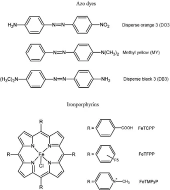
In this context, in the present study the commercial ironporphyrins 5,10,15,20-tetrakis-(4-carboxyphenyl) porphyrin iron(III) chloride (FeTCPP) 5,10,15,20-tetrakis -(N-methyl-4-pyridyl)porphyrin iron(III) penta-c h l o r i d e ( F e T M P y P ) a n d 5 , 1 0 , 1 5 , 2 0 -t e t ra k i s -(2,3,4,5,6-penta fluorophenyl))porphyrin iron(III) chloride (FeTFPP) were investigated as P450 models for the oxidation of the mono azo dyes Disperse Black 3 (DB3), Disperse Orange 3 (DO3) and Methyl Yellow (MY) (Figure 1) using various oxidants.
Experimental
Materials and equipments
All the compounds used in this study were commercially available from Aldrich or Sigma and were of analytical grade purity unless otherwise stated. Hydrogen peroxide (30 wt.% solution in water) was stored at 5 °C and titrated periodically for purity assessment. Tert-butyl hydroperoxide (TBHP) (70 wt.% solution in water) was acquired from Acros Oganics. Iodosylbenzene (PhIO) was synthesized by hydrolysis of iodosylbenzenediacetate using a procedure adapted from the literature.29 The solid was
carefully dried under reduced pressure and kept at 5 °C; its purity was periodically controlled by iodometric titration.30
The free base porphyrins were purchased from MidCentury. Iron insertion into the free base porphyrins was carried out using the method of Adler et al.31Acetonitrile (ACN) was
HPLC grade.
UV-Vis absorption spectra were obtained on a Hewlett-Packard 8452 diode array spectrophotometer. The spectra were recorded in quartz cells with 2 mm path length (Hellma).
column temperature was set at 50 ºC for 5 min, followed by a linear increase to 270 ºC at a rate of 10 ºC min-1, and
maintenance of this temperature for 4 min. The temperature of the injection port was 260 ºC, and the GC-MS interface was kept at 300 ºC. The helium carrier gas flow rate was 1.0 mL min-1. The reaction products were identified by
comparison of their retention times with known reference compounds, and by comparing their mass spectra to fragmentation patterns observed in the NIST spectral library stored in the computer software (version 1.10 beta, Shimadzu) of the mass spectrometer.
The high-performance liquid chromatography (HPLC) analyses were accomplished on a Shimadzu liquid chromatograph equipped with an LC-10AS solvent pump, an SPD-M 10A VP spectrophotometric detector coupled to a CTO-10A VP column oven and an SCL-10A VP system controller. The products were analyzed by using a Lichrospher 100RP-18 column with a particle size of 5 µm (125 mm × 4 mm), supplied by Merck. The analytical column was protected by a Lichrospher pre-column (4 mm × 4 mm). The mobile phase consisted of a mixture of ultrapure water (A) and acetonitrile (B). The analysis started with 30% of eluent A for 5 min, and then the concentration of the latter was increased linearly up to 70% in the following 6 min. This composition was held for further 4 min before being returned to 30% of eluent A for 2 min, to give a total run time of 22 min. The flow rate was 1.0 mL min-1, the detection was carried out at 320 nm, and
the column temperature was maintained at 32 ºC.
The HPLC-MS/MS analyses were performed on a Shimadzu LC-20A HPLC apparatus equipped with a diode array detector (CBM20A, Shimadzu, Kyoto, Japan) coupled to an UltrOTOFq (Bruker Daltonics, Billerica, MA, USA), ESI-qTOF (electrospray ionization-quadrupole time-of-flight) mass spectrometer operating in the positive ionization mode.
Oxidation reactions
Reactions were carried out in a 3 mL vial covered with a screw cap. A solution of disperse dye in ACN (1.25 µmol) and the oxidant (7.5 µmol) was added to the vial containing one of the ironporphyrins (0.125 µmol) in ACN/H2O 2:1 (2 mL). This catalyst/dye/oxidant molar
ratio (1:10:60) was the optimized condition to obtain higher product yields. All used ironporphyrins are soluble in the solvent mixture ACN/H2O 2:1. Reactions were accomplished
under magnetic stirring, at room temperature, for 4 h. At the end of the reaction, magnetic stirring was interrupted, and an aliquot of the reaction was analyzed by HPLC and GC-MS. The oxidation products were identified by comparing
their retention times with those of authentic standards. Conversion yields were calculated on the basis of the added dyes and were determined by means of a calibration curve. Other products were identified by mass spectrometry. The oxidation percentage was computed with the aid of the UV-Vis spectroscopy and calculated on the basis of the difference between the final and initial absorbance values at the maximum absorption wavelength of the dye.
Control reactions were conducted in all the different conditions, in the absence of the ironporphyrins (blank reactions). Results confirmed that no products from dye oxidation were obtained in these conditions.
Results and Discussion
Dye oxidation monitored by UV-Vis spectroscopy
The complexes investigated as catalysts for the oxidation of DO3, DB3 and MY dyes were the commercial ironporphyrins FeTCPP, FeTFPP and FeTMPyP (Figure 1), which have been reported to be good catalysts for the oxidation of hydrocarbons, drugs and other organic molecules.20,31-33 The reactions were carried out in
ACN/H2O (2:1, v:v), taking the solubility of the dyes and
the catalysts into account.
The oxidation reactions were followed by UV-Vis spectroscopy, by monitoring the main absorption band of the DB3 (λmax at 420 nm), DO3 (λmax at 430 nm) and
MY (λmax at 410 nm) dyes, which is due to the n →π*
transition and is known as the azo band. The other less intense bands observed in the UV region are assigned to the electronic π→π* transition of the aromatic ring.9,34,35
The UV-Vis spectra of azo dyes are a “finger print” of these compounds, changes in these spectra indicate alterations in the structure of these molecules.
Figure 2 depicts the catalytic results obtained for the reactions using TBHP as oxidant and different ironporphyrins. Although FeTFPP is considered a good catalyst for the oxidation of various substrates,31,33,36 it was
not able to promote the oxidation of the studied dyes. On the other hand, the ironporphyrins bearing ionic groups, FeTMPyP and FeTCPP, virtually promoted complete disappearance of the main absorption band, indicating almost 100% dye conversion. This high conversion was also observed by us in a previous work, which used FeTMPyP supported on montmorillonite as the catalyst and the same products were observed in both systems.20 These results
between the catalyst and the substrate probably favor the oxygen transfer from the active species to the substrate.37
Other oxidants were also evaluated, namely PhIO and H2O2 (Figure 3). As a simple oxygen donor, PhIO favors the
formation of the high-valent oxoferryl porphyrin π-cation radical, •+PFeIV=O, as the catalytic species,19,38 responsible
for the relatively high dye conversions (Figure 3). Although H2O2 is an environmentally clean oxidant, it leads to
low conversion yields (Figure 3). This is probably due to the decomposition of this oxidant catalyzed by the metalloporphyrin which occurs in a similar way to the reaction catalyzed by the enzyme catalase,39,40 previously
verified for other similar catalytic systems involving this oxidant.24
When alkyl peroxides such as TBHP are employed as an oxidant, the heterolytic cleavage of the O−O bond is expected to occur upon peroxide coordination to the
metalloporphyrin central metal ion, giving rise to the active species oxoferryl porphyrin π-cation radical, •+PFeIV=O
(Figure 4a). However, the major problem regarding the use of ROOH with ironporphyrins is the avoidance of the thermodynamically favored homolysis of the peroxide bond. Homolytic O−O bond cleavage generates a less reactive intermediate, PFeIV−OH, and culminates in an
alkoxyl radical RO•, which is able to abstract a hydrogen
atom from a saturated C−H bond but is unable to produce epoxides from an olefinic substrate (Figure 4b).41 The
relatively high conversion yields achieved in the reactions using TBHP suggest that the heterolytic cleavage of the O−O bond takes place during the oxidation of these dyes (Figure 3).
Besides the disappearance of the azo band, the UV-Vis spectrum of the reaction medium relative to the catalytic system FeTMPyP/TBHP displayed a new absorption band at ca. 330 nm (Figure 5). In order to identify the oxidation products responsible for this absorption, the reaction mixtures were analyzed by HPLC and GC-MS. The relative product yields were determined on the basis of the calibration curves constructed by using the dye standards.
Table 1 lists the retention times obtained from the HPLC and GC-MS analyses of each dye standard as well as those verified for the oxidation products present in the reaction mixtures when the FeTMPyP/TBHP catalytic system was employed. The molecular ion and the fragmentation profiles detected during the GC-MS analysis allowed the identification of the eluted products by comparison with the mass spectra of the NIST library, with over 95% similarity (Table 1).
As for the oxidation of MY, the products 4-methylamino-azobenzene (main product, relative yield of 56%), 4-aminoazobenzene (relative yield of 26%), resulting from the mono- and bis-demethylation of the terminal amine, respectively, and 4-nitroazobenzene (relative yield of 14%), a result of the additional oxidation of the demethylated amine (Table 1), were identified. These products are in agreement with those expected from the Figure 2. Conversion percentages for the dye oxidation reactions by
TBHP in the presence of the ironporphyrins, as monitored by UV-Vis spectroscopy at λ of the maximum absorption band for each dye. Standard conditions as defined in the text.
Figure 3. Conversion percentages for the dye oxidation reactions by different oxidants in the presence of FeTMPyP, as monitored by UV-vis spectroscopy at λ of the maximum absorption band for each dye. Standard conditions as defined in the text.
oxidation of tertiary amines catalyzed by P450 enzymes.11,21
Therefore, these catalytic systems can be considered good biomimetical models of P450 enzymes for MY oxidation.
Concerning DO3 and DB3, the main oxidation product was 4,4’-dinitroazobenzene, originated from the oxidation of the terminal amine in the same way that the reaction occurred in the case of MY and in accordance with what is observed for in vivo systems catalyzed by P450. These results indicate that similar intermediate species are probably responsible for the in vivo oxidation, and that in these biomimetical systems, the high-valent oxoferryl porphyrin π-cation radical, •+PFeIV=O,41,42 operates. This
highly electrophylic species is able to transfer the oxygen atom to the di-amine, furnishing hydroxylamine. The latter is easily oxidized to the nitro compound, as already reported by Porta et al.43 for the oxidation of aromatic
amines catalyzed by ironporphyrins.
For the DO3 dye, the product 4-nitroaniline was attained (Table 1, entry 6). This compound had also been observed during the in vitro oxidation of this dye catalyzed by lignolytic enzymes/H2O2 for the fungus Pleurotus ostreatus.44,45
Reactions carried out in the absence of oxygen (argon atmosphere) gave similar results as compared to the ones conducted in air atmosphere. This attested the absence of radical mechanisms stemming from the homolytic cleavage of the peroxide O−O bond in these oxidations41,46 and
confirmed that the heterolytic cleavage was predominant in these conditions.
The fact that the high product yields obtained from the oxidation of the terminal amine groups while the azo bond was left intact evidences that the reduction in the azo absorption band in the UV-Vis spectra of these compounds may not be associated with the azo bond cleavage, but that it
Table 1. Retention times (tR) of the compounds present in the reaction mixture obtained from the GC-MS and HPLC analyses of the oxidation of the dyes by TBHP in ACN/H2O (2:1, v:v); molecular ion and product fragments (m/z) obtained from the GC-MS of the same solutions
entry Compound (conversion / %) tR / min Molecular
ion / MI Fragments, m/z (relative percentage) HPLC CG-MS
1 DB3 11.0 30.1 240 240 (100), 148 (10), 120 (89), 105 (14), 92 (46), 77 (17), 65 (23), 42 (58)
2 P1 10.0 – – nd
3 P2 10.3 – – nd
4 4,4'-dinitroazobenzene (82%) 10.8 28.6 272 227 (25), 150 (77), 122 (100), 92 (27), 76 (56), 50 (22)
5 P4 11.3 – – nd
6 DO3 12.6 29.0 242 242 (20), 212 (22), 120 (35), 92 (100), 65 (51)
7 4-nitroaniline (17%) 3.6 19.3 138 138 (75), 122 (12), 108 (67), 92 (41), 80 (21), 65 (100) 8 4,4'-dinitroazobenzene (79%) 10.8 28.4 272 272 (22), 150 (68), 122 (100), 92 (50), 76 (63) 9 MY 12.4 25.7 225 225 (89), 148 (25), 120 (100), 105 (27), 91 (14), 77 (50), 42 (58) 10 4-methylamino-azobenzene (56%) 9.6 25.4 211 211 (46), 134 (30), 106 (100), 77 (57), 65 (19), 51 (17) 11 4-aminoazobenzene (26%) 10.3 24.3 197 197 (32), 120 (38), 92 (100), 77 (39), 65 (48), 51 (15) 12 4-nitroazobenzene (14%) 11.0 24.1 227 227 (20), 122 (15), 105 (35), 77 (100), 51 (23) nd: not detected.
Figure 5. UV-Vis spectra of the ACN/H2O (2:1, v:v) solutions of the DB3, DO3 and MY azo dyes (0.625 mmol L-1) in the presence of FeTMPyP
may be due to the oxidation of a substituent in the aromatic ring thereby causing a shift of the azo bond absorption. This was reported in the literature for other dyes.47
On the basis of the catalytic results obtained in our studies, our group proposes a reaction scheme that represents the oxidation of these azo dyes by TBHP in the presence of the ironporphyrins (Figure 6).
Conclusions
The investigated catalytic systems are efficient for the oxidation of azo dyes by PhIO and TBHP. The high
conversion yields obtained by using PhIO indicate that the reaction goes through the mechanism involving the high-valent oxoferryl porphyrin π-cation radical, •+PFeIV=O.
This is probably the catalytic species that participates in the oxidation with TBHP and is formed through the heterolytic cleavage of the peroxide O−O bond. The oxidation products for all the three dyes are the result of terminal amine oxidation, while the azo bond is left intact. Similar products have been verified during DO3 oxidation in biological systems, showing that the studied catalytic systems are good biomimetical models. Therefore, the systems have potential application for evaluation of the toxicity of this class of dyes.
Supplementary Information
Supplementary information associated with this work contains HLPC and GC-MS chromatograms and mass spectra. These data are available free of charge at http://jbcs.sbq.org.br as a PDF file.
Acknowledgements
The authors thank CNPq, CAPES and FAPESP for financial support, Dr. Cynthia Maria de Campos Prado Manso for linguistic advice, Izabel Cristina C. Turatti (Faculdade de Ciências Farmacêuticas de Ribeirão Preto, USP) for the GC-MS analysis and José Carlos Tomaz (Faculdade de Ciências Farmacêuticas de Ribeirão Preto, USP) for the HPLC-MS analysis.
References
1. El-Desoky, H. S.; Ghoneim, M. M.; Zidan, N. M.; Desalination
2010, 264, 143.
2. Liu, G.; Wang, J.; Lu, H.; Jin, R.; Zhou, J.; Zhang, L.; J. Hazard. Mater.2009, 171, 222.
3. Ayed, L.; Mahdhi, A.; Cheref, A.; Bakhrouf, A.; Desalination
2011, 274, 272.
4. Rauf, M. A.; Meetani, M. A.; Hisaindee, S.; Desalination2011,
276, 13.
5. Wang, Y.; Bingtao, T.; Shufen, Z.; Dyes Pigm.2011, 91, 294. 6. Crabtree, H. C.; Hart, D.; Thomas, M. C.; Witham, B. H.;
McKenzie, I. G.; Smith, C. P.; Mutat. Res. Lett.1991, 264, 155. 7. Pacheco, M. J.; Santos, V.; Ciríaco, L.; Lopes, A.; J. Hazard.
Mater.2011, 186, 1033.
8. Franciscon, E.; Piubeli, F.; Fantinatti-Garboggini, F.; de Menezes, C. R.; Silva, I. S.; Cavaco-Paulo, A.; Grossman, M. J.; Durrant, L. R; Enzyme Microb. Technol.2010, 46, 360. 9. Saratale, R. G.; Saratale, G. D.; Chang, J. S.; Govindwar, S. P.;
J. Taiwan Inst. Chem. Eng.2011, 42, 138. Figure 6. Proposed scheme for methyl yellow (MY) oxidation by TBHP
10. Platt, K. L.; Edenharder, R.; Aderhold, S.; Muckel, E.; Glatt, H.; Mutat. Res., Genet. Toxicol. Environ. Mutagen. 2010,
703, 90.
11. Hunger, K.; Chimia1994, 48, 520.
12. Zbaida, S.; Stoddart, A. M.; Levine, W. G.; Chem. Biol. Interact.
1989, 69, 61.
13. Chen, C.-H.; Chang, C.-F.; Liu, S.-M.; J. Hazard. Mater.2010,
177, 281.
14. Mansuy, D.; Ann. Pharm. Fr. 2011, 69, 62.
15. Hannemann, F.; Bichet, A.; Ewen, K. M.; Bernhardt, R.;
Biochim. Biophys. Acta, Gen. Subj. 2007, 1770, 330.
16. Kotrbová, V.; Mrázová, B.; Moserová, M.; Martínek, V.; Hodek, P.; Hudeček, J.; Frei, E.; Stiborová, M.; Biochem. Pharmacol.
2011, 82, 669.
17. Montellano, P. R. O.; Cytochrome P450: Structure, Mechanism and Biochemistry, 3rd ed.; KluwerAcademic/Plenum
Publishers: New York, USA, 2005.
18. Tani, F.; Matsuura, M.; Nakayama, S.; Naruta, Y.; Coord. Chem. Rev.2002, 226, 219.
19. Mansuy, D.; C. R.Chim.2007, 10, 392.
20. Barros, V. P.; Faria, A. L.; MacLeod, T. C. O.; Moraes, L. A. B.; Assis, M. D.; Int. Biodeterior. Biodegrad. 2008, 61, 337. 21. Emmert III, F. L.; Thomas, J.; Hon, B.; Gengenbach, A. J.;
Inorg. Chim. Acta2008, 361, 2243.
22. Zidane, Y.; Ourari, A.; Bataille, T.; Hapiot, P.; Hauchard, D.;
J. Electroanal. Chem.2010, 641, 64.
23. Biswas, A. N.; Pariyar, A.; Bose, S.; Das, P.; Bandyopadhyay, P.;
Catal. Commun.2010, 11, 1008.
24. Faria, A. L.; Mac Leod, T. O. C.; Barros, V. P.; Assis, M. D.;
J. Braz. Chem. Soc.2009, 20, 895.
25. Pálfi, T.; Wojnárovits, L.; Takács, E.; Radiat. Phys. Chem. 2011,
80, 462.
26. Hodges, G.; Smith, J. R. L.; Oakes, J.; Stud. Surf. Sci. Catal.
1997, 110, 653.
27. Serra, A. C.; Docal, C.; Gonsalves, A. M. d’A. R.; J. Mol. Catal. A: Chem.2005, 238, 192.
28. Hager, M.; Holmberg, K.; Gonsalves, A. M. d’A. R.; Serra, A. C.; Colloids Surf., A2001, 183,247.
29. Saltzman, H.; Sharefkin, J. G.; Organic Syntheses Collective, vol. 5; John Wiley & Sons: New York, USA, 1973.
30. Lucas, H. J.; Kennedy, E. R.; Organic Syntheses Collective, vol. 3; John Wiley & Sons: New York, USA, 1955.
31. Zucca, P.; Rescigno, A.; Sanjust, E.; Chin. J. Catal. 2012, 32, 1663.
32. Neves, C. M. B.; Simões, M. M. Q.; Santos, I. C. M. S.; Domingues, F. M. J.; Neves, P. M. S.; Paz, F. A. A.; Silva, A. M. S.; Cavaleiro, J. A. S.; Tetrahedron Lett. 2011, 52, 2898. 33. Luz, R. C. S.; Damos, F. S.; Tanaka, A. A.; Kubota, L. T.;
Gushikem, Y.; Talanta2008, 76, 1097.
34. Zucca, P.; Sollai, F.; Garau, A.; Rescigno, A.; Sanjust, E.; J. Mol. Catal. A: Chem. 2009, 306, 89.
35. Li, J.; Jiang, P.; Wei, C.; Shi, J.; Dyes Pigm.2008, 78, 219. 36. Rau, H.; Azocompounds; Elsevier: Amsterdam, 1990. 37. Mac Leod, T. C. O.; Faria, A. L.; Barros, V. P.; Queiroz, M. E. C.;
Assis, M. D.; J. Mol. Catal. A: Chem. 2008, 296, 54. 38. Santos, A. C. M. A.; Smith, J. R. L.; Assis, M. D.; J. Porphyrins
Phthalocyanines2005, 9, 326.
39. Groves, J. T.; J. Inorg. Biochem. 2006, 100, 434.
40. Horst, F.; Rueda, E. H.; Ferreira, M. L.; Enzyme Microb. Technol.2006, 38, 1005.
41. Vlasits, J.; Furtmüller, P .G.; Jakopitsch, C.; Zamocky, M.; Obinger, C.; Biochim. Biophys. Acta, Proteins Proteomics2010,
1804, 799.
42. Nam, W.; Han, H. J.; Oh, S.-Y.; Lee, Y. J.; Choi, M.-H.; Han, S.-Y.; Kim, S.; Woo, S. K.; Shin, W.; J. Am. Chem. Soc.2000,
122, 8677.
43. Porta, F.; Colonna, F.; Arciprete, F.; Banfi, S.; Coppa, F.; J. Mol. Catal. A: Chem.1996, 113, 359.
44. Zhao, X.; Hardin, I. R.; Dyes Pigm.2007, 73, 322.
45. Zhao, X.; Hardin, I. R.; Hwang, H.-M.; Int. Biodeterior. Biodegrad.2006, 57, 1.
46. Meunier, B.; Metal-Oxo and Metal-Peroxo Species in Catalytic Oxidations; Springer: New York, USA, 2000.
47. Wu, T.; Lin, T.; Zhao, J.; Hidaka, H.; Serpone, H.; Environ. Sci. Technol.1999, 33, 1379.
Submitted: December 5, 2012
Published online: May 10, 2013
Supplementary Information
Printed in Brazil - ©2013 Sociedade Brasileira de Química0103 - 5053 $6.00+0.00S
I
*e-mail: [email protected]
Iron Porphyrins as Biomimetical Models for Disperse Azo Dye Oxidation
Valéria P. Barros* and Marilda D. Assis
Departamento de Química, Faculdade de Filosofia Ciências e Letras de Ribeirão Preto, Universidade de São Paulo, Av. Bandeirantes 3900, 14040-901 Ribeirão Preto-SP, Brazil
Figure S7. Mass spectrum of 4-nitroaniline.
Figure S10. Mass spectrum of 4-aminoazobenzene. Figure S9. Mass spectrum of 4-methylamino-azobenzene. Figure S8. Mass spectrum of 4,4’-dinitroazobenzene.