Greater expression of the human leukocyte antigen-G
(HLA-G) and interleukin-17 (IL-17) in cervical intraepithelial
neoplasia: analytical cross-sectional study
Aumento da expressão do antígeno leucocitário humano-G (HLA-G) e interleucina-17
(IL-17) em neoplasia intraepitelial cervical: estudo transversal analítico
Lidyane Neves Miranda
I, Fernanda Priscila Santos Reginaldo
II, Daliana Maria Berenice Oliveira Souza
III, Christiane Pienna
Soares
IV, Tarsia Giabardo Alves Silva
V, Keyla Borges Ferreira Rocha
VI, Carlos André Nunes Jatobá
VI, Eduardo Antonio Donadi
VII,
Joanlise Marco Leon Andrade
VIII, Ana Katherine Silveira Gonçalves
IX, Janaína Cristiana Oliveira Crispim
XDepartment of Toxicology and Clinical Analysis, School of Pharmaceutical Sciences, Universidade Federal do Rio Grande do Norte, Natal
(UFRN), Rio Grande do Norte, Brazil
ABSTRACT
CONTEXT AND OBJECTIVE: Impaired local cell immunity seems to contribute towards the pathogen-esis and progression of cervical intraepithelial neoplasia (CIN), but the underlying molecular mechanisms promoting its progression remain unclear. Identiication of new molecular markers for prognosis and di-agnosis of early-stage CIN may aid in decreasing the numbers of CIN cases. Several novel immunoregula-tory molecules have been discovered over the past few years, including the human leukocyte antigen G (HLA-G), which through interaction with its receptors exerts important tolerogenic functions. Several lines of evidence suggest that T-helper interleukin-17 (IL-17)-producing cells (Th17 cells) may play a role in antitumor immunity. However, recent reports have implicated Th17 cells and their cytokines in both pro and anti-tumorigenic processes. The aim of the study was to evaluate the roles of HLA-G and Th17 in the immunopathogenesis of CIN I.
DESIGN AND SETTING: Analytical cross-sectional study with a control group using 58 cervical specimens from the iles of a public university hospital providing tertiary-level care.
METHODS: We examined HLA-G and IL-17 expression in the cervical microenvironment by means of im-munohistochemistry, and correlated these indings with clinical and pathological features.
RESULTS: There was a greater tendency towards HLA-G and IL-17 expression in specimens that showed CIN I, thus suggesting that these molecules have a contribution towards cervical progression.
CONCLUSION: These indings suggest that HLA-G and IL-17 expression may be an early marker for assess-ing the progression of cervical lesions.
RESUMO
CONTEXTO E OBJETIVO: A deiciência na imunidade celular localizada parece contribuir para a pato-gênese e progressão das neoplasias intraepiteliais cervicais (NIC), no entanto, ainda não está totalmente esclarecido o mecanismo molecular fundamental nesse processo de progressão. A identiicação de novos marcadores moleculares de prognóstico e diagnóstico das NIC em estágios precoces pode ajudar a dimi-nuir a quantidade de casos de NIC. Várias novas moléculas com função imunorregulatória foram desco-bertas nos últimos anos, inclusive o antígeno leucocitário humano G (HLA-G), que, através de interação com os receptores, tem importantes funções tolerogênicas. Diversas linhas de evidência sugerem que as células T-ajudantes produtoras de interleucina-17 (IL-17, células Th17), podem desempenhar um papel na imunidade antitumoral. Porém, recentes relatos implicaram as células Th17 e suas citocinas tanto em processos pro- quanto anti-tumorigênicos. O objetivo do estudo foi avaliar o papel do HLA-G e Th17 na imunopatogênese das NIC I.
TIPO DE ESTUDO E LOCAL: Estudo transversal analítico com grupo controle em 58 espécimes cervicais dos arquivos de um hospital universitário público com assistência prestada no nível terciário.
MÉTODOS: Avaliamos a expressão de HLA-G e IL-17 por imunoistoquímica no microambiente cervical, associando esses achados com as características clínico-patológicas.
RESULTADOS: Houve tendência aumentada da expressão de HLA-G e IL-17 em espécimes que apresenta-ram NIC I, sugerindo que essas moléculas têm contribuição na progressão cervical.
CONCLUSÃO: Estes resultados sugerem que a expressão do HLA-G e da IL-17 pode ser um marcador precoce para avaliar a progressão das lesões cervicais.
IBSc. Master’s Student, Department of Toxicology and
Clinical Analysis, School of Pharmaceutical Sciences, Universidade Federal do Rio Grande do Norte (UFRN), Natal, Rio Grande do Norte, Brazil.
IIBSc. Postgraduate Student, Department of Toxicology
and Clinical Analysis, School of Pharmaceutical Sciences, Universidade Federal do Rio Grande do Norte (UFRN), Natal, Rio Grande do Norte, Brazil.
IIIMSc. Pharmacist, Department of Toxicology and
Clinical Analysis, School of Pharmaceutical Sciences, Universidade Federal Rio Grande do Norte (UFRN), Natal, Rio Grande do Norte, Brazil.
IVPhD. Associate Professor, Department of Clinical
Analysis, School of Pharmaceutical Sciences, Universidade Estadual Paulista “Júlio de Mesquita Filho” (Unesp), Araraquara, São Paulo, Brazil.
VPhD. Researcher, Department of Clinical Analysis,
School of Pharmaceutical Sciences, Universidade Estadual Paulista “Júlio de Mesquita Filho” (Unesp), Araraquara, São Paulo, Brazil.
VIPhD. Assistant Professor, Department of Pathology,
School of Medicine, Universidade Federal do Rio Grande do Norte (UFRN), Natal, Rio Grande do Norte, Brazil.
VIIPhD. Titular Professor, Division of Clinical Immunology,
Faculdade de Medicina de Ribeirão Preto (FMRP), Ribeirão Preto, São Paulo, Brazil.
VIIIPhD. Associate Professor, Department of Statistics,
Universidade Federal do Rio Grande do Norte (UFRN), Natal, Rio Grande do Norte, Brazil.
IXPhD. Associate Professor, Department of Obstetrics
and Gynecology, School of Medicine, Universidade Federal do Rio Grande do Norte (UFRN), Natal, Rio Grande do Norte, Brazil.
XPhD, Associate Professor, Department of Toxicology
and Clinical Analysis, School of Pharmaceutical Sciences, Universidade Federal do Rio Grande do Norte (UFRN), Natal, Rio Grande do Norte, Brazil.
KEY WORDS:
HLA-G antigens. Interleukin-17. Cervix uteri.
Immunohistochemistry. Cervical intraepithelial neoplasia.
PALAVRAS-CHAVE:
INTRODUCTION
Impaired local cell immunity contributes towards the pathogen-esis and progression of cervical intraepithelial neoplasia (CIN). he mechanism of progression from CIN to cancer has not been well explained, but intensive research has been conducted in an attempt to discover which molecules of the immune system are involved in this process, since they are known to have a very
important role.1
Human leukocyte antigen G (HLA-G) is a non-classical class I molecule, which can be present both in membrane-bound and in soluble form, and it has been well recognized as a tolero-genic molecule, inhibiting both innate and adaptive immune
responses.2 Under physiological conditions, HLA-G expression
has limited distribution, occurring particularly in
cytotropho-blast cells, where it contributes towards fetal-maternal tolerance.3
However, HLA-G expression may be induced under several path-ological conditions, including malignant lesions, allograts and
inlammatory and autoimmune disorders.4
Recently, studies have provided evidence that the tolerogenic protein HLA-G shows aberrant expression in a variety of can-cers, and it has been suggested that this is a mechanism for tumor escape from immunosurveillance. Within the context of cervi-cal cancer, HLA-G expression has been correlated with disease progression in patients with cervical cancer. However, the role of HLA-G in cervical premalignant and malignant lesions has not
been deined clearly.5
h17 cells have been characterized as interleukin (IL)-17-producing CD4(+) T cells that also produce IL-21, IL-22, and
IL-26.6 Greater numbers of IL-17-producing cells have also been
found both in peripheral blood and in tumor tissues from
can-cer patients at advanced stages.7 Although these data suggest that
T-helper 17 (h17) cells potentially have an impact on tumors, the nature and role of h17 cells in the progression of cervical cancer remain unknown.
he presence of HLA-G in CIN patients has been correlated with a worse prognosis and less chance of survival, but the cer-vical expression of HLA-G and IL-17 has not been evaluated. In the present study, the possible role of HLA-G and IL-17 in the pathogenesis and progression of cervical lesions was investigated. We measured HLA-G and IL-17 expression and correlated their levels in CIN I and chronic cervicitis (CC) patients with the clini-cal and pathologiclini-cal features, by means of immunohistochemistry. his study may help in understanding the possible roles of coex-pression of HLA-G and IL-17 in the progression of cervical lesions.
OBJECTIVES
he aim of this study was to assess the expression of human leukocyte antigen-G and interleukin-17 in cervical intraepithe-lial neoplasia.
METHODS
Patients
he study protocol was approved under No. 526/11 by the Ethics Committee of Hospital Universitário Onofre Lopes (HUOL), Universidade Federal do Rio Grande do Norte (UFRN). Cervical biopsies obtained from 58 patients were selected from the archives of the Pathology Department, UFRN School of Medicine, Brazil, during the years 2006-2009. Out of these patients, 35 were con-irmed as presenting CIN I and 23 had CC. Clinical and patho-logical information about the patients, such as age, histories of smoking and alcohol consumption, contraceptive method, edu-cation level, age at irst intercourse, ethnicity, number of sexual partners during lifetime, parity and number of abortions, was obtained from the patients’ medical records.
Histology
Cervical biopsies are routinely performed in our gynecology unit. Fity-eight cervical specimens were obtained. All biopsy material was prepared using hematoxylin and eosin (HE) stain-ing for analysis and was classiied by a sstain-ingle pathologist as
pre-scribed by Richart for diagnosing CIN.8
he CIN terminology divides cervical cancer precursors into three groups: CIN I corresponds to lesions previously diag-nosed as mild dysplasia; CIN II corresponds to moderate
dyspla-sia; and CIN III to both severe dysplasia and carcinoma in situ,
since pathologists could not reproducibly distinguish between the two. At the time of introducing the CIN system, it was thought to deine a spectrum of histological abnormalities that
shared common etiology, biology and natural history.9 In cases
of chronic cervicitis, round cells (including lymphocytes, plasma cells and histiocytes) predominate in the inlammatory iniltrate and are associated with varying amounts of granulation tissue and stromal ibrosis. he diagnosis of chronic cervicitis should be reserved for cases in which there is deinite clinical and
histo-logical evidence of a signiicant chronic inlammatory process.10
he 20 cervical biopsies from healthy women were kindly provided by the gynecology department of Faculdade de Medicina de Ribeirão Preto (FMRP).
Immunohistochemistry
immersed in 10 mM sodium citrate bufer (pH 6.2). Endogenous peroxidase blocking was performed using 3% hydrogen peroxide. Nonspeciic binding was performed using 3% low-fat dried milk diluted 1:100 in phosphate-bufered saline (PBS). he slides were incubated with the primary monoclonal antibody in a humidi-ied chamber at 4 ºC overnight. Next, MACH 4 Universal HRP Polymer Detection (Biocare Medical, California, USA) was added and incubated for 30 minutes. Finally, the samples were incu-bated with 3,3-diaminobenzidine (DAB, Gibco; Gaithersburg, Maryland, USA), lightly counterstained with Harris hematox-ylin for 15 seconds, exhaustively rewashed with tap water, air-dried and mounted using Permount mounting medium (Merck, Darmstadt, Germany).
To validate the anti-HLA-G monoclonal antibody (mAb) and the immunohistochemical method, we systematically ana-lyzed parain-embedded sections of trophoblastic tissue (posi-tive control). To validate the anti-IL-17, we systematically ana-lyzed parain-embedded sections of laryngeal tissue (positive control). he baseline expression of these molecules was eval-uated by means of twenty cervical biopsies on healthy patients without cytological abnormalities, which were obtained from the Department of Gynecology, FMRP. he negative control was pre-pared by omitting the primary antibody.
he immunohistochemical evaluation of protein expression was done through analysis by a pathologist. he indings were classiied according to the quantity of labeled cells and the inten-sity of their expression pattern. An average of 10 ields at 400 x magniication was used for microscopic evaluation of immu-nostaining, on each histological section. For quantiication, the expression of the markers used was scored as follows: 0 for no expression; 1 for 1-30% positive cells; 2 for 31-70% positive cells; and 3 for 71-100% positive cells. his classiication system was
based on earlier work by Xie et al.11
Statistical analysis
he chi-square test or Fisher’s exact test was used to test for asso-ciations between: 1) qualitative clinical or demographic variables (education level, ethnicity, smoking habit, alcohol use and con-traceptive method used) and expression of HLA-G (present or absent) and IL-17 (present or absent); and 2) histopathological
indings and expression levels (Tables 1 and 2) relating to HLA-G
(0%, 1-30%, 31-70% and 71-100%) and IL-17 (0%,1-30%, 31-70% and 71-100%).
he means of the quantitative clinical or demographic vari-ables (age, age at irst intercourse, number of sexual partners during lifetime, parity and number of abortions) were compared between the CC and CIN I groups by means of two-sample t tests. Logistic regression analysis was performed to assess the efect of HLA-G and IL-17 expression (present or absent) on the odds
of presenting CIN I (in comparison with CC), ater adjustment for relevant clinical and demographic variables. hese included age, age at irst intercourse, number of sexual partners during lifetime, parity and oral contraceptive used.
he statistical analyses were conducted using the GraphPad InStat sotware (San Diego, California, USA) or R version 2.12.2. Tests yielding P-values < 0.05 were considered signiicant.
RESULTS
Characteristics of the study population
he clinical and pathological variables of the participants
studied are shown according to histological group in Table 3.
he average ages of the CC and CIN I patients were 38.21 years (standard deviation, SD = 14.43) and 33.02 years (SD = 10.61), respectively. In both groups, the majority of the participants were non-Caucasian and had completed high school. he CC and CIN I patients were comparable regarding their age at irst sexual intercourse and the number of sexual partners during their lifetimes, but CIN I cases were more likely to have had a higher number of sexual partners during their lifetimes. In rela-tion to cigarette smoking and alcohol use, both groups showed a few users. CC patients were likely to be regular users of con-traceptive methods.
Histology
In the present study, we used immunohistochemical staining to analyze the expression and cell location of HLA-G and IL-17 in 58 cervical specimens. From the histopathological indings, the cervical tissue samples were stratiied as CIN I patients (n = 35) or CC patients (n = 23).
Table 1. Quantitative distribution of expression of human
leukocyte antigen G (HLA-G) in cervical precursor lesions
Low expression High expression 0 negative 1 (1-30%) 2 (31-70%) 3 (71-100%)
n (%) n (%) n (%) n (%)
CC (n = 23) 8 (34.81) 4 (17.39) 6 (26.06) 5 (21.74) CIN I (n = 35) 6 (17.14) 14 (40.00) 11 (31.43) 4 (11.43)
Chi-square test for independent samples, P = 0.1604. CC = chronic cervicitis; CIN = cervical intraepithelial neoplasia.
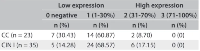
Table 2. Quantitative distribution of expression of IL-17 in
cervical precursor lesions
Low expression High expression 0 negative 1 (1-30%) 2 (31-70%) 3 (71-100%)
n (%) n (%) n (%) n (%)
CC (n = 23) 7 (30.43) 14 (60.87) 2 (8.70) 0 (0) CIN I (n = 35) 5 (14.28) 24 (68.57) 6 (17.15) 0 (0)
Expression of HLA-G and IL-17 in cervical precursor lesions To explore whether HLA-G and IL-17 might be involved in the CIN cases, we irst examined whether HLA-G and IL-17 were present in cervical specimens. In the whole group, HLA-G molecules were detected in 44 cases (75.86%). Among speci-mens that presented HLA-G expression, 29 out of 35 (82.86%) exhibited CIN I, and 15 out of 23 (65.22%) exhibited CC. Similarly, IL-17 molecules were detected in 46 cases (79.31%). Considering only the patients who presented IL-17 expres-sion, 30 out of 35 patients (85.71%) exhibited CIN I and 16 out of 23 (69.56%) exhibited CC. Absence of HLA-G and IL-17
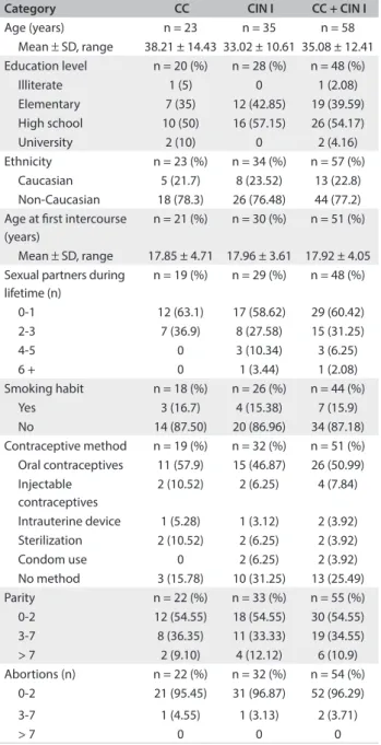
Table 3. Clinical and pathological variables observed in
patients with cervical lesions
Category CC CIN I CC + CIN I
Age (years) n = 23 n = 35 n = 58 Mean ± SD, range 38.21 ± 14.43 33.02 ± 10.61 35.08 ± 12.41 Education level n = 20 (%) n = 28 (%) n = 48 (%)
Illiterate 1 (5) 0 1 (2.08) Elementary 7 (35) 12 (42.85) 19 (39.59) High school 10 (50) 16 (57.15) 26 (54.17) University 2 (10) 0 2 (4.16) Ethnicity n = 23 (%) n = 34 (%) n = 57 (%)
Caucasian 5 (21.7) 8 (23.52) 13 (22.8) Non-Caucasian 18 (78.3) 26 (76.48) 44 (77.2) Age at irst intercourse
(years)
n = 21 (%) n = 30 (%) n = 51 (%)
Mean ± SD, range 17.85 ± 4.71 17.96 ± 3.61 17.92 ± 4.05 Sexual partners during
lifetime (n)
n = 19 (%) n = 29 (%) n = 48 (%)
0-1 12 (63.1) 17 (58.62) 29 (60.42) 2-3 7 (36.9) 8 (27.58) 15 (31.25) 4-5 0 3 (10.34) 3 (6.25) 6 + 0 1 (3.44) 1 (2.08) Smoking habit n = 18 (%) n = 26 (%) n = 44 (%)
Yes 3 (16.7) 4 (15.38) 7 (15.9) No 14 (87.50) 20 (86.96) 34 (87.18) Contraceptive method n = 19 (%) n = 32 (%) n = 51 (%) Oral contraceptives 11 (57.9) 15 (46.87) 26 (50.99) Injectable
contraceptives
2 (10.52) 2 (6.25) 4 (7.84)
Intrauterine device 1 (5.28) 1 (3.12) 2 (3.92) Sterilization 2 (10.52) 2 (6.25) 2 (3.92) Condom use 0 2 (6.25) 2 (3.92) No method 3 (15.78) 10 (31.25) 13 (25.49) Parity n = 22 (%) n = 33 (%) n = 55 (%) 0-2 12 (54.55) 18 (54.55) 30 (54.55) 3-7 8 (36.35) 11 (33.33) 19 (34.55) > 7 2 (9.10) 4 (12.12) 6 (10.9) Abortions (n) n = 22 (%) n = 32 (%) n = 54 (%)
0-2 21 (95.45) 31 (96.87) 52 (96.29) 3-7 1 (4.55) 1 (3.13) 2 (3.71)
> 7 0 0 0
CC = chronic cervicitis; CIN = cervical intraepithelial neoplasia; SD = standard deviation.
Table 4. Association between human leukocyte antigen G (HLA-G)
and interleukin-17 (IL-17) status and histopathological indings
Histopathological indings
HLA-G expression
n (%) Positive Negative
CC 23 15 (65.22) 8 (34.78)
CIN I 35 29 (82.86) 6 (17.14)
Histopathological indings
IL-17 expression
n (%) Positive Negative
CC 23 16 (69.56) 7 (30.44)
CIN I 35 30 (85.71) 5 (14.29)
CC = chronic cervicitis; CIN = cervical intraepithelial neoplasia.
expression was observed in the control group, as shown in
Figures 1 and 2, and in Table 4.
Ater adjustment for other covariables, the occurrences of HLA-G were signiicant (P-value = 0.04), with odds ratio (OR) estimated as 6.61 (95% conidence interval, CI: 1.22-49.55). his indicated that the odds of having CIN I (compared with CC) was 6.61 times greater (or 661% greater) for women who expressed HLA-G than for those who did not. Age was the only other sig-niicant predictor, ater adjustment for other covariables, with an OR of 0.87 (95% CI: 0.75- 0.96, P-value = 0.02), thus indicating a protective efect. In other words, the older the patient was, the smaller the odds of having CIN I also were. None of the other
covariables were signiicant (Table 5).
HLA-G expression was primarily detected in the epithe-lial cells, ibroblasts and lymphocytes, and a standard dial-type cytoplasmic membrane was maintained. HLA-G was strongly expressed in trophoblastic slices that were used as positive con-trols, while HLA-G expression was not found in any specimens obtained from healthy controls.
DISCUSSION
Cervical squamous intraepithelial lesions are a precancerous
stage of cervical cancer.12 he mechanism that promotes the
pro-gression of cervical lesions has not been clearly explained, but the
immune response appears to be an important factor.5 herefore,
identiication of new molecular markers to improve clinical diag-nosing of early-stage cervical lesions is still necessary and may enable more efective evaluation of patients with early-stage lesions, thereby resulting in slower progression of these lesions.
In the cervical context, Guimarães et al. reported that HLA-G
expression was low in cervical cancer specimens.13 On the other
hand, Zheng et al. reported that HLA-G is abundantly expressed
in premalignant and malignant cervical intraepithelial lesions.5
as sex, ethnicity, number of partners during lifetime, parity and oral contraceptive use. his suggests that the HLA-G molecule could be associated with the progression of cervical lesions.
In addition, polymorphic sites of HLA-G genes in cervi-cal lesions and cancer have been studied. In high-grade and
invasive cervicovaginal cancer patients, the 14 base pair (bp) In/In polymorphism seems to be associated with greater
development of invasive cervical cancer.14 On the other
hand, spontaneous demethylation events in the HLA-G pro-moter do not play a primary role in promoting escape from immunosurveillance, in relation to development of
precan-cerous cervical lesions.15
To our knowledge, the present study is the irst to explore the HLA-G and IL-17 expression proile in the cervical micro-environment in CIN I patients, in whom more than half of the biopsies (75.86%) exhibited HLA-G expression. he presence of HLA-G, which was signiicantly associated with CIN and age, was not inluenced by other variables. hus, the data shown here certainly contribute towards shedding some light on the physiopathology of CIN, in which HLA-G expression in cer-vical cells may act in conjunction with other factors, such as immunosuppression induced by HPV infection, thereby result-ing in the more severe cervical disease observed in CIN III and cervical cancer patients.
Since the irst description of HLA-G expression, its asso-ciation with malignant lesions has been intensively studied. he data available so far have shown that the vast majority of tumors may express varying degrees of HLA-G isoforms, thus relecting a potential immune escape mechanism. It is also worth mentioning that HLA-G expression is highly dependent on tumor microenvironment factors, particular when IL-10 and
a hypoxic factor are present.16
he role of IL-17 in the tumor microenvironment is still controversial. Information about the behavior of cytokines in CIN cases is scarce. Previous studies have shown that patients with uterine cervical cancer (UCC) have a higher proportion of h17 cells. Notably, in UCC patients, increased h17 prevalence has been correlated with clinical stage, lymph node metastases
and vasoinvasion.7
Our study found that IL-17 expression was greater in CIN I cases. IL-17 molecules were detected in 46 cases (79.31%) and, when considering only the patients that presented IL-17 expres-sion, 30 out of 35 (85.71%). Recently, in patients with ovarian cancer, Lan et al. showed that the IL-17 levels were signiicantly greater in ovarian cancer cases than in normal ovarian tissues
(P < 0.001).17 Moreover, ovarian tumor antigen-speciic CD4(+)
T cells secrete high levels of IL-17.18 However, the exact role of
IL-17 in tumor immunopathogenesis remains undeined. It has been reported that expression of interleukin-17 in tumor cells suppresses tumor progression through enhanced antitumor immunity or promotes tumor progression through an increase in
inlammatory angiogenesis.19
Many studies have discussed the role of HLA-G and IL-17
in various types of cancer.16,20 However, this is the irst study
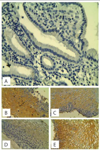
Figure 1. Human leukocyte antigen G (HLA-G) expression in cervical
epithelium was analyzed by means of immunohistochemistry. Labeling was accomplished using anti-HLA-G 5A6G7 mAb (EXBIO, Czech Republic).
A) Absence of expression of human leukocyte antigen G (HLA-G) in normal cervix. B) Slight human leukocyte antigen G (HLA-G) staining in chronic cervicitis (CC). C) Moderate human leukocyte antigen G (HLA-G) staining in chronic cervicitis (CC). D) Strong human leukocyte antigen G (HLA-G) staining in chronic cervicitis (CC). E) Slight human leukocyte antigen G (HLA-G) staining in cervical intraepithelial
neoplasia (CIN) I.
F) Moderate human leukocyte antigen G (HLA-G) staining in cervical intraepithelial neoplasia (CIN) I.
G) Strong human leukocyte antigen G (HLA-G) staining in cervical intraepithelial neoplasia (CIN) I.
A
B
D
F
C
E
to correlate the expression of two molecules that are impor-tant in the early stages of the cervical lesion. Consistently, our study demonstrated that coexpression of HLA-G and IL-17 was implicated in the pathogenesis and progression of cervical lesions (CC and CIN I), correlating these indings with clini-cal and pathological features.
CONCLUSION
Taken together, our data suggest that in CIN I patients, the increased HLA-G levels could be correlated with progression of cervical lesions, and that presence of IL-17 may be a useful indi-cator for representing the severity of tissue injury. hus, the data suggest that h17 cells are mediators during the immunological process of CIN development, thereby indicating that HLA-G and IL-17 potentially have a role in the development and progression of cervical lesions.
REFERENCES
1. Burd EM. Human papillomavirus and cervical cancer. Clin Microbiol
Rev. 2003;16(1):1-17.
2. Carosella ED, Favier B, Rouas-Freiss N, Moreau P, Lemaoult J. Beyond
the increasing complexity of the immunomodulatory HLA-G
molecule. Blood. 2008;111(10):4862-70.
3. Rouas-Freiss N, Gonçalves RM, Menier C, Dausset J, Carosella ED.
Direct evidence to support the role of HLA-G in protecting the fetus
from maternal uterine natural killer cytolysis. Proc Natl Acad Sci U S A.
1997;94(21):11520-5.
4. Carosella ED, Moreau P, Lemaoult J, Rouas-Freiss N. HLA-G: from
biology to clinical beneits. Trends Immunol. 2008;29(3):125-32.
5. Zheng N, Wang CX, Zhang X, et al. Up-regulation of HLA-G expression
in cervical premalignant and malignant lesions. Tissue Antigens.
2011;77(3):218-24.
6. Bettelli E, Carrier Y, Gao W, et al. Reciprocal developmental pathways
for the generation of pathogenic efector TH17 and regulatory T cells.
Nature. 2006;441(7090):235-8.
7. Zhang Y, Ma D, Zhang Y, et al. The imbalance of Th17/Treg in patients with
uterine cervical cancer. Clin Chim Acta. 2011; 412(11-12):894-900.
8. Richart RM. The incidence of cervical and vaginal dysplasia after
exposure to DES. JAMA. 1986;255(1):36-7.
9. Wright TC, Ronnett BM, Kurman RJ, Ferenczy A. Precancerous lesions
of the cervix. In: Kurman RJ, Ellenson LH, Ronnett BM, editors.
Blaustein’s pathology of the female genital tract. New York: Springer;
2011. p. 193-252.
10. Wright TC, Ronnett BM, Ferenczy A. Benign diseases of the cervix. In:
Kurman RJ, Ellenson LH, Ronnett BM, editors. Blaustein’s pathology of
the female genital tract. New York: Springer; 2011. p. 155-91.
11. Xie X, Clausen OP, Boysen M. Bag-1 expression as a prognostic
factor in tongue squamous cell carcinomas. Laryngoscope.
2004;114(10):1785-90.
Table 5. Logistic regression analyses on cervical intraepithelial
neoplasia (CIN) I status. Model adjusted according to
interleukin-17 (IL-17), human leukocyte antigen G (HLA-G), age, age at irst intercourse, number of partners during lifetime, parity and oral contraceptive use
Independent variable OR 95% CI P-value
IL-17 4.46 0.73-33.07 0.11 HLA-G 6.61 1.22-49.55 0.04
Age 0.87 0.75-096 0.02
Age at irst intercourse 1.10 0.91-1.35 0.35 Number of partners during lifetime 1.25 0.66-3.46 0.58 Parity 1.54 0.98-2.72 0.09 Oral contraceptive use 0.80 0.18-3.53 0.77
OR = odds ratio; CI = conidence interval.
Figure 2. Interleukin-17 (IL-17) expression in the cervical
epithelium was analyzed by means of immunohistochemistry. Labeling was accomplished using anti-IL-17 (Ebioscience, San Diego, California, USA).
A) Absence of expression of interleukin-17 (IL-17) in normal cervix. B) Slight interleukin-17 (IL-17) staining in chronic cervicitis (CC). C) Moderate interleukin-17 (IL-17) staining in chronic cervicitis (CC). D) Slight interleukin-17 (IL-17) staining in cervical intraepithelial neoplasia (CIN) I.
E) Moderate interleukin-17 (IL-17) staining in cervical intraepithelial neoplasia (CIN) I.
A
B
D
C
12. Lukovic L, Milasin J. Sister chromatid exchanges in patients
with carcinoma in situ of cervix uteri. Cancer Genet Cytogenet.
1992;59(1):84-5.
13. Guimarães MC, Soares CP, Donadi EA, et al. Low expression of
human histocompatibility soluble leukocyte antigen-G (HLA-G5) in
invasive cervical cancer with and without metastasis, associated with
papilloma virus (HPV). J Histochem Cytochem. 2010;58(5):405-11.
14. Ferguson R, Ramanakumar AV, Koushik A, et al. Human leukocyte
antigen G polymorphism is associated with an increased risk of
invasive cancer of the uterine cervix. Int J Cancer. 2012;131(3):E312-9.
15. Gillio-Tos A, Bicalho Mda G, Fiano V, et al. Case-control study of HLA-G
promoter methylation status, HPV infection and cervical neoplasia in
Curitiba, Brazil: a pilot analysis. BMC Cancer. 2012;12:618.
16. Singer G, Rebmann V, Chen YC, et al. HLA-G is a potential tumor
marker in malignant ascites. Clin Cancer Res. 2003;9(12):4460-4.
17. Lan C, Huang X, Lin S, et al. High density of IL-17-producing cells is
associated with improved prognosis for advanced epithelial ovarian
cancer. Cell Tissue Res. 2013;352(2):351-9.
18. Cannon MJ, Goyne HE, Stone PJ, et al. Modulation of p38 MAPK
signaling enhances dendritic cell activation of human CD4+ Th17
responses to ovarian tumor antigen. Cancer Immunol Immunother.
2013;62(5):839-49.
19. Kryczek I, Banerjee M, Cheng P, et al. Phenotype, distribution,
generation, and functional and clinical relevance of Th17 cells in the
human tumor environments. Blood. 2009;114(6):1141-9.
20. Wägsäter D, Löfgren S, Hugander A, Dimberg J. Expression of
interleukin-17 in human colorectal cancer. Anticancer Res.
2006;26(6B):4213-6.
Acknowledgements: We are grateful to Elizabeth Maia de Oliveira, who provided technical help, and Francisco Pignataro Lima (departmental
chair of pathology), who provided general support. We want to thank
the National Council for Scientiic and Technological Development
(Conselho Nacional de Desenvolvimento Cientíico e Tecnológico, CNPq)
for the grant for this research.
Sources of funding: None
Conlict of interest: None
Date of irst submission: May 14, 2013
Last received: December 20, 2013
Accepted: February 12, 2014
Address for correspondence: Janaína Cristiana de Oliveira Crispim
Departamento de Análises Clínicas e Toxicológicas
Faculdade de Ciências Farmacêuticas, Universidade Federal do Rio
Grande do Norte
Rua General Cordeiro de Farias, s/no
Petrópolis — Natal (RN) — Brasil
CEP 59012-570
Tel. (+55 84) 3342-9829