From the Departments of Radiology1 and Thoracic Surgery2 of the Cancer Hospital – “A. C. Camargo”; Paulista University (UNIP) and LIM 623.
Received for publication on June 10, 2002.
COMPUTED TOMOGRAPHY-GUIDED BIOPSY OF
MEDIASTINAL LESIONS: FINE VERSUS CUTTING
NEEDLES
André Piovesan de Farias1, Daniel Deheinzelin2, Riad N.Younes2,3 and Rubens Chojniak1
FARIAS AP de et al. – Computed tomography-guided biopsy of mediastinal lesions: fine versus cutting needles. Rev. Hosp.
Clín. Fac. Med. S. Paulo 58(2):69-74, 2003.
PURPOSE: To report the experience of a radiology department in the use of computed tomography guided biopsies of
mediastinal lesions with fine and cutting needles, describing the differences between them. The results of adequacy of the sample and histologic diagnoses are presented according to the type of needle used.
METHODS: We present a retrospective study of mediastinal biopsies guided by computed tomography performed
from January 1993 to December 1999. Eighty-six patients underwent mediastinal biopsy in this period, 37 with cutting needles, 38 with fine needles, and 11 with both types (total of 97 biopsies).
RESULTS: In most cases, it was possible to obtain an adequate sample (82.5%) and specific diagnosis (67.0%).
Cutting-needle biopsy produced a higher percentage of adequate samples (89.6% versus 75.5%, P = 0.068) and of specific diagnosis (81.3% versus 53.1%, P = 0.003) than fine-needle biopsy. There were no complications that required intervention in either group.
CONCLUSION: Because they are practical, safe, and can provide accurate diagnoses, image-guided biopsies should
be considered the procedure of choice in the initial exploration of patients with mediastinal masses. In our experience, cutting needles gave higher quality samples and diagnostic rates. We recommend the use of cutting needles as the preferred procedure.
DESCRIPTORS: Mediastinal masses. Tomography, computed axial. Needle biopsy.
INTRODUCTION
Image-guided percutaneous needle biopsy is a reliable technique for the diagnosis of thoracic diseases, particu-larly for the assessment of mediastinal lesions. Computed tomography (CT) is established as the best imaging method for needle biopsy guidance1;
CT provides detailed images for even small lesions located in any part of the mediastinum, which allows for accu-rate planning, avoiding inadvertent puncture of vascular structures or the lung.
In most cases, the lesions are suc-cessfully reached, avoiding invasive procedures like bronchoscopy, medi-astinoscopy, or even thoracotomy. The major concern has been the inherent risk of pneumothorax, reported to oc-cur in as many as 10% to 23%2,3 of
procedures, although this complica-tion usually requires treatment in only
1% of cases.
There are variations in needle bi-opsy technique among institutions, the most important difference being the type of needle used. While fine-needle biopsy (FNA) uses cytological tech-niques for analysis, cutting-needle bi-opsy provides larger tissue samples that are suitable for histologic study.
METHODS
We reviewed all biopsies of medi-astinal masses performed between January 1993 and December 1999. Eighty-six patients underwent medias-tinal biopsy during the study period, 38 with fine needles, 37 with cutting needles, and 11 with both types (total of 97 biopsies). Some authors recom-mend cutting needles according to the suspected diagnosis1. In our practice,
the size and the relationship of the le-sion to adjacent structures determine the type of needle chosen. We use cut-ting needles when the lesion is larger than 20 mm and there is a low possi-bility of puncture of the lung or vas-cular structures.
The study included 43 women and 43 men, ranging in age from 1 to 95 years (median = 34 years). We did not repeat biopsies in the same patient. Masses from all parts of mediastinum were included. The size of the lesions ranged from 12 to 140 mm (median = 35 mm), and the distance from the skin ranged from 18 to 140 mm (median = 50 mm).
The biopsies were performed with the CT scanners GE Pace Plus and GE Pro Speed. Lesions were localized with contiguous scans 5 or 10 mm thick. The best path was displayed on the computer monitor, and electronic cur-sors were used to measure the distance from the skin and from a body marker, information that allowed calculation of the degree of inclination needed. The location for needle entry was marked on the patient’s skin by corre-lating the light beam from the CT scner. After preparation of skin with an-tiseptic solution, local anesthesia was performed on this site using 1% lido-caine. Children under 5 years of age underwent general anesthesia. The bi-opsy needle was then introduced, and new scans were obtained to modify or confirm the needle position.
The fine-needle biopsies were
per-formed with 22-gauge Chiba needles. The aspiration sample was smeared onto glass slides and immersed in 90% alcohol. A new puncture was per-formed when the sample obtained was judged insufficient. One to five aspi-rations were performed in each indi-vidual case and were then sent to the pathology department.
The cutting-needle biopsies were performed with an 18-gauge Tru-Cutâ
(MD Tech) needle. When the tissue was obtained, it was placed in formalin and sent to the pathology department for final histological diagnosis.
For pathologic analysis, the sample was stained in hematoxylin-eosin and classified as adequate or inadequate by the pathologist. The results of cytol-ogy and histolcytol-ogy of the adequate sample were designated by the pa-thologist as the specific diagnosis, or when this was not possible, as positive or negative for malignancy.
The rates of adequate sampling and specific diagnosis for the 2 differ-ent needle types were compared using the chi-square test, and the results were considered significant when P <0.05.
RESULTS
Adequate sampling for diagnosis was obtained in 80 biopsies (82.5%). The final diagnoses were 65 specific
diagnoses, and 7 classified as positive and 8 negative for malignancy. The specific diagnoses of these procedures are listed in table 1.
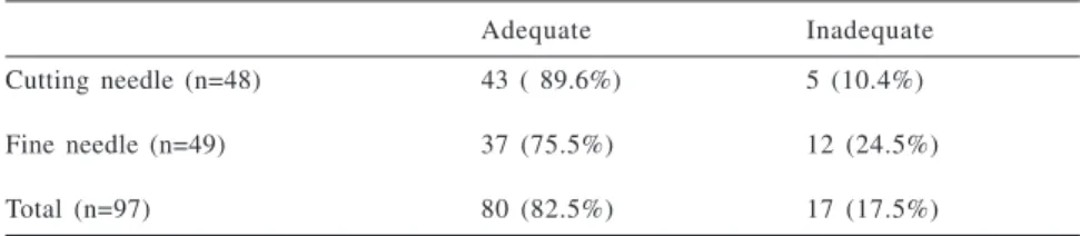
In the group undergoing cutting-needle biopsy (n = 48), there were 43 (89.6%) with an adequate sample and 5 with inadequate samples (10.4%). The sample tissue provided for 39 (81.3%) specific diagnoses, as well as for 2 classified as positive and 2 nega-tive for malignancy. In the group with specific diagnoses, lymphoma was the most frequent diagnosis (18 cases), fol-lowed by lung cancer, teratoma, semi-noma, normal thymus, and mesenchy-mal neoplasms. In the 2 cases classi-fied as positive for malignancy, the di-agnoses were later confirmed as lym-phoma and metastatic adenocarci-noma. For the 2 biopsies classified as negative for malignancy, further inves-tigation established the diagnosis of ganglioneuroma and lymphoma. In 5 cases of inadequate sampling, the pa-tients underwent thoracotomy, which revealed 3 cases of lymphoma, 1 bron-chogenic cyst, and 1 mesothelioma.
The fine-needle biopsies (n = 49) produced 37 (75.5%) adequate sample punctures. There were 26 (53.1%) spe-cific diagnoses. Lymphoma was the most common histologic diagnosis, followed by lung cancer and others (thymoma, liposarcoma, seminoma, and metastasis). All 5 cases classified
Table 1 - Distribution of specific diagnoses in 80 fine- or cutting-needle biopsies of mediastinal tumors.
Lymphoma 2 2 Thymic hyperplasia 2
Lung carcinoma 1 0 Neuroendocrine tumor 1
Metastasis (lung and breast cancer) 7 Fibrosis 2
Thymoma 3 Liposarcoma 1
Seminoma 2 Mesenchymal neoplasm 1
Teratoma 2 Hürthle cell tumor 1
as positive for malignancy were con-firmed as lymphoma or lung cancer. There were 6 cases classified as nega-tive for malignancy, with 3 of them later confirmed as benign tumors, while the other 3 were actually malig-nant disease (lymphoma). In the 12 cases of inadequate sampling, subse-quent investigation led to diagnosis of 5 cases of lymphoma, 3 of lung can-cer, and 1 of bronchogenic cyst. In 3 other cases of this category, clinical diagnosis of benign masses was sus-pected, and they are still being fol-lowed up with no evidence of malig-nancy.
The results of all punctures, in-cluding those for which specific diag-nosis was not possible and those of in-adequate sampling, are described ac-cording to the needle type and shown in tables 2 and 3. Cutting-needle bi-opsy was more successful. There was a statistically significant difference in the rate of specific diagnosis (P = 0.003), and there were more adequate sample punctures (P = 0.068), al-though these did not reach statistical significance.
There were no complications that required any kind of intervention in
the patients of both groups. However, minor complications such as small pneumothorax and hematoma could be underestimated in our series, be-cause this is a retrospective study, and this information could be incom-pletely recorded in the radiology re-ports or subsequent clinical evalua-tions.
DISCUSSION
The potential for malignancy and the possibility of severe complica-tions, such as respiratory or circulatory compression, mandate the fast and sometimes urgent diagnosis and treat-ment for mediastinal masses. Explora-tory thoracotomy, mediastinoscopy, and anterior mediastinotomy are still widely used, despite potential morbid-ity and even mortalmorbid-ity associated with these procedures. With dedicated and experienced pathologists, percutane-ous biopsies can provide adequate di-agnosis in most cases, independent of the type of needle used, whether fine or cutting. Either needle type is an ad-equate alternative that is well tolerated by the patients.
These procedures are cost effective because they shorten the period from admission to diagnosis, decrease the number of surgical operations, and shorten the time of hospital stay, which reduces overall treatment costs4.
Unfortunately, some contra-indications exist that preclude the use of percutaneous biopsies in every case. The absolute contraindications in-clude uncontrollable cough and suspi-cion of hydatid cyst, whereas relative contraindications include bleeding diathesis, vascular lesions, pulmonary hypertension, uncooperative patient, and advanced emphysema4.
Fluoroscopy, CT, and more re-cently ultrasound can guide the punc-tures5,6. The last method has the
advan-tage of real-time imaging, but is lim-ited in the evaluation of parts of me-diastinum hidden by bone or air. Com-puted tomography can show more de-tails than fluoroscopy, and provides complete evaluation of the thoracic cavity.
Our success rate for adequate sam-pling (82.5%) and specific diagnosis (67.0%) was similar to the rates re-ported in the literature, ranging from 72% to 100%4,7,11. Our high rate of
ma-lignancies is probably due to the pa-tient population referred to our cancer hospital.
As expected, we found more cases of adequate sampling in the cutting-needle group (89.6% versus 75.5%). The tissue samples obtained with these needles also resulted in more specific diagnosis than in the fine-needle group (81.3% versus 53.1%).
Fine-needle biopsy has long been recognized as useful because it is eas-ily performed and provides adequate samples from deeply seated lesions. Fine-needle biopsy has a high sensi-tivity and specificity for metastatic dis-ease11,12. Morrisey et al.10 reported a
slightly higher sensitivity of cutting-needle biopsy (96%) for diagnosis of metastatic carcinoma comparing to the
Table 3 - Specific diagnosis: Comparison between cutting- and fine-needle biopsies.
Specific diagnosis Nonspecific diagnosis
Cutting needle (n = 48) 39 (81.3%) 9 (18.8%)
Fine needle (n = 49) 26 (53.1%) 23 (46.9%)
Total (n = 97) 65 (67.0%) 32 (33.0%)
P = 0.003
Table 2 - Adequacy of results with cutting- vs fine-needle biopsy.
Adequate Inadequate
Cutting needle (n=48) 43 ( 89.6%) 5 (10.4%)
Fine needle (n=49) 37 (75.5%) 12 (24.5%)
Total (n=97) 80 (82.5%) 17 (17.5%)
90% sensitivity of FNA. Fine-needle biopsy is also useful for cystic masses13 and germ cell tumors8.
How-ever, cytopathology with FNA samples requires a considerable amount of ex-perience from the pathologist in order to obtain an accurate diagnosis based on few aspirated cells.
On the other hand, the quality of the tissue sample provided by cutting-needle biopsy is superior compared to that by FNA. Because more tissue is available, histologic diagnosis can be made and further pathologic investiga-tions can be performed if necessary9.
This technique is also useful for other histologic types, such as lymphoma, thymoma, and benign tumors. With the sample obtained by FNA, a patholo-gist may suspect these diagnoses but may not be able to differentiate among them. Recent reports describe the higher accuracy of cutting-needle bi-opsy for the diagnosis of lym-phoma14,15, with the possibility of also
concurs with reports from the litera-ture10. The absence of complications in
our series also can be associated with the low number of punctures through the lung parenchyma or pleura, which can be avoided in most cases by us-ing CT guidance.
In conclusion, because they are minimally invasive, safe, cost effective, and have a high rate of success in pro-viding adequate tissue sample, percu-taneous needle biopsies guided by CT should be considered as the procedure of choice in the investigation of me-diastinal masses. Both types of needle produced satisfactory results, but the higher quality sample of cutting nee-dles provided more histologic diag-noses. The use of the cutting-needle bi-opsy can lead to more specific diag-nosis without an increased incidence of complications; therefore, we recom-mend cutting-needle biopsy as the pre-ferred method whenever possible. determining the histologic subtype in
most cases16, which is fundamental
in-formation required to determine the subsequent treatment modality.
A disadvantage of cutting-needle biopsies is local pain due to the larger gauge. Nowadays, the number of punc-tures is reduced, and the rapid sam-pling with the biopsy gun decreases the likelihood of pain.
Our results confirm the advantages of cutting-needle biopsy for histologic diagnosis, but in some cases, we rec-ommend the use fine needles instead. Fine needles are preferable when there is pulmonary parenchyma between the tumor and the chest wall, when the le-sions are located adjacent to great ves-sels, and when highly vascular lesions are suspected. In these cases, and if metastatic carcinoma is suspected, FNA should be the initial method1,11,17.
The safety of these procedures was confirmed by the low incidence of complications in both groups, which
RESUMO
FARIAS AP de e col. - Biópsia de mas-sas mediastinais guiadas por Tomo-grafia Computadorizada: agulhas finas versus cortantes. Rev. Hosp. Clín. Fac. Med. S. Paulo 58(2): 69-74, 2003.
OBJETIVO: Apresentar a
experi-ência de um serviço de radiologia na prática de punções biópsias de massas
mediastinais guiadas por tomografia computadorizada com agulhas finas ou cortantes, descrevendo as diferen-ças entre elas. Os resultados referentes a material suficiente e diagnóstico histológico são apresentados de acor-do com o tipo de agulha utilizaacor-do.
MÉTODOS: Apresentamos um
estu-do retrospectivo de biópsias medias-tinais guiadas por tomografia
computa-dorizada realizadas em nosso hospital no período de janeiro de 1993 a dezem-bro de 1999. Oitenta e seis pacientes fo-ram submetidos a biópsia mediastinal neste período, sendo 37 realizadas com agulhas cortantes, 38 com agulhas finas e 11 com ambas, (total de 97 biópsias).
RESULTADOS: Na maioria dos
específi-co (67 %). As agulhas específi-cortantes apresen-taram maior porcentagem de material suficiente (89.6% versus 75,5%, P=0,068) e de diagnóstico específico (81,3% versus 53,1%, p=0,003 ) do que as agulhas finas. Não houveram com-plicações que requisessem intervenção em nenhum dos grupos.
CONCLUSÃO: Pela praticidade,
segurança e grande probabilidade de diagnóstico acurado sem procedimen-tos mais invasivos, as biópsias guiadas por imagem devem ser consideradas como a primeira etapa na investigação de massas mediastinais. Pela nossa ex-periência as agulhas cortantes
forne-cem material de maior qualidade e maior taxa de diagnóstico. Nós reco-mendamos o uso das agulhas cortan-tes como procedimento preferencial.
DESCRITORES: Massa medias-tinal. Biópsia por agulha. Tomogra-fia computadorizada.
REFERENCES
1 . PROTOPAPAS Z, WESTCOTT JL - Transthoracic hilar and mediastinal biopsy. Radiol Clin North Am 2000; 38 (2):281-291.
2 . MOINUDDIN SM, LEE LH, MONTGOMERY JH - Mediastinal needle biopsy. AJR 1984; 143:531-532.
3 . BELFIORE G, CAMERA L, MOGGIO G, SALVATORE M et al. - Middle mediastinum lesions: preliminary experience with CT-guided fine-needle aspiration biopsy with a suprasternal approach. Radiology 1997; 202:870-873.
4 . ZAFAR N, MOINUDDIM S - Mediastinal needle biopsy. Cancer 1995; 76:1065-1068.
5 . HEILO A - Tumors in the mediastinum: US-guided histologic core needle biopsy. Radiology 1993; 189:143-146. 6 . WERNECKE K, VASSALO P, VON BASSEWITZ DB et al.
-Mediastinal tumors: biopsy under US guidance. Radiology 1989; 172: 473-476.
7 . BRESSLER EL, KIRKHAM JA - Mediastinal masses: alternative approach to CT-guided needle biopsy. Radiology 1994; 191:391-396.
8 . HERMAN SJ, HOLUB RV, CHAMBERLEIN DW et al. - Anterior mediastinal masses: utility of transthoracic needle biopsy. Radiology 1991; 180:167-170.
9 . PARKER SH, HOPPER KD, CARTER TE et al. - Image-directed percutaneous biopsies with a biopsy gun. Radiology 1989; 171: 663-669.
10. MORRISEY B, ADAMS H, CRANE MD et al. - Percutaneous needle biopsy of the mediastinum: review of 94 procedures. Thorax 1993; 48:632-637.
12. PROTOPAPAS, WESTCOTT JL - Transthoracic needle biopsy of mediastinal lymph nodes for staging lung cancers. Radiology 1996; 199: 489-496.
13. KUHLMAN JE, FISHMAN EK, WANG KP et al. - Mediastinal cysts: Diagnostic by CT and needle aspiration. AJR 1988; 150:75-78.
14. BEM-YEHUDA D, POLLIACK A, OKON E et al. - Image guided core needle biopsy in malignant lymphoma: Experience with 100 patients that suggests the technique is reliable. J Clin Oncol 1996; 14:2431-2434.
15. PAPA VI, HUSSAIN HK, REZNEK RH et al. - Role of image guided core needle biopsy in the management of patients with lymphoma. J Clin Oncol 1996; 14:2427-2430.
16. MOULTON JS, MOORE PT - Coaxial percutaneous biopsy technique with automated biopsy devices: Value in improving accuracy and negative predictive value. Radiology 1993; 186:515-522.