*Correspondence: J. Olivero-Verbel. Environmental and Computa-tional Chemistry Group. School of Pharmaceutical Sciences. University of Cartagena. Zaragocilla Campus. Cartagena - 130015 – Colombia. E-mail: [email protected]
A
vol. 52, n. 2, apr./jun., 2016 http://dx.doi.org/10.1590/S1984-82502016000200007
Direct effect of
p,p'-
DDT on mice liver
Bárbara Arroyo-Salgado, Jesús Olivero-Verbel
*, Angélica Guerrero-Castilla
Environmental and Computational Chemistry Group, School of Pharmaceutical Sciences, Zaragocilla Campus, University of Cartagena, 130015, Cartagena, Colombia
Contact with the pesticide dichlorodiphenyltrichloroethane (p,p′-DDT) can be the cause of various harmful efects in humans, wildlife, and the environment. This pesticide is known to be persistent, lipophilic, resistant to degradation, and bioaccumulive in the environment and to be slowly released into bloodstream. Growing evidence shows that exposure to DDT is linked to type 2 diabetes mellitus. Individuals exposed to elevated levels of DDT and its metabolite have an increased prevalence of diabetes and insulin resistance. To evaluate these possible relationships, experiments were performed on eight-week-old female mice, divided into three groups (n = 10 per group): Group 1 received a vehicle-control intraperitoneal (i.p.) injection of sesame oil; Groups 2 and 3 received an i.p. dose of 50 and 100 µg/g p,p′-DDT respectively, dissolved in sesame oil. All groups were treated once daily for four days. Real-time PCR analysis of several genes was undertaken. Additionally, biochemical parameters and histopathological changes were measured. NQO1, HMOX1, NR1I3 and NR3C1were up-regulated in DDT-exposed animals compared to the vehicle control group, while only SREBP1 was down-regulated in the 100 µg/g group. MTTP and FABP5, not previously reported for DDT exposure, but involved in regulation of fatty acid luxes, could also function as biomarkers cross-talking between these signaling pathways. These results suggest that beyond epidemiological data, there is increasing molecular evidence that DDT may mimic diferent processes involved in diabetes and insulin resistance pathways.
Uniterms: Pesticides.Persistent organic pollutant. Dichlorodiphenyltrichloroethane/exposure.Type 2 diabetes mellitus/exposure to dichlorodiphenyltrichloroethane/experimental study. Insulin/resistance/ exposure to dichlorodiphenyltrichloroethane. Gene expression.
O contato com o praguicida diclorodifeniltricloroetano (p, p’-DDT) pode ser a causa de vários efeitos nocivos sobre os seres humanos, animais silvestres e o meio ambiente. Sabe-se de sua característica de bioacumulação, ser altamente persistente no meio ambiente, lipofílico, resistente à degradação e lentamente liberado na corrente sanguínea. Existe uma evidência crescente de que a exposição ao DDT pode ser ligada a Diabetes mellitus tipo 2. Os indivíduos expostos a níveis elevados de DDT e seu metabólito apresentam maior prevalência de diabetes e resistência à insulina. A im de obter informações sobre essas possíveis relações, camundongos fêmeas de oito semanas de idade foram divididos em três grupos (n = 10 por grupo): Grupo 1 recebeu um veículo de óleo de gergelim via i.p.; os Grupos 2 e 3 receberam, via i.p., 50 e 100 µg/g de p, p’-DDT, respectivamente, dissolvidos em óleo de gergelim. Todos os grupos foram tratados uma vez ao dia durante quatro dias. Além da análise de PCR em Tempo Real de vários genes, os parâmetros bioquímicos e alterações histopatológicas também foram medidos. A expressão gênica do mRNA dos genes NQO1, HMOX1, NR1I3 e NR3C1 foi maior nos animais expostos ao DDT, em comparação ao grupo controle, enquanto a expressão gênica do SREBP1 diminuiu na concentração de 100 µg/g. Os genes MTTP e FABP5 envolvidos na regulação do luxo de ácidos graxos, embora não estudados quanto à exposição ao DDT, também podem funcionar como biomarcadores de resposta cruzada entre essas vias de sinalização. Esses resultados sugerem que, além de dados epidemiológicos, há cada vez mais evidências moleculares de que o DDT poderia, de fato, imitar diferentes processos que envolvem as rotas de diabetes e de resistência à insulina.
INTRODUCTION
Organochlorine pesticides (OC) were widely used
in agriculture and pest control, but their use was banned during the 1970s and 1980s because of their high toxicity and environmental persistence. However, Asia, Latin America, and Africa are still producing and using OC because of their high efectiveness as pesticides and their low cost (Lucena et al., 2007).
Actually, abundant OC exists in the environment, with DDT (1,1,1-trichloro-2,2-bis[4-chlorophenyl]ethane), methoxychlor[1,1,1-trichloro-2,2-bis(p-methoxyphenyl)
ethane, MXC], tris(4-chlorophenyl)methanol (TCPM),
hexachlorocyclohexanes (HCHs) and related compounds taking priority. Dichlorodiphenyltrichloroethane (DDT)
is a synthetic chemical that includes p,p′-DDT, p,p
′-dichlorodiphenyldichloroethylene (p,p′-DDE), and p,p
′-dichlorodiphenyldichloroethane (p,p′-DDD or p,p′-TDE).
Technical grade DDT is a mixture of three isomers of DDT,
the main components of which are p,p′-DDT (85%),
o,p′-DDT (15%), and o,o′-DDT (trace amounts).
DDT has been extensively used worldwide to
control malaria, typhus, body lice, and bubonic plague (ATSDR, 2002), but was banned in the United States by the Environmental Protection Agency in 1972, as a consequence of its potentially harmful efects on humans, wildlife, and the environment (Porta et al., 1999).
However, it continues to be found frequently at high
concentrations in human serum.
The half-life of DDT in the environment usually
ranges from 2 to 15 years. It is known to bioaccumulate
and to be highly persistent in the environment as well
as resistant to degradation. High levels of DDT are still
present in the environment and in the food chain. Even p,p-DDE, its main metabolite, is frequently found all over
the world in food, adipose tissues, and breast milk, among
others (Arrebola et al., 2012).
Some studies suggest that DDT-induced toxicity is associated with estrogen receptor (ER). DDT metabolites p,p′-DDT, o,p′-DDT and DDE promote estrogenic and
anti-androgenic properties (Sohoni, Sumpter, 1998), the
production of reactive oxygen species (ROS) (Jin et al.,
2014), toxicity and carcinogenicity (Everett, Frithsen, Player, 2011; La Merrill et al., 2014). Moreover, perinatal DDT exposure contributes to the development of insulin resistance and metabolic syndrome in adult female mice
(La Merrill et al., 2014).
Data from several studies suggest a possible
association between DDT and type 2 diabetes mellitus
(T2DM) (Everett, Frithsen, Player, 2011). DDT has been found to be associated with increased odds of T2DM (Cox et
al., 2007). However, there is no conclusive epidemiological
evidence for such relationship (Bloomgarden, 2000).
Some existing epidemiological studies have demonstrated associations between DDT and DDE exposures and diabetes
in humans (Rylander, Rignell-Hydbom, Hagmar, 2005; Cox
et al., 2007; Patel, Bhattacharya, Butte, 2009; Lee et al.,
2010a; Lee et al., 2010b).
Prevalence of obesity and T2DM has increased
at alarming rates in countries where DDT is still in
use (Danaei et al., 2011). T2DM is classified as a
complex disease, where interactions between genetic
and environmental factors may underlie the etiology of
diabetes. T2DM has become a global epidemic in recent decades. Some 382 million individuals worldwide are estimated to have diabetes, with this number projected to increase to 592 million by 2035. The vast majority of those afected will develop T2DM (Sargis, 2014).
Adult rodents exposed to DDT had impaired insulin
secretion, glucose intolerance, and elevated gluconeogenesis (Kacew, Singhal, 1974; Yau, Mennear, 1976). Rats and monkeys exposed to acute DDT also presented increased
hepatic cholesterol circulation and triglyceride synthesis
(Sanyal et al., 1982). There is evidence that DDT may alter
adipogenesis and adipokine-cytokine production in vitro
(Moreno-Aliaga, Matsumura, 2002) and that persistent organic pollutants (POPs) may induce abdominal obesity, impair insulin sensitivity, reduce glucose uptake, and cause
dyslipidemia and hepatosteatosis (Ruzzin et al., 2010;
Ibrahim et al., 2011). This pesticide could also contribute to
other risk factors of diabetes, such as obesity, by distressing
neural circuits that regulate feeding behavior or by altering
differentiation of adipocytes (Thayer et al., 2012). It is
possible that DDT and a high-fat diet may induce hepatic de novo lipogenesis (Rolo, Palmeira, Wallace, 2002), although
the exact role of DDT/DDE on these processes requires additional studies (Lee et al., 2010a)
The liver is the tissue most sensitive to xenobiotic exposure and constitutes the main target of DDT toxicity.
Currently, there is little information on the intracellular mechanisms related to the efects of DDT on health and its relationship to diabetes. However, DDT and related OC exert estrogenic efects through a variety of molecular mechanisms involving ER-alpha (ERα), cellular-signaling systems such as activator protein (AP-1), p38, and extracellular signal-regulated kinase 1 and 2 (ERK1/2), activation of the p38 mitogen-activated protein kinase (MAPK) signaling cascade (Frigo et al., 2005), and apoptosis.
Recently, Collotta, Bertazzi, Bollati (2013) argued
that DDT could impact DNA methylation and microRNA
p,p′-DDT was found to alter adipocyte differentiation
and induce the expression of CCAAT/enhancer binding protein-α (C/EBPα) and peroxisome proliferator-activated receptor-γ (PPARγ) through the modiication of transcription factors regulating these events (Moreno-Aliaga, Matsumura, 2002).
In addition, exposure to the pesticide DDT have
been reported to cause changes in the expression of
genes such as heme oxygenase decycling 1 (HMOX1), epoxide hydrolase 1 (EPHX1), gamma-glutamylcysteine synthetase (GCLM), and glutathione peroxidase 2 (GPX2),
among others. Exposure to this pesticide is also associated
with oxidative stress, one of the intracellular imbalances
that is associated with mitochondrial dysfunction and
insulin resistance (Kiyosawa et al., 2008a). Recently Jin et
al. (2014) showed that p,p’-DDT increased ROS content, and this was accompanied by the activation of the Janus
kinase/signal transducer and activator of transcription 3 (JAK/STAT3) pathway. In addition, Shen and Novak
(1997) showed that DDT at physiological concentrations
of 10 nM elevated signal transducer and activator of
transcription 1 (STAT1) phosphorylation.
The aim of the present research was to establish
the efect of an acute dose of DDT in female BalBc mice, speciically in terms of tissue damage and gene expression proile. We measured markers of oxidative stress, insulin signaling, apoptosis and changes in lipid metabolism. This study suggests that, beyond epidemiological data, there
is increasing molecular evidence that DDT could in fact
mimic diferent processes leading to diabetes. The present
research hypothesizes that persistent pesticides such as DDT contribute to pathological mechanisms associated with the development of diabetes.
MATERIAL AND METHODS
Experimental animals
Female BALBc mice were purchased from the Colombian National Institute of Health. Handling of
animals was in compliance with the guidelines for the
care and use of animals for scientiic purposes. All mice were housed in conventional plastic cages at 23 °C ± 2 °C,
60±10% humidity, 12 h light/12 h dark photoperiod, and ad libitum drinking water and standard diet. Body weight
was measured with a scale to the nearest 0.01 g (Oxaus,
NavigatorTM, USA) during the entire experiment.
Experimental Groups and Protocol
Eight-week-old female mice were utilized in
the experiments. Animals were divided into three groups (n = 10 per group) (Figure 1). Group 1 received a vehicle-control intraperitoneal (i.p.) injection
consisting of sesame oil for four days; Groups 2 and 3 received 50 mg/kg p,p′-DDT and 100 mg/kg p,p′-DDT respectively dissolved in sesame oil i.p. This treatment was given during four consecutive days. p,p′-DDT (p,p′-
dichlorodiphenyltrichloroethane, CAS Nº 50-29-3) was purchased from Supelco Analytical (Bellefonte, PA,
USA). The doses and time used for the present study were derived from the results of our preliminary experiments.
Animals were observed daily for manifestations of
adverse efects and clinical symptoms. At the end of the experiment, the animals were anesthetized with sodium
pentobarbital and euthanized by cervical dislocation. Necropsy was carried out and portal blood was obtained in sodium citrate. Serum was isolated by centrifugation
(2000×g for 10 min) and kept at -80 °C until use. After
blood collection, the livers were rapidly excised, and
sections were immediately stored in RNAlater®
(Sigma-Aldrich Co. USA) at -80 °C for gene expression analysis. A portion of the liver was fixed with 10% buffered
formalin.
Biochemical Assays
Aspartate aminotransferase (AST) and alanine
transferase (ALT) activities were measured in plasma employing commercial kits (Biosystems, Barcelona, Spain). The activities of enzymes were expressed as U/L.
Changes in hepatic mRNA expression by real-time polymerase chain reaction (PCR)
Total RNA was extracted from liver tissue using
the RNeasy Mini Kit (Qiagen Inc., Valencia, CA,
USA). The concentration of RNA was determined by
spectrophotometry (A260), and its purity was assessed by measuring the A260/A280 ratio (1.9-2.0) with a Nanodrop
spectrophotometer ® (Thermo Scientiic, Wilmington,
DE, USA). The integrity of RNA was checked by visual inspection of 28S and 18S ribosomal RNA on an agarose gel. Aliquots of RNA samples were stored at −80 °C. First-strand cDNA was synthesized from 1 μg of total
RNA by reverse transcription with a QuantiTect Reverse
Transcription Kit (Qiagen Inc., Valencia, CA, USA),
according to the manufacturer’s instructions. The resultant
cDNA was used as a template in a 20 μL PCR reaction
containing 10 pmol, each with forward and reverse gene
specific primers. The analysis of real-time PCR was
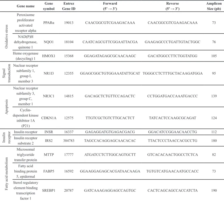
performed utilizing SYBR ® Green PCR Master Mix (Qiagen Inc., Valencia, CA, USA) in a Step One® 7000 thermocycler from Applied Biosystems, employing a PCR initial heat activation step by 10 min at 95 °C, followed by 40 cycles of 95 °C for 15 s and 60 °C for 1 min. In total, 12 genes were analyzed, including one housekeeping gene. Gene names, accession numbers, forward and reverse primer sequences, as well as amplicon sizes, are listed in Table I.
The selection of gene biomarkers was aimed to
TABLE I - Oligonucleotide sequences for real-time PCR
Gene name Gene symbol
Entrez Gene ID
Forward (5’ → 3’)
Reverse (5’ → 3’)
Amplicon Size (pb) O xi da ti ve s tre ss Peroxisome proliferator activated receptor alpha
PPARα 19013 CAACGGCGTCGAAGACAAA CAACGGCGTCGAAGACAAA 73
NAD(P)H dehydrogenase,
quinone 1
NQO1 18104 CAATCAGCGTTCGGAATTACGA GAAGAGCCCTGATTGTACTGGC 76
Heme oxygenase
(decycling) 1 HMOX1 15368 GGAGATAGAGCGCAACAAGC GACATGGCCTTCTGGTATGG 105
S igna l tra ns duc ti
on Nuclear receptor
subfamily 1, group I, member 3
NR1I3 12355 GGAGCGGCTGTGGAAATATTGCAT TGGGCCTCTTTGCTACAAGATGGA 95
A popt os is Nuclear receptor subfamily 3, group C, member 1
NR3C1 14815 GACAGCTCTGTTCCAGACTC CCTGGATGACCAAATGACCC 139
Cyclin-dependent kinase
inhibitor 1A (P21)
CDKN1A 12575 TTGTCGCTGTCTTGCACTCT TATCACTCCAAGCGCAGAT 124
Ins ul in re ce pt
or Insulin receptor INSR 16337 GAGAGGATGTGAGACGACG GGACATCCGGAACAACCTG 112
Insulin receptor
substrate 2 IRS2 384783 TAGCCACAGGAGCAACACAC TTACTCCCTAACCACGCCTG 180
F at ty a ci d m et abol is m Microsomal triglyceride transfer protein
MTTP 17777 ATGATCCTCTTGGCAGTGCTT GTCACACAACTGGCCTCTCA 82
Fatty acid binding protein
5, epidermal
FABP5 16592 GGAAGGAGAGCACGATAACAAGA TGTGTCATGAACAATGCCACC 73
Sterol regulatory element binding transcription
factor 1
include those that may be involved in cross-talking
between insulin signaling and DDT exposure. These
comprised signal transduction, apoptosis, insulin receptor, and fatty acid metabolism, all supported by data from epidemiological studies linking DDT exposure and prevalence of diabetes, cardiovascular diseases, and elevated markers of liver toxicity.
Changes in gene expression were normalized
against the reference endogenous control gene, β-actin (ACTB). The comparative CT method (2-ΔΔCt) was used
for the relative quantiication of each gene. Following ampliication, melting curve analysis was performed to verify the correct product according to its speciic melting
temperature. All experiments were run in duplicate and negative controls contained no cDNA.
Histopathology
Fixed tissues were embedded in parain, and ive μm sections were cut and mounted on glass slides, followed by staining with hematoxylin and eosin (H&E). Sections were examined by a pathologist without knowledge of
the treatment group to which each animal belonged. The histopathology of each tissue section was scored according to reported guidelines.
Statistical Analysis
Morphometric and biochemical parameters are
presented as mean ± standard error (SE). ANOVA was used to compare means for variables between control
and treated groups using Tukey as the post-test. The signiicance for all cases was set at P<0.05. The real-time
PCR data are presented as mean ± SE for ten samples. Statistical analysis was performed using GraphPad Prism
software (v4.0; GraphPad Software Inc., San Diego, CA,
USA).
RESULTS
General characteristics observed in animals from different groups
During treatment animals did not show signs of any
demonstrative behavioral changes or adverse efects, and
no deaths were recorded during the entire experiment.
Biochemical Assays
No significant differences were noted for the
hepatosomatic index (Figure 2A). Enzymatic activities
ALT and AST are presented in Figure 2 (B-C). DDT-treated mice did not show signiicant signs of liver toxicity under working conditions.
Expression of mRNA for genes involved in liver function
The mRNA expression profile of selected genes
is shown in Table II. Compared to vehicle-control,
significant up-regulation of mRNA expression was
observed in the DDT-treated group (100 mg/kg) for
oxidative stress responsive genes such as NAD(P)
H dehydrogenase, quinone 1 (NQO1) (10.05 ± 3.00), peroxisome proliferator-activated receptor alpha (PPARα) (2.38 ± 0.57) and HMOX1 (2.21 ± 0.43), as well as in genes involved in fatty acid metabolism, including microsomal triglyceride transfer protein (MTTP) (1.95 ± 0.30) and fatty acid binding protein 5, epidermal (FABP5) (1.95 ± 0.28). Statistically signiicant diferences were not noted for PPARα. On the other hand, sterol regulatory element binding transcription factor 1 (SREBP1) (0.50 ±
0.07) was previously shown to be down-regulated at the largest tested concentration. Nuclear receptor subfamily
1, group I, member 3 or constitutive androstane receptor (CAR), a gene related to signal transduction/transcription, was induced during both treatments (2.69 ± 0.57 and 3.11 ± 0.64). Regarding insulin receptor (INSR) and insulin receptor substrate 2 (IRS2), no changes were observed (1.19 ± 0.16; 1.17 ± 0.20 and 0.88 ± 0.14, respectively). Finally, for nuclear receptor subfamily 3, group C, member 1 (NR3C1) (1.71± 0.21) significant up-regulation of
mRNA expression was observed in the DDT-treated
group (100 mg/kg). Animals treated with DDT at 50 mg/kg did not present signiicant changes in gene expression.
Histopathology
Histopathological indings from liver sections are presented in Figure 2. Liver histopathology (H&E) shows
normal parenchymal architecture in the control group
(Figure 2 D), with the hepatic central vein surrounded by hepatocytes organized in rows in a circumferential manner,
separated by liver capillary sinusoids showing a mild dilatation. These presented minor changes in treated mice
(E-F), mostly represented by mild and moderate dilatation
of liver capillary sinusoids.
DISCUSSION
to several pathways associated with oxidative stress, insulin signaling, apoptosis, and changes in lipid
metabolism.
In this study, female BalBc adult mice were acutely
liver enzymes when compared to the control group. Gene expression data showed that DDT increased hepatic NQO1 expression compared to the control group. NQO1 is a gene induced by a wide variety of
chemicals, including oxidants, xenobiotics, electrophiles, and phenolic compounds (Zhu, Li, 2012). This gene is
involved in the body’s defense system against oxidative
stress (Gaikwad et al., 2001) and its up-regulation may be an adaptive mechanism to protect against oxidative
and inlammatory stress. NQO1 may also play a role in lipid metabolism and insulin resistance (Zhu, Li, 2012),
since its expression levels have been positively correlated
with adiposity, glucose tolerance, and markers of liver dysfunction, suggesting a possible involvement of NQO1
in the metabolic complications of human obesity (Palming et al., 2007).
In the model presented here, hepatic mRNA levels of HMOX1 were signiicantly induced by p,p’-DDT, similar
to what was observed with o,p’- DDT (Kiyosawa et al.,
2008b). HMOX1 is also elevated in response to oxidative stress, and its up-regulation mediates signal transduction networks involving AP-1, nuclear factor-kappa B (NF-κB) and nuclear factor E2-related factor-2 (Nrf2), as well as upstream kinases related to the maintenance of cellular redox homeostasis (Farombi, Surh, 2006).
Results demonstrated hepatic expression of CAR
was markedly increased for both treatments. Pesticide
exposure can lead to alterations in the endogenous
levels of hormones, and subsequently compromise their hormone signaling. For example, DDT and its metabolite DDE activate nuclear receptor subfamily 1, group I, member 2 or pregnane X receptor (PXR) and TABLE II - Hepatic expression of genes involved in insulin signaling measured in hepatic tissue of DDT-exposed mice
Gene name Gene symbol Control DDT
50 µg/g
DDT 100 µg/g
O
xi
da
ti
ve
s
tre
ss Peroxisome
proliferator activated receptor alpha
PPARα 1.06 ± 0.11 1.18 ± 0.18 2.38 ± 0.57
NAD(P)H dehydrogenase,
quinone 1 NQO1 1.24 ± 0.26 2.29 ± 0.53 10.05 ± 3.00*
S
igna
l
tra
ns
duc
ti
on Heme oxygenase (decycling) 1 HMOX1 1.12 ± 0.18 1.24 ± 0.21 2.21 ± 0.43*
Nuclear receptor subfamily 1, group I,
member 3
NR1I3 1.39 ± 0.33 2.69 ± 0.57 3.11 ± 0.64*
A
popt
os
is
Nuclear receptor subfamily 3, group C,
member 1
NR3C1 1.06 ± 0.11 1.12 ± 0.15 1.71 ± 0.21*
Cyclin-dependent kinase inhibitor 1A
(P21) CDKN1A 1.43 ± 0.34 0.89 ± 0.22 1.12 ± 0.36
Ins
ul
in
re
ce
pt
or Insulin receptor INSR 1.03 ± 0.08 1.07 ± 0.12 1.19 ± 0.16
Insulin receptor
substrate 2 IRS2 1.11 ± 0.17 1.17 ± 0.20 0.88 ± 0.14
F
at
ty a
ci
d m
et
abol
is
m Fatty acid binding
protein 5, epidermal FABP5 1.18 ± 0.22 1.20 ± 0.18 1.95 ± 0.30
Microsomal triglyceride transfer
protein
MTTP 1.05 ± 0.11 1.05 ± 0.15 1.95 ± 0.28*
Sterol regulatory element binding transcription factor 1
SREBP1 1.10 ± 0.14 0.95 ± 0.11 0.50 ± 0.07*
CAR in rodents (Tebourbi, Sakly, Rhouma, 2011). On the contrary, (Kiyosawa et al., 2008a), using C57BL/6
mice treated with 300 µg/g o,p’-DDT and comparing the
results of their study to those of Sprague-Dawley rat data,
reported that mouse CAR mRNA levels decreased. This
may be a result of the activation of mouse-PXR, while ER-mediated efects were insigniicant in rats, probably due to the inhibitory efects of CAR on ER activities.
PPARα and MTTP had a weak increase in gene
expression. Common fatty acids have been shown
to activate PPARα and to regulate the expression
of genes of various lipid oxidation pathways and
transport (Hsu, Huang, 2007), and its activation leads
to the concomitant activation of genes encoding for classical peroxisomal straight chain fatty acid-oxidation
system, microsomal cytochrome P450 CYP4A isoforms CYP4A1and CYP4A3, and some of the genes involved in the mitochondrial-oxidation, among others (Reddy, Hashimoto, 2001). Several studies have suggested an association between MTTP and diabetic dyslipidemia
(Rubin et al., 2008). In particular, high-fat diets as well
as diabetic conditions are associated with elevated hepatic
and intestinal MTTP expressions (Lin et al., 1994). DDT also induced a down-regulation of hepatic expression
of SREBP-1, opposite to what has been reported for this gene in DMT2 (Knip et al., 2005). Studies have
shown that SREBP-1 overexpression may lead to lipid
metabolism disorder and cause lipid accumulation and a
fatty liver. However, there are many studies on high and low expression of SREBP-1c in rats and humans. It may be due to SREBP down-regulation of beta cells, afecting
signaling pathways for insulin secretion stimulated by
glucose and glucolipotoxicity during T2DM (Shao et al.,
2010).
Findings presented here showed transcriptional changes are clearly evidenced in the absence of histopathological damage in mice exposed to moderate doses of DDT. These results are in agreement with those
of other authors, who showed that rodents exhibited
negligible histopathology with rapid o,p’-DDT metabolism
(Kiyosawa et al., 2008a; Kiyosawa et al., 2008b).
Since toxicogenomic profiling may precede clinical
chemistry, histopathology, clinical, or even ultrastructural changes (Heinloth et al., 2007; Miyawaki et al., 2011;
Wang, Papoutsi, Wiesmann, 2011), gene expression
measurements are useful to gain insight into the early
signaling perturbations that presage toxicologic efects
(Ruepp et al., 2002).
Numerous epidemiological studies have suggested
that DDT exposure is likely to contribute to the increase of T2DM (Lee et al., 2010a; Everett, Frithsen, Player, 2011)
Due to the lipophilic nature of DDT, it is trapped and stored within the adipose tissue. Therefore, the
concentration of these compounds in the adipose tissue
may exceed that found in blood. Interestingly, there is the
possibility that patients with diabetes may retain more
of these pollutants than healthy individuals (Everett, Frithsen, Player, 2011), although there is no conclusive epidemiological evidence for such a relationship. Thus, future studies of efects of DDT on adipose tissues will be necessary. However, it is now clear that legacy OC insecticides afect multiple pathways that involve glucose homeostasis (Swaminathan, 2013) and collectively contribute to hyperglycemia (Rahimi, Abdollahi, 2007).
Diabetes may be produced by an immunotoxic
efect of POPs via their binding with ER. This mechanism would induce a chronic low-grade inlammation process, decreased mitochondrial function, fatty acid oxidation, and increased lipolysis, which are related to the insulin
resistance syndrome (Guilherme et al., 2008), as well as
another mechanism involving tissue speciic up- or down-regulation of gene expression, which might promote
glucose intolerance and induce diabetes-independent
oxidative stress and mitochondrial dysfunction (Wallace, 2005). In fact, recent studies have shown that oxidative stress has a role in the development of insulin resistance, which is characterized by hyperinsulinemia (Henriksen, Diamond-Stanic, Marchionne, 2011). One of the major sources of ROS is xenobiotics, and as presented here, oxidative stress is an important mechanism that may link DDT to diabetes, afecting cell functions, metabolism, gene expression proile, and other pathological conditions (Young, Woodside, 2001). The research on oxidative
stress and its connection with other signal transduction pathways could also disclose new scenarios to better evaluate the toxicological response to xenobiotics
(Miyawaki et al., 2011), in particular, the role of DDT on regulation of glucose metabolism.
CONCLUSIONS
The findings of this study suggests that acute
exposure to DDT in mice did not promote adverse efects on weight and on hepatic markers of toxicity or liver histopathology. However, there were changes at the
molecular level represented in the activation of several
genes known to participate in insulin resistance pathways, such as CAR, SREBP, NQO1, NR3C1, HMOX1 and MTTP. This conclusion could be particularly relevant in
view of the recently emerging evidence showing that DDT
ACKNOWLEDGEMENTS
The authors express their gratitude to Colciencias
and the University of Cartagena (Grant 110745921616, 2008).
CONFLICT OF INTERESTS
The authors declare that there is no conlict of
interest.
REFERENCES
ARREBOLA, J.P.; CUELLAR, M.; CLAURE, E.; QUEVEDO, M.; ANTELO, S.R.; MUTCH, E.; RAMIREZ, E.; FERNANDEZ, M.F.; OLEA, N.; MERCADO, L.A. Concentrations of organochlorine pesticides and polychlorinated biphenyls in human serum and adipose tissue from Bolivia. Environ. Res., v.112, p.40-47, 2012.
AGENCY FOR TOXIC SUBSTANCES & DISEASES REGISTRY. ATSDR. Public health statement for DDT, DDE, AND DDD, Sep., 2002. Retrieved from Available at http: [http://www.atsdr.cdc.gov/phs/phs. asp?id=79&tid=20]. Accessed on: June 17, 2014.
BLOOMGARDEN, Z.T. European Association for the study of diabetes Annual Meeting, 1999: treatment modalities. Diabetes Care, v.23, n.7, p.1012-1017, 2000.
COLLOTTA, M.; BERTAZZI, P.A.; BOLLATI, V. Epigenetics and pesticides. Toxicology, v.307, p.35-41, 2013.
COX, S.; NISKAR, A.S.; NARAYAN, K.M.; MARCUS, M. Prevalence of self-reported diabetes and exposure to organochlorine pesticides among Mexican Americans: Hispanic health and nutrition examination survey, 1982-1984. Environ. Health Perspect., v.115, n.2, p.1747-1752, 2007.
DANAEI, G.; FINUCANE, M.; LU, Y.; SINGH, G.; COWAN, M.; PACIOREK, C.; LIN, J.; FARZADFAR, F.; KHANG, Y.-H.; STEVENS, G.; RAO, M.; ALI, M.; RILEY, L.; ROBINSON, C.; EZZATI, M.; GLOBAL BURDEN OF METABOLIC RISK FACTORS OF CHRONIC DISEASES COLLABORATING, G. National, regional, and global trends in fasting plasma glucose and diabetes prevalence since 1980: systematic analysis of health examination surveys and epidemiological studies with 370 country-years and 2·7 million participants. Lancet, v.378, p.31-40, 2011.
EVERETT, C.J.; FRITHSEN, I.; PLAYER, M. Relationship of polychlorinated biphenyls with type 2 diabetes and hypertension. J. Environ. Monit., v.13, n.2, p.241-251, 2011.
FAROMBI, E.O.; SURH, Y.J. Heme oxygenase-1 as a potential therapeutic target for hepatoprotection. J. Biochem. Mol. Biol., v.39, n.5, p.479-491, 2006.
FRIGO, D.E.; VIGH, K.A.; STRUCKHOFF, A.P.; ELLIOTT, S.; BECKMAN, B.S. Xenobiotic-induced TNF-α expression and apoptosis through the p38 MAPK signaling pathway. Toxicol. Lett., v.155, n.2, p.227-238, 2005.
GAIKWAD, A.; LONG, D.J.; STRINGER, J.L.; JAISWAL, A.K. In vivo role of NAD (P) H: quinone oxidoreductase 1 (NQO1) in the regulation of intracellular redox state and accumulation of abdominal adipose tissue. J. Biol. Chem., v.276, n.25, p.22559-22564, 2001.
GUILHERME, A.; VIRBASIUS, J.V.; PURI, V.; CZECH, M.P. Adipocyte dysfunctions linking obesity to insulin resistance and type 2 diabetes. Nat. Rev. Mol. Cell. Biol. v.9, n.5, p.367-377, 2008.
HEINLOTH, A.N.; BOORMAN, G.A.; FOLEY, J.F.; FLAGLER, N.D.; PAULES, R.S. Gene expression analysis ofers unique advantages to histopathology in liver biopsy evaluations. Toxicol. Pathol., v.35, n.2, p.276-283, 2007.
H E N R I K S E N , E . J . ; D I A M O N D - S TA N I C , M . K . ; MARCHIONNE, E.M. Oxidative stress and the etiology of insulin resistance and type 2 diabetes. Free. Radic. Biol. Med., v.51, n.5, p.993-999, 2011.
HSU, S.-C.C.; HUANG, C.-J.J. Changes in liver PPARalpha mRNA expression in response to two levels of high-salower-oil diets correlate with changes in adiposity and serum leptin in rats and mice. J. Nutr. Biochem., v.18, n.2, p.86-96, 2007.
IBRAHIM, M.; FJÆRE, E.; LOCK, E.-J.; NAVILLE, D.; AMLUND, H.; MEUGNIER, E.; LE MAGUERESSE BATTISTONI, B.; FRØYLAND, L.; MADSEN, L.; JESSEN, N.; LUND, S.; VIDAL, H.; RUZZIN, J. Chronic consumption of farmed salmon containing persistent organic pollutants causes insulin resistance and obesity in mice. PLoS One, v.6, p.e25170, 2011.
KACEW, S.; SINGHAL, R.L. Role of cyclic adenosine 3’:5’-monophosphate in the action of 1,1,1-trichloro-2,2-bis-(p-chlorophenyl)ethane (DDT)on hepatic and renal metabolism. Biochem. J., v.142, n.1, p.145-152, 1974.
KIYOSAWA, N.; KWEKEL, J.; BURGOON, L.; DERE, E.; WILLIAMS, K.; TASHIRO, C.; CHITTIM, B.; ZACHAREWSKI, T. Species-speciic regulation of PXR/ CAR/ER-target genes in the mouse and rat liver elicited by o, p’-DDT. BMC Genomics., v.9, p.487, 2008a.
KIYOSAWA, N.; KWEKEL, J.C.; BURGOON, L.D.; WILLIAMS, K.J.; TASHIRO, C.; CHITTIM, B.; ZACHAREWSKI, T.R. o,p’-DDT elicits PXR/CAR-, not ER-, mediated responses in the immature ovariectomized rat liver. Toxicol. Sci., v.101, n.2, p.350-363, 2008b.
KNIP, M.; VEIJOLA, R.; VIRTANEN, S.M.; HYOTY, H.; VAARALA, O.; AKERBLOM, H.K. Environmental triggers and determinants of type 1 diabetes. Diabetes, v.54, p.S125-136, 2005.
LA MERRILL, M.; KAREY, E.; MOSHIER, E.; LINDTNER, C.; LA FRANO, M.R.; NEWMAN, J.W.; BUETTNER, C. Perinatal exposure of mice to the pesticide DDT impairs energy expenditure and metabolism in adult female ofspring. PloS One, v.9, p.e103337, 2014.
LEE, D.-H.; STEFFES, M.; SJÖDIN, A.; JONES, R.; NEEDHAM, L.; JACOBS, D. Low dose of some persistent organic pollutants predicts type 2 diabetes: a nested case-control study. Environ. Health Perspect., v.118, n.9, p.1235-1242, 2010a.
LEE, D.-H.H.; STEFFES, M.W.; SJÖDIN, A.; JONES, R.S.; NEEDHAM, L.L.; JACOBS, D.R. Low dose organochlorine pesticides and polychlorinated biphenyls predict obesity, dyslipidemia, and insulin resistance among people free of diabetes. PloS One, v.6, n.1, p.e15977, 2010b.
LIN, M.C.; ARBEENY, C.; BERGQUIST, K.; KIENZLE, B.; GORDON, D.A.; WETTERAU, J.R. Cloning and regulation of hamster microsomal triglyceride transfer protein. The regulation is independent from that of other hepatic and intestinal proteins which participate in the transport of fatty acids and triglycerides. J. Biol. Chem., v.269, n.46, p.29138-29145, 1994.
LUCENA, R.A.; ALLAM, M.F.; JIMÉNEZ, S.S.; VILLAREJO, M.L. A review of environmental exposure to persistent organochlorine residuals during the last ifty years. Curr. Drug Saf., v.2, n.2, p.163-172, 2007.
MIYAWAKI, I.; MATSUMOTO, I.; HORIE, H.; INADA, H.; KIMURA, J.; FUNABASHI, H.; SEKI, T. Toxicological approach for elucidation of clobazam-induced hepatomegaly in male rats. Regul. Toxicol. Pharmacol., v.60, n.3, p.323-331, 2011.
MORENO-ALIAGA, M.; MATSUMURA, F. Effects of 1,1,1-trichloro-2,2-bis(p-chlorophenyl)-ethane (p,p’-DDT) on 3T3-L1 and 3T3-F442A adipocyte differentiation. Biochem. Pharmacol., v.63, n.5, p.997-1007, 2002.
PALMING, J.; SJÖHOLM, K.; JERNÅS, M.; LYSTIG, T.C.; GUMMESSON, A.; ROMEO, S.; LÖNN, L.; LÖNN, M.; CARLSSON, B.; CARLSSON, L.M. The expression of NAD(P)H:quinone oxidoreductase 1 is high in human adipose tissue, reduced by weight loss, and correlates with adiposity, insulin sensitivity, and markers of liver dysfunction. J. Clin. Endocrinol. Metab., v.92, n. 6, p.2346-2352, 2007.
PATEL, C.J.; BHATTACHARYA, J.; BUTTE, A.J. An Environment-Wide Association Study (EWAS) on type 2 diabetes mellitus. PloS One, v.5, p.2009.
PORTA, M.; MALATS, N.; JARIOD, M.; GRIMALT, J.O.; RIFÀ, J.; CARRATO, A.; GUARNER, L.; SALAS, A.; SANTIAGO-SILVA, M.; COROMINAS, J. M.; ANDREU, M.; REAL, F.X. Serum concentrations of organochlorine compounds and K-ras mutations in exocrine pancreatic cancer. PANKRAS II Study Group. Lancet, v.354, p.2125-2129, 1999.
RAHIMI, R.; ABDOLLAHI, M. A review on the mechanisms involved in hyperglycemia induced by organophosphorus pesticides. Pestic. Biochem. Phys., v.88, n.2, p.115-121, 2007.
REDDY, J.K.; HASHIMOTO, T. Peroxisomal β-oxidation and peroxisome proliferator-activated receptor α: an adaptive metabolic system. Annu. Rev. Nutr., v.21, p.193-230, 2001.
RUBIN, D.; SCHNEIDER-MUNTAU, A.; KLAPPER, M.; NITZ, I.; HELWIG, U.; FÖLSCH, U.R.; SCHREZENMEIR, J.; DÖRING, F. Functional analysis of promoter variants in the microsomal triglyceride transfer protein (MTTP) gene. Hum. Mutat., v.29, n.1, p.123-129, 2008.
RUEPP, S.U.; TONGE, R.P.; SHAW, J.; WALLIS, N. Genomics and proteomics analysis of acetaminophen toxicity in mouse liver. Toxicol. Sci., v. 65, n.1, p.135-150, 2002.
RUZZIN, J.; PETERSEN, R.; MEUGNIER, E.; MADSEN, L.; LOCK, E.-J.; LILLEFOSSE, H.; MA, T.; PESENTI, S.; SONNE, S.; MARSTRAND, T.; MALDE, M.; DU, Z.-Y.; CHAVEY, C.; FAJAS, L.; LUNDEBYE, A.-K.; BRAND, C.; VIDAL, H.; KRISTIANSEN, K.; FRØYLAND, L. Persistent organic pollutant exposure leads to insulin resistance syndrome. Environ. Health Perspect., v.118, n.4, p.465-471, 2010.
RYLANDER, L.; RIGNELL-HYDBOM, A.; HAGMAR, L. A cross-sectional study of the association between persistent organochlorine pollutants and diabetes. Environ. Health Glob., v.4, p.28, 2005.
SANYAL, S.; AGARWAL, N.; DUDEJA, P.K.; MAHMOOD, A.; SUBRAHMANYAM, D. Efect of a single oral dose of DDT on lipid metabolism in protein-calorie malnourished monkeys. Indian J. Biochem. Biophys., v.19, n.2, p.111-114, 1982.
SARGIS, R.M. The hijacking of cellular signaling and the diabetes epidemic: mechanisms of environmental disruption of insulin action and glucose homeostasis. Diabetes Metab. J., v.38, n.1, p.13-24, 2014.
SHAO, S.; LIU, Z.; YANG, Y.; ZHANG, M.; YU, X. SREBP-1c, Pdx-1, and GLP-1R involved in palmitate-EPA regulated glucose-stimulated insulin secretion in INS-1 cells. J. Cell Biochem., v.111, n.3, p.634-642, 2010.
SHEN, K.; NOVAK, R.F. DDT stimulates c-erbB2, c-met, and STATS tyrosine phosphorylation, Grb2-Sos association, MAPK phosphorylation, and proliferation of human breast epithelial cells. Biochem. Biophys. Res. Commun., v.231, n.1, p.17-21, 1997.
SOHONI, P.; SUMPTER, J.P. Several environmental oestrogens are also anti-androgens. J. Endocrinol., v.158, n.3, p.327-339, 1998.
SWAMINATHAN, K. Pesticides and human diabetes: a link worth exploring? Diabet. Med., v.30, n.11, p.1268-1271, 2013.
TEBOURBI, O.; SAKLY, M.; RHOUMA, K.B.Pesticides in the
modern world - pests control and pesticides exposure and
toxicity assessment. Rijeka, Croatia: Intech Open Access Publisher, 2011. cap 15, p.297-332.
THAYER, K.; HEINDEL, J.; BUCHER, J.; GALLO, M. Role of environmental chemicals in diabetes and obesity: a national toxicology program workshop review. Environ. Health Perspect., v.120, n.6, p.779-789, 2012.
WALLACE, D.C. A mitochondrial paradigm of metabolic and degenerative diseases, aging, and cancer: a dawn for evolutionary medicine. Annu. Rev. Genet., v.39, p.359-407, 2005.
WANG, T.; PAPOUTSI, M.; WIESMANN, M. Investigation of correlation among safety biomarkers in serum, histopathological examination, and toxicogenomics. Int. J. Toxicol., v.30, n.3, p.300-312, 2011.
YAU, D.T.; MENNEAR, J.H. The inhibitory efect of DDT on insulin secretion in mice. Toxicol. Appl. Pharmacol., v.39, n.1, p.81-88, 1976.
YOUNG, I.S.; WOODSIDE, J.V. Antioxidants in health and disease. J. Clin. Pathol., v.54, n.3, p.176-186, 2001.
ZHU, H.; LI, Y. NAD(P)H: quinone oxidoreductase 1 and its potential protective role in cardiovascular diseases and related conditions. Cardiovasc. Toxicol., v.12, n.1, p.39-45, 2012.