ed inside the carotid canal due to technical problems. Consequently, a clinical picture of Raeder’s para-trigeminal neuralgia took place. This is the first case report in the literature with theses characteristics. A review of the anatomic pathways and further considerations about the possible pathophysiological mech-anisms involved are presented.
KEY WORDS: Raeder’s paratrigeminal neuralgia, headache, cephalalgia, neuroendovascular procedures.
S í n d rome de Raeder após embolização de aneurisma gigante de artéria carótida intracaver-nosa: considerações fisiopatológicas
RESUMO - Apresentamos o caso de uma mulher de 47 anos submetida a obliteração endovascular de um aneurisma gigante de carótida interna cavernosa à esquerda, no qual o balão distal foi inflado, tal como usual, dentro do segmento cavernoso da artéria carótida interna, diferente do proximal, o qual foi infla-do dentro infla-do canal carotídeo deviinfla-do a problemas técnicos. Conseqüentemente, um quadro clínico de neu-ralgia paratrigeminal de Raeder se instalou. Este é o primeiro relato na literatura com estas característi-cas. Uma revisão das vias anatômicas e maiores considerações a respeito de possíveis mecanismos fisiopa-tológicos envolvidos são apresentados.
PALAVRAS-CHAVE: neuralgia paratrigeminal de Raeder, cefaléia, procedimentos neuroendovasculares.
Department of Neurosurgery - Hospital Nossa Senhora das Graças, Curitiba PR, Brazil:1Neurosurgeon;2Resident in Neurosurgery; 3Neurosurgeon Chief of the Residency Program.
Received 5 November 2004, received in final form 21 December 2004. Accepted 12 March 2005.
Dr. Daniel Monte Serrat Prevedello - Rua Alcides Munhoz 433 - 80810-040 Curitiba PR - Brasil. E-mail: [email protected] Oculosympathetic paresis associated with
ipsi-lateral continuous head pain was first described b y a Norwegian neurologist, J.G. Raeder, in 19181 - 3.
In the author's original reports, a skull base infil-trative neoplasm in the middle cranial fossa, close to the petrous apex and Gasserian ganglion, cau-sed the symptoms1. Raeder's paratrigeminal
neu-ralgia became a frequently used terminology at that time, correlating, after autopsy cases, the sym-ptoms described by Raeder to expanding lesions involving the cavernous sinus and stru c t u res adja-cent to the trigeminal branches. The picture con-sisting of orbital pain plus oculosympathetic pal-sy (ptosis and miosis) had, in consequence, an im-portant clinical localizing value before the devel-opment of modern imaging methods. Advances
in micro n e u ro s u rg e ry and better understanding of the microanatomy of the cavernous sinus4 , 5,
pe-t rous apex6 , 7, orbit8and infratemporal fossa9,
asso-ciated with the introduction of modern staining techniques, re t rograde axonal mapping1 0 , 1 1a n d
i m m u n o c y t o c h e m i s t ry1 2yielded more complete
i n f o rmation about the skull base innervation and its relation with pathophysiologic events involved in the genesis of Raeder’s neuralgia.
damaging of the left internal carotid art e ry in the petrous segment, leading to Raeder’s syndrome.
CASE
A 47 years old woman presented incapacitating ver-tigo with no other symptom. Magnetic resonance imag-ing (MRI) revealed a left paraselar-expandimag-ing lesion demonstrated by angiography to be a giant aneurysm of the cavernous segment of the left internal caro t i d a rt e ry (Fig 1). Occlusion tests of left internal carotid ar-t e ry showed absence of neurological deficiar-ts and a good c rossover perfusion through anastomoses of the circ l e of Willis.
One week after the diagnosis, an endovascular trap-ping of the cavernous segment of the left internal caro-tid with detachable balloons, under general anesthe-sia, was accomplished. The proximal balloon had to be positioned at the level of the carotid canal, due to tech-nical difficulties in placing it closer to the lesion. Flow absence within the aneurysm and patency of collateral c i rculation were again confirmed before ending the pro-cedure.
Immediately after the pro c e d u re the patient re f e rre d intense pain around the left periorbital area and deep face in relation to the nasal fossa. The pain persisted in spite of appropriate medication, reaching degree 9 in the analogical visual scale for pain1 3 , 1 4. In addition to
pain, she had ptosis of the left superior eyelid and mio-sis of the left pupil, which was brought out by darken-ing the room, leaddarken-ing the right normal pupil to dilate. T h e re was no deficit of the extraocular motor system and the facial and trigeminal nerves were clinically in-tact. Pharmacologic testing with 1% hydro x y a m p h e t a-mine confirmed the lesion to be postganglionic, caus-ing no dilation of the left pupil.
A new MRI and angiography (Fig 2) demonstrated
a thrombosed aneurysm inside the left cavernous sinus without any increase in its volume comparing to the pre operative exam and a good perfusion of the ipsilateral hemisphere through posterior circulation. No associat-ed lesion could be demonstratassociat-ed.
Pain and oculosympathetic paresis remained unchan-ged for exactly two months. Subsequently, sings and symptoms pro g ressively subsided up to a normal
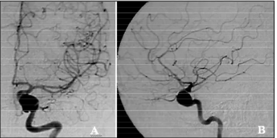
clini-Fig 1. Angiography showing a giant aneurysm of the cavernous segment of the left internal caro t i d artery. A) frontal view. B) side view.
demonstrated17,18. It is believed that these
neuro-peptides, present in large vessels and capillaries, a re related to an integrated system with a substan-tial role in cerebral blood flow autoregulation un-der normal and pathological conditions1 5.
Experi-mental evidence demonstrates the vascular re a c-tivity under the action of these modulators19.
R e t rograde axonal mapping in laboratory sho w s that most of these fibers originate from the tri-geminal ganglion, while other studies demonstrate that the stimulation of stru c t u res innervated by these terminations causes the expression of gene c-fos, marker of neuronal activation, in the neu-rons of the spinal trigeminal nucleus2 0, there f o re
demonstrating that these fibers are trigeminal a ff e rents in nature. These mechanisms are part of what Moskowitz denominated trigeminovascular system20.
Scientific evidence suggests that such innerv a-tions are present in humans and they would be in-volved in the pathophysiology of headaches. A mechanism of central activation is postulated, with a n t i d romic conduction, in which neuro p e p t i d e s a re delivered on terminals of the trigeminovascu-lar system, leading to an aseptic neurogenic inflam-mation. Subsequently, through anterograde con-duction mechanisms there would be an activation of trigeminal projections to the thalamus, result-ing in perception of the painful sensation in the specific head segment21.
Internal carotid art e ry is subdivided into sever-al segments. One of them, the petrous segment, c rosses through the temporal bone inside the c a rotid canal, where a bony framework intimate-ly wraps it up2 2. In the same way, sympathetic fibers
a re found inside the carotid canal, lying on the surface of the internal carotid artery23.
s t ru c t u res inside the head travel along branches of the carotid arteries to their targets25.
Postgan-glionic sympathetic fibers assemble in a network around the common carotid artery and follow its branches. Following the bifurcation, fibers along branches of external carotid art e ry bring sympa-thetic innervation to the sweat glands, erector pili muscles and subcutaneous capillary blood vessels of the face. Intern a l l y, within the carotid canal, so-me postganglionic fibers organize to form the deep petrosal nerve, which gives sympathetic i n n e rvation mainly to the lacrimal gland2 5. The
cav-e rnous scav-egmcav-ent of thcav-e intcav-ernal carotid art cav-e ry con-tains postganglionic fibers which send eff e re n t s that follow the abducens, trochlear and oculomo-tor nerves and the ophthalmic art e ry itself. Ultima-tely they get to the ciliary ganglion and long cil-i a ry nerv e s1 , 2 3to reach the pupillary dilator
mus-cle and tarsal musmus-cle of Muller26.
The cell bodies of the preganglionic neuro n s of the parasympathetic system are located within the brain stem and in segments S2 to S4 of the spinal cord. Parasympathetic preganglionic nuclei to the head include the Edinger- Westphal nucle-us and superior and inferior salivary nuclei. The orbit receives parasympathetic eff e rents from the E d i n g e r- Westphal and from the superior salivary nuclei. The inferior salivary nucleus sends fibers to the parotid gland27,28.
The nervus intermedius of the facial nerve con-tains fibers from the superior salivary nucleus, which synapse in the pterygopalatine and subman-dibular ganglia to innervate the lacrimal gland and submandibular-sublingual glands re s p e c t i v e-l y2 7 , 2 8. The great petrosal nerve arises from the
to lie in a groove on its anterior surface beneath the temporal lobe and dura of the middle cranial fossa. It runs beneath the trigeminal ganglion, an-t e romedially across an-the foramen lacerum. There , it is joined by the deep petrous nerve (sympathet-ic) and becomes the nerve of the pterygoid canal. It passes through the pterygopalatine fossa to en-ter the posen-terior aspect of the pen-tery g o p a l a t i n e ganglion. Fibers are then distributed with branch-es of the maxillary division mainly through the zy-gomatic-orbital nerve, which terminates on the lateral orbital wall to the lacrimal gland6.
E d i n g e r- Westphal nucleus sends fibers that inte-grate the oculomotor nerve, which after passing t h rough the cavernous sinus enters the orbit t h rough the superior orbital fissure and splits into superior and inferior divisions. The inferior divi-sion gives rise to the inferior oblique nerve, which c a rries the general visceral motor fibers (parasym-pathetic) to the ciliary ganglion. Short ciliary ner-ves, from the ganglion, innervate the ciliary body, sphincter pupillae and uveal tract glands27,28.
All three systems described above (sympathet-ic, parasympathetic and trigeminovascular) gen-erate a rich nervous plexus located in the lateral wall of the cavernous sinus, the so called “lateral sellar nerve plexus”, demonstrated by Bleys et al.2 9.
Anastomoses between the lateral sellar plexus and fibers from the Gasserian ganglion carry trige-minal somatic aff e rent fibers along the wall of the i n t e rnal carotid arteries. These connections are responsible for the conduction of nociceptive sen-sation mainly from petrous, lacerum and cavern o u s segments. Multiple anastomoses from the lateral sellar plexus, connected to the sympathetic caro t i d n e rves and fibers coming from the ptery g o p a l a-tine ganglion form a mixed plexus with sympathe-tic, parasympathetic and aff e rent somatic fibers2 9.
Alterations in the venous drainage of the caver-nous sinus triggered by an activation of the trige-minovascular system (related to the lateral sellar plexus inside the cavernous sinus) may cause peri-c a rotid aseptiperi-c neurogeniperi-c inflammation, playing an important role in the genesis of pain and ocu-losympathetic dysfunction in attacks of cluster- t y-pe migraine30.
It is postulated, in the present case, that place-ment of a proximal balloon at the level of the ca-rotid canal caused compression of sympathetic ner-ves located around the internal carotid art e ry aga-inst the bony wall of the carotid canal, causing sympathetic dysfunction, as well as activation of nociceptive terminals from the trigeminovascular system (Fig 3). Connections between such stru c t u-Fig 3. Anatomical illustration demonstrating sympathetic, parasympathetic and trigeminal fibers at
ar plexus and the pterygopalatine ganglion . Re-lief obtained after anesthetic blockade of the gan-glion for certain types of headache, such as clus-t e r-like migrane, may be explainned by a possible reduction of input to the trigeminovascular sys-tem and its connections32.
In conclusion, this re p o rt supports Bley’s obser-vations, which identified a nerve plexus in re l a t i o n to the internal carotid art e ry wall, with aff e re n t i n n e rvation from trigeminal fibers2 9. Multiple
con-nections between sympathetic and parasympa-thetic systems may explain the character of pain i rradiation when these nerve fibers are stimulat-ed. Furt h e rm o re, Raeder's neuralgia would corre-spond to a clinical manifestation of a dysfunction in the trigeminovascular system involving the inter-nal carotid art e ry and multiple connections of the lateral sellar plexus in the cavernous sinus.
Acknowledgement -We thank the medical stu-dent, Endrigo Monte Serrat Prevedello for contributing with a didactic illustration to this manuscript (Fig 3).
REFERENCES
1. Goadsby PJ. Raeder’s syndrome: paratrigeminal paralysis of the ocu-lopillary sympathetic system. J Neurol Neuro s u rg Psychiatry 2002; 72:297-299.
2. Solomon S, Lustig JP. Benign Raeder's syndrome is probably a mani-festation of carotid artery disease. Cephalalgia 2001;21:1-11. 3. Salvesen R. Raeder's syndrome. Cephalalgia 1999;19(Suppl 25)S42-S45 4. Rhoton A L J r, Inoue T. Micro s u rgical approaches to the cavernous sinus.
Clin Neurosurg 1991;37:391-439.
5. Rhoton A L J r, Hardy DG, Chambers SM. Micro s u rgical anatomy and dissection of the sphenoid bone, cavernous sinus and sellar re g i o n . Surg Neurol 1979;12:63-104.
6. Rhoton A L J r. The anterior and middle cranial base. Neuro s u rg e r y 2002;51(Suppl 4)S273-S302.
logue scale (TAS), a new scale. Cephalalgia 2000;20:323.
15. Buki A, Horvath Z, Kallo I, Liposits Z, Lengvari I, Doczi TP. Peptiderg i c innervation of human cerebral blood vessels and saccular aneurysms. Acta Neuropathol (Berl) 1999;98:383-388.
16. Edvinsson L, Jansen I, Cunha e Sa M, Gulbenkian S. Demonstration of neuropeptide containing nerves and vasomotor responses to perivas-cular peptides in human cerebral arteries. Cephalalgia 1994;14:88-96. 17. Edvinsson L. Sensory nerves in man and their role in primary
headaches. Cephalalgia 2001;21:761-764.
18. Edvinsson L, Goadsby PJ. Neuropeptides in the cerebral circ u l a t i o n : relevance to headache. Cephalalgia 1995;15:272-276.
19. Edvinsson L, Jansen Olesen I, Kingman TA, McCulloch J, Uddman R. Modification of vasoconstrictor responses in cerebral blood vessels by lesioning of the trigeminal nerve: possible involvement of CGRP. Cephalalgia 1995;15:373-383.
20. Moskowitz MA. Trigeminovascular system. Cephalalgia 1992;12:127. 21. Moskowitz MA. The trigeminovascular system. In Olesen J, (ed). The
headaches. New York: Raven Press, 1993:97-104.
22. Bouthillier A, van Loveren HR, Keller JT. Segments of the internal carotid artery: a new classification. Neurosurgery 1996;38:425-433. 23. Von Overbeeke JJ, Dujovny M, Dragovic L, Ausman JI. Anatomy of the
sympathetic pathways in the carotid canal. Neuro s u rgery 1991;29: 838-843.
24. Loewy AD, Araujo JC, Kerr FW. Pupillodilator pathways in the brain stem of the cat: anatomical and electrophysiological identification of a central autonomic pathway. Brain Res 1973;60:65-91.
25. Von Overbeeke JJ, Dujovny M, Dragovic L, Ausman JI. Anatomy of the sympathetic pathways in the carotid canal. Neuro s u rgery 1991;29:838-843.
26. Ruskell GL. Accommodation and the nerve pathway to the ciliary mus-cle: a review. Ophthalmic Physiol Opt 1990;10:239-242.
27. Ruskell GL. The orbital branches of the pterygopalatine ganglion and their relationship with internal carotid nerve branches in primates. J Anat 1970;106:323-339.
28. Ruskell GL. Orbital passage of pterygopalatine ganglion eff e rents to paranasal sinuses and nasal mucosa in man. Cells Tissues Org a n s 2003;175:223-228.
29. Bleys RLAW, Janssen LM, Groen GJ. The lateral sellar nerve plexus and its connections in humans. J Neurosurg 2001;95:102-110.
30. Dodick DW, Rozen TD, Goadsby PJ, Silberstein SD. Cluster headache. Cephalalgia 2000;20:787-803.
31. Piovesan EJ, Kowacs PA, Tatsui CE, Lange MC, Ribas LC, Werneck LC. R e f e r red pain after painful stimulation of the greater occipital nerve in humans: evidence of convergence of cervical aff e rences on trigem-inal nuclei. Cephalalgia 2001;21:107-109.