Arq Neuropsiquiatr 2005;63(3-A):681-684
State University of Campinas (UNICAMP) Campinas SP, Brasil: 1Neurology and Neurosurgery Department; 2Radiology Department. Received 15 October 2004, received in final form 27 December 2005. Accepted 14 March 2005.
Dr. Guilherme Borges - Rua Visconde do Rio Claro 322 - 13083-650 Campinas SP - Brasil. E-mail: [email protected]
IMAGING FEATURES AND TREATMENT OF AN
INTRADURAL LUMBAR CYSTIC SCHWANNOMA
Guilherme Borges
1, Leonardo Bonilha
1, Marcílio Proa Jr.
1, Yvens Barbosa Fernandes
1,
Ricardo Ramina
1, Veronica Zanardi
2, Jose Ribeiro Menezes
2ABSTRACT - Spinal schwannomas are frequently observed among patients treated in a reference neuro-s u rg e ry center. Cyneuro-stic neuro-spinal neuro-schwannomaneuro-s, however, are very neuro-scantly found. Due to itneuro-s indolent behavior and benign course, the diagnosis of schwannomas may pose a challenge to the care giver, and the imag-ing findimag-ings can be misleadimag-ing. In this article, we illustrate an example of a pauci-symptomatic 55 year- o l d male patient whose complaint was solely a non specific lumbar pain. Investigation revealed a large cystic lesion comprising the lower lumbar intradural space. He was then treated with micro n e u ro s u rgical tech-nique involving complete removal of the tumor and re c o n s t ruction of the duramater. Histological and immunohystochemical diagnosis were consistent with cystic schwannoma. The patient presented with com-plete re c o v e ry of his symptom. In this article we aim to emphasize the clinical presentation and tre a t m e n t of lumbar spine schwannomas, and to illustrate the imaging findings within this uncommon case.
KEY WORDS: schwannoma, cystic tumor, nerve sheath tumor, neurinoma.
Achados imagenológicos e tratamento de um schwannoma cístico intradural lombar
RESUMO - O schwannoma cístico intra-espinal é tumor muito raro e poucos casos estão descritos na litera-tura: são usualmente assintomáticos e somente diagnosticados quando atingem grande tamanho causan-do compressão radicular. Com o intuito de ilustrar as armadilhas existentes re f e rentes ao diagnóstico e tratamento desse tipo de tumor, nós relatamos um caso raro, focando nos passos da investigação e tera-pêutica. É descrito o caso de um paciente de 55 anos que apresentava apenas queixas de dor lombar. A investigação revelou uma lesão cística extensa na região intradural lombar inferior. O tumor foi totalmente ressecado por técnica micro neurocirúrgica, sendo a dura-máter re c o n s t ruída. O diagnóstico patológico e imuno-histoquímico evidenciou tratar-se de um schwannoma cístico. Neste artigo, nós procuramos enfa-tizar as características clínicas e tratamento de schwannomas lombares, ilustrando os achados imagenológi-cos desse caso incomum.
PALAVRAS-CHAVE: schwannoma, tumor cístico, tumor de bainha nervosa, neurinoma.
Schwannomas are benign tumors of the
periph-eral nervous system, arising from Schwann cells1.
Schwannomas can occur virtually in any body loca-tion where peripheral or cranial nerves are pre
s-e n t2, and they correspond to 8% of primary
intra-cranial and 29% of primary spinal tumors1. Wi t h i n
the spinal region, Prevedello et al. have investi-gated the prevalence of diff e rent extramedullary intradural spinal tumors and observed that
schwa-nnomas comprised 66% of all cases3. The lumbar
region is one of the most common sites for occur-rence of spinal schwannomas, as demonstrated by Conti et al., who studied the distribution of spinal schwannomas and re p o rted that 48% lie within
the lumbar spine4. While schwannomas occurr i n g
within the lumbar spine are not rare, large and
p redominantly cystic schwannomas occurring in the lumbar spine have scantly been demonstrat-ed, even though it is well defined that benign schwannomas can eventually display degenerative changes defined by cyst formation, calcification, h e m o rrhage and hyalinization1. Intere s t i n g l y, larg e
schwannomas within the lumbar spine may go unnoticed for a large time span mainly due to the indolent growth of the tumor and the paucity of symptoms generated. With a more malleable stru c-t u re, a cysc-tic schwannoma may pose a challenge to the diagnosis.
682 Arq Neuropsiquiatr 2005;63(3-A)
clinical picture, imaging findings and tre a t m e n t of this unique case. This re p o rt was approved by the Committee of Ethics and Research of our Ins-titution (UNICAMP).
CASE
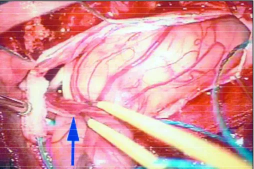
A 55-year-old man without remarkable pre v i o u s medical history was admitted in our Hospital due to a 12-month history of lumbar pain with a poorly defined i rradiation to both lower limbs. His general physical exa-mination was normal and did not exhibit signs compat-ible with neuro f i b romatosis. His neurological physical examination failed to disclose signs of radicular irr i t a-tion, motor or sensory deficits. Magnetic resonance ima-ging (MRI) (Fig 1) depicted an extensive lesion (6.0x1.8x 1.8 cm) extending from the 4thto 5thlumbar vertebrae almost completely constituted by a large cyst. Gadoli-nium contrasted MRI depicted a ring-like enhancement of the cyst. Tumor was totally removed by micro n e u ro-surgical technique (Fig 2). The remaining thin duramater was not large enough for primary repair, and an artifi-cial graft (DuraGenT M) was successfully used to accom-plish a water tight seal dura-mater closure. Histological examination of the excised specimen revealed typical schwannoma cell nuclei (Fig 3A). Immunohystochemical study included staining for S-100 protein and NGFR (nerve growth factor receptor), which were both posi-tive diffusely across the tumor cells (Fig 3B and 3C). The immunohystochemical analysis alongside with the his-tological observations confirmed the diagnosis of a cys-tic schwannoma. After an unremarkable postoperative period, the patient experienced a complete re m i s s i o n of preoperative symptoms. At a follow-up visit per-f o rmed 12 months aper-fter surg e ry, the patient was asymp-tomatic.
DISCUSSION
Schwannomas are slow growing benign tumors. They are usually encapsulated, and rarely
under-go malignant transform a t i o n1. Schwannomas arise
f rom the Schwann cells of the nerve sheath, and they comprise the most common tumor type aff e
c-ting the peripheral nerv e s1. The most common
lo-cation of schwannomas are around peripheral ner-ves in the extradural space. Intracranial schwanno-mas have also been observed, and they usually ari-se from the facial, trigeminal, or vestibular
ner-ves5,6. Conspicuously, schwannomas are more
fre-quently observed in patients with neuro f i b ro m a t
o-sis type 21, schwannomas can be observed in rather
Fig 1. Pre-operative sagittal MRI scan of the lumbar spine: In A) T2-weighted image showing an intradural cystic lesion; In B) T1-weighted image (gadolinium injection) showing the enhanced thin ring-like cystic tumor capsule.
Arq Neuropsiquiatr 2005;63(3-A) 683
r a re locations7. Women and men are equally aff e c
t-ed by schwannomas1, and the literature is pro l i
f-ic in showing cases of schwannomas affecting a l a rge age range2, albeit there is a predefined pre d
i-lection for occurrence in between the fourth to sixth decades of life1.
Benign schwannomas can occasionally display degenerative changes that are encompassed by cyst formation, calcifications, hemorrhage and hya-linization. When multiple degenerative changes a re encountered, schwannomas fit into the cate-g o ry of “ancient schwannomas”, which are extre-mely benign in course, rarely demanding any form of tre a t m e n t8. Specifically, the cystic degeneration
of schwannomas occurring in isolation, i.e., with-out additional features of ancient schwannomas, is scantly encountered. Cystic degenerations have
been observed in the orbital re g i o n9, in the
olfac-tory groove10, in the tentorial hiatus and
posteri-or cavernous sinus1 1, in the presacral re g i o n1 2 , 1 3,
within the pancre a s1 4, in the maxillary sinus1 5,
with-in the spwith-inal cord1 6 - 1 8, and intraventricular1 9. Cystic
schwannomas have also been observed surro u n
d-ing cranial nerves such as the vestibular nerv e2 0,
the vagus nerv e8and within the jugular foramen2 1.
Only seven cases of cystic lumbar nerve sheath tu-mors have been described in the literature point-ing out its resonance magnetic imagpoint-ing pre s e n t a-tion22,23.
As schwannomas are benign tumors with a slow g rowth rate, the diagnosis of extracranial schwan-nomas my pose a challenge to the care giver when few symptoms are observed. In the case we re p o rt , t h e re is a striking contrast with the paucity of sym-ptoms and the size of the tumor. This highlights the importance of suspecting of a cystic lumbar n e rve sheath tumor when symptoms associated to lumbar spinal cord, or nerve roots compression are e n c o u n t e red. More o v e r, schwannomas should be included in the diff e rential diagnosis of a cystic mass in the spinal region.
D i ff e rently from cystic calcified schwannomas, i.e., “ancient schwannomas”, cystic schwannomas possibly behave in a similar fashion to solid sch-wannomas. There f o re, the treatment of a cystic schwannoma in that setting should involve radi-cal surgiradi-cal excision of the tumor, which, as demon-strated in this case re p o rt, can be perf o rmed with-out inflicting any harm to the patient.
In conclusion, cystic schwannomas are rare be-nign tumors of the lumbar region. Clinical
symp-toms are usually due to the compression of lum-bar spine or nerve roots structures, but given the slow growth of the tumor, few symptoms can be o b s e rved up until the tumor has reached a larg e mass. The treatment of cystic and non-calcified schwannomas involves safe radical excision of the lesion.
REFERENCES
1. Kleihues P, Cavenee WK. Pathology and genetics of tumours of the nervous system. Lyon: World Health Organization, 1997.
2. B o rges G, Guerre i ro MM, Piovesana AM. Infratentorial malignant neuri-noma: report of a case. Arq Neuropsiquiatr 1986;44:206-209. 3. P revedello DM, Koerbel A, Tatsui CE, et al. Prognostic factors in the
treatment of the intradural extramedullary tumors: a study of 44 cas-es. Arq Neuropsiquiatr 2003;61:241-247.
4. Conti P, Pansini G, Mouchaty H, Capuano C, Conti R. Spinal neurino-mas: re t rospective analysis and long-term outcome of 179 consecutive-ly operated cases and review of the literature. Surg Neurol 2004;61: 34-43.
5. Ramina R, Maniglia JJ, Meneses MS, et al. Acoustic neurinomas: diag-nosis and treatment. Arq Neuropsiquiatr 1997;55:393-402.
6. F o rd LC, Cruz RM, Rumore GJ, Klein J. Cervical cystic schwannoma of the vagus nerve: diagnostic and surgical challenge. J Otolaryngol 2003;32:61-63.
7. Ramina R, Maniglia JJ, Fernandes YB, et al. Jugular forâmen tumors: diagnosis and treatment. Neurosurg Focus, 2004:17:E5.
8. Hide IG, Baudouin CJ, Murray SA, Malcolm AJ. Giant ancient schwan-noma of the pelvis. Skeletal Radiol 2000;29:538-542.
9. Tokugawa J, Nakao Y, Mori K, Maeda M. Orbital cystic neurinoma. Acta Neurochir (Wien ) 2003;145:605-606.
10. Shenoy SN, Raja A. Cystic olfactory groove schwannoma. Neurol India 2004;52:261-262.
11. Du R, Dhoot J, McDermott MW, Gupta N. Cystic schwannoma of the anterior tentorial hiatus: case report and review of the literature. Pediatr Neurosurg 2003;38:167-173.
12. Andonian S, Karakiewicz PI, Herr HW. Presacral cystic schwannoma in a man. Urology 2003;62:551.
13. Ogose A, Hotta T, Sato S, Takano R, Higuchi T. Presacral schwannoma with purely cystic form. Spine 2001;26:1817-1819.
14. Tan G, Vitellas K, Morrison C, Frankel WL. Cystic schwannoma of the pancreas. Ann Diagn Pathol 2003;7:285-291.
15. Sarioglu S, Ozkal S, Guneri A, et al. Cystic schwannoma of the maxil-lary sinus. Auris Nasus Larynx 2002;29:297-300.
16. Palma L, Mariottini A. Cystic ectopic schwannoma extending anteri-orly from the pontomedullary cistern to the thoracic spinal cord: case illustration. J Neurosurg Spine 2003;98:113.
17. Bauer M, Brock M, Cervos-Navarro J, Prosenc N, Marx P. [Intraspinal neurinoma in the thoracolumbar junction presenting unusual symp-toms: the diff e rential diagnosis of lumbago]. Dtsch Med Wo c h e n s c h r 1994;119:628-630.
18. Shen WC, Lee SK, Chang CY, Ho WL. Cystic spinal neurilemmoma on magnetic resonance imaging. Neuroradiology 1992;34:447-448. 19. Barbosa MD, Rebelo O, Barbosa P, Goncalves J, Fernandes R. Cystic
intraventricular schwannoma: case report and review of the literature . Neurocirugia (Astur) 2001;12:56-60.
20. Muzumdar DP, Goel A, Pakhmode CK. Multicystic acoustic neurino-ma: report of two cases. J Clin Neurosci 2002;9:453-455.
21. Carvalho GA, Tatagiba M, Samii M. Cystic schwannomas of the jugu-lar foramen: clinical and surgical remarks. Neuro s u rgery 2000;46: 560-566.
22. Parmar H, Patkar D, Gadani S, Shah J. Cystic lumbar nerve sheath tumours: MRI features in five patients. Australas Radiol 2002;45: 123-127.
23. Shiono T, Yoshikawa K, Iwasaki N. Huge Lumbar spinal cystic neu-rinomas with unusual MR findings. AJNR 1995(Suppl)16:S881-S882.