Validity and reliability of methods for
the detection of secondary caries around
amalgam restorations in primary teeth
Abstract: Secondary caries has been reported as the main reason for res-toration replacement. The aim of this in vitro study was to evaluate the performance of different methods – visual inspection, laser luorescence (DIAGNOdent), radiography and tactile examination – for secondary caries detection in primary molars restored with amalgam. Fifty-four primary molars were photographed and 73 suspect sites adjacent to amalgam restorations were selected. Two examiners evaluated indepen-dently these sites using all methods. Agreement between examiners was assessed by the Kappa test. To validate the methods, a caries-detector dye was used after restoration removal. The best cut-off points for the sample were found by a Receiver Operator Characteristic (ROC) analysis, and the area under the ROC curve (Az), and the sensitivity, speciicity and ac-curacy of the methods were calculated for enamel (D2) and dentine (D3) thresholds. These parameters were found for each method and then com-pared by the McNemar test. The tactile examination and visual inspec-tion presented the highest inter-examiner agreement for the D2 and D3 thresholds, respectively. The visual inspection also showed better per-formance than the other methods for both thresholds (Az = 0.861 and Az = 0.841, respectively). In conclusion, the visual inspection presented the best performance for detecting enamel and dentin secondary caries in primary teeth restored with amalgam.
Descriptors: Dental caries; Dental amalgam; Diagnosis; Primary tooth. Mariana Minatel Braga(a)
Ana Paula Sturion Chiarotti(b)
José Carlos Pettorossi Imparato(c)
Fausto Medeiros Mendes(c)
(a) PhD; (c)PhD, Assistant Professor – School
of Dentistry, University of São Paulo, São Paulo, SP, Brazil.
(b) MSc, Hermínio Ometto University Center,
University of Araras (Uniararas), Araras, SP, Brazil.
Corresponding author:
Mariana Minatel Braga Departamento de Ortodontia e Odontopediatria
Faculdade de Odontologia da USP Av. Prof. Lineu Prestes, 2227 CEP: 05508-000
São Paulo - SP - Brazil E-mail: [email protected]
Received for publication on Sep 18, 2008 Accepted for publication on Dec 18, 2008
Introduction
The replacement of restorations has been a frequent approach among clinicians, and secondary caries has been the major reason for explain-ing that.1 Since operative treatment is adopted with no control of the etiologic factors of dental caries, new caries lesions can develop around restorations, leading to the re-restoration of teeth.2
Furthermore, secondary caries detection is not a simple task for clini-cians. In general, its detection at early stages is dificult.3 Discoloration next to the restoration or ditched amalgam margins are not predictive of secondary caries.4 These doubts can lead clinicians to making wrong decisions concerning the replacement of restorations.
and bitewing radiographs.1,4-6 Nevertheless, objec-tive methods have also been studied in order to im-prove detection, and some perspectives in detection and quantiication of secondary caries have been raised.5-8
Laser luorescence (LF) is a recently proposed option for caries detection. DIAGNOdent (KaVo, Biberach, Germany) is a LF-based device, capable of distinguishing caries lesions from sound tissues based on the difference of luorescence exhibited by the two different structures when exposed to a red and infrared spectrum. The luorescence is trans-formed into a numerical scale, and the deeper the lesion, the higher the value displayed by the device.9
Even though the use of LF has been extensively studied for the detection of occlusal caries,10 few studies have been conducted about its use in the detection of secondary caries. Besides, these stud-ies were performed in permanent teeth.6,7,11,12 Since permanent teeth are signiicantly different from pri-mary teeth,13 the method should also be tested and validated in primary teeth.
Therefore, the aim of this in vitro study was to evaluate the performance of four different methods – visual inspection, LF, radiography and tactile ex-amination – in detecting secondary caries in prima-ry molars restored with amalgam.
Material and Methods
Ethical concern and sample preparation The experimental protocol was approved by the Local Ethics Committee. Fifty-four primary extract-ed or exfoliatextract-ed molars presenting the occlusal sur-face restored with amalgam were donated by a bank of human teeth and had their surfaces photographed. One or two suspected sites were selected adjacent to the restorations (n = 73). The specimens were cleaned with a toothbrush with pumice/water slurry and stored in saline solution until the examinations.
Caries detection methods
All selected sites were assessed by two examiners, using four different methods for caries detection: vi-sual inspection, LF, radiography and tactile exami-nation. The examiners were previously trained and performed the evaluations independently, unaware
of each other’s results.
Visual inspection
The teeth were positioned about 30 cm far from the examiner’s eyes and air dried with a 3-in-1 sy-ringe. The examiners judged the teeth according the scoring system:14
0. No or slight change in enamel translucency after prolonged air drying (> 5s)
Opacity or discoloration hardly visible on wet surface, but distinctly visible after air drying Opacity or discoloration distinctly visible with-out air drying
Localized enamel breakdown in opaque or dis-colored enamel
Cavitation in opaque or discolored enamel ex-posing the dentine
LF
An LF device (DIAGNOdent) was used. The de-vice was calibrated against a ceramic standard and then re-calibrated after every 10th tooth. Tooth was air-dried for 3 s,15 and the tip A was placed on a previously selected site and rotated around a verti-cal axis. The LF was verti-calibrated on the center of the buccal surface of every tooth prior to examination of the suspected site. Three measurements were per-formed in each site. Peak values were registered and the mean value was calculated.
Radiography
Groups of three teeth each were ixed on an X-ray ilm placed in a bitewing holder and radiographs were taken. The aim of this procedure was to obtain radiographs comparable to typical bitewings. The focus-to-ilm distance was 40 cm.
The Spectro 70 X unit (Dabi Atlante, Ribeirão Preto, SP, Brazil) and Kodak Ektaspeed ilms were used at 70 kV and 8 mA, with an exposure time of 0.3 seconds. The radiographs were processed in a standardized manner in order to obtain an accept-able contrast.
The radiographs were examined on a backlit screen, without magniication. For the evaluation, the examiners used the following criteria:7
deinitely not caries
1.
2.
3.
4.
probably not caries questionable probably caries deinitely caries
Tactile examination
The tactile examination was performed by prob-ing gently the suspected site with a blunt explorer probe to avoid damage to the dental tissues. Addi-tionaly, this examination was the last one to be per-formed in order to avoid interference in the results of the other methods in case of any damage.
The examiners evaluated the teeth regarding the presence of ditches and presence of softened dental tissue, using the following scores:6
0. no ditches
ditches hardly visible ditches visible (< 0.2 mm) ditches visible (> 0.2 mm)
Validation
The restorative material was removed carefully by one of the examiners using a tungsten carbide bur in a high-speed hand-piece under water refrigeration. Any contact with the cavity walls and margins was avoided. The remnants of restorative material in the cavity were removed using a sharp excavator.
Then a caries-detector dye (Vide Cárie – Inodon, Porto Alegre, RS, Brazil) was applied. The dye was applied in the cavities using a small brush, follow-ing the manufacturer’s instructions. It was removed after 10 seconds by a 5-second water spray and air-dried for 5 seconds.
The suspected sites were classiied according to the color of the stained tissues by both examiners and scored following a scale adapted from Yazici et al.16 (2005):
0. white (sound)
red (carious) - limited to the enamel red (carious) - involving dentine
Statistical analysis
A Receiver Operator Characteristic (ROC) anal-ysis was performed for enamel (D2) and dentine (D3) thresholds, and the area under the ROC curve (Az) and the best cut-off points were obtained.
Us-2. 3. 4. 5.
1. 2. 3.
1. 2.
ing these cut-off points, sensitivity, speciicity and accuracy (percentage of correct diagnosis in sound and decayed teeth) of each method were calculated at each threshold. The McNemar test was used to assess statistically signiicant differences between the methods.
The inter-examiner reproducibility was assessed by Kappa coeficients, considering D2 and D3 thresholds. The level of signiicance for all the tests was p < 0.05.
Results
The Az for the visual inspection was higher than those for the other methods, at both thresholds (D2 = 0.86; D3 = 0.84). At the D2 threshold, LF and tactile examination did not show statistically signif-icant differences in Az. The same was observed be-tween tactile examination and radiography, which presented the worst performances in enamel. At the D3 threshold, except for visual inspection, all the other methods showed similar Az values (Table 1).
The best cut-off points found for the sample are presented in Table 1. Considering sensitivity, the vi-sual inspection also showed the highest values at the D2 threshold, and values similar to those presented by the tactile examination at the D3 threshold. Ra-diography presented the lowest sensitivity at both depths (Table 1).
No signiicant differences were observed in spec-iicity for all the tested methods at both thresholds, except for the tactile examination, which showed lower speciicity than the other methods at the D3 threshold (Table 1).
The visual inspection and the radiography showed, respectively, the highest and the lowest ac-curacy in detecting enamel secondary caries. All methods presented similar accuracies at the dentine thresholds. The visual inspection showed higher ac-curacy at the D2 threshold than at the D3 threshold. The other methods presented higher accuracy at the D3 threshold than at the D2 threshold.
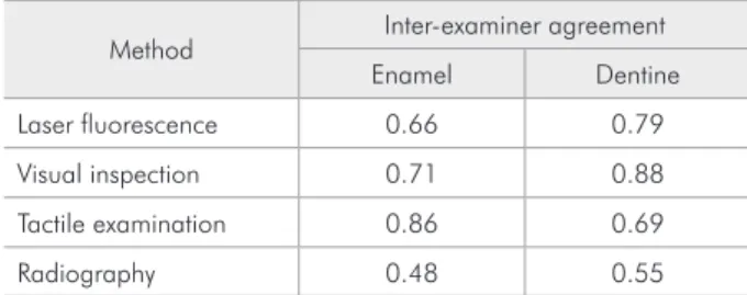
reproduc-ibility at the enamel (0.48) and dentine thresholds (0.55) (Table 2).
Discussion
Our study aimed at testing the conventional methods, as well as the LF method, for detecting secondary caries lesions around amalgam restora-tions in primary teeth. Although LF is an objective method, the readings can be inluenced by the pres-ence of calculus and dental plaque,9,15,17 time of dry-ing,15,17 the professional’s experience and training18,19 and storage of the specimens for in vitro studies.20 Nevertheless, when cut-off points are adjusted for each examination separately, no signiicant differ-ences have been found.21
The cut-off points will determine a limit between health and disease. Low cut-off points for LF have been related to high sensitivity values.17,22
In vitro studies generally have found lower cut-off points than in vivo studies. This fact is prob-ably related to time and conditions of storage of the specimens.20,23 Previous studies on secondary caries detection have also shown the difference mentioned above between cut-off points obtained by in vitro and in vivo studies. This difference has been report-ed as being around 10 arbitrary units.7,11
In our study, the observed cut-off points were lower than those found in previous studies, both at the enamel and dentine thresholds.7,11 Considering that the teeth used in our study were obtained from a bank of human teeth, and that it was impossible to
determine the storage time precisely, we assumed that the low cut-off values may have been a result of that.
Regarding the cut-off points for the conventional methods tested, an inversion was observed between the cut-off points found for radiography and for the tactile examination, in that higher cut-off points were found in enamel than in dentine. This fact con-tributes to emphasize the lower reliability of the test-ed methods.24 Hence, tactile examination and radi-ography would not be the most appropriate methods to detect secondary caries at the dentine threshold, which disagrees with previous indings related to ra-diographic examination,7,11,25 but agrees with previ-ous results related to tactile examination.26
Despite the limitation of the visual index used in detecting early secondary caries due to the dif-iculty in distinguishing discolorations originated from the restoration or from demineralizations,6 the visual inspection presented the highest sensitivity
Method Best cut-off point AZ Sensitivity Specificity Accuracy Enamel (D2)
Laser Fluorescence 1 0.73 a 0.58 a 0.84 a 0.65 a Visual Inspection 0 0.86 b 0.78 b 0.86 a 0.80 b Tactile Examination 2 0.70 a, c 0.41 c 0.93 a 0.56 a Radiography 2 0.59 c 0.27 d 0.90 a 0.46 c
Dentine (D3)
Laser Fluorescence 1.6 0.68 a 0.56 a 0.84 a 0.69 a Visual Inspection 1 0.84 b 0.75 b 0.80 a 0.77 a Tactile Examination 1 0.75 a 0.74 b 0.62 b 0.68 a Radiography 1 0.65 a 0.48 a 0.72 a 0.59 a
Different letters in the same column express statistically significant differences.
Table 1 - Sensitivity, specificity and accuracy of the laser fluorescence, visual inspection, radiography and tactile examination methods for detecting secondary caries lesions in primary teeth at enamel (D2) and dentine (D3) thresholds, and their performance expressed as the area under the ROC curve (Az).
Table 2 - Inter-examiner agreement for the laser fluores-cence, visual examination, radiography and tactile examina-tion methods for detecting secondary caries lesions at the enamel and dentine thresholds.
among the tested methods for both thresholds. On the other hand, some previous studies had shown that the LF performance was superior to that of the visual examination in detecting secondary caries le-sions in permanent teeth.6,8 The scoring system used for the visual inspection aims at estimating lesion depth.14 The thinness of the primary enamel13 can explain the best performance in detecting second-ary enamel lesions in primsecond-ary teeth. The worse per-formance at the dentine threshold probably relects the major dificulty in detecting dentinal caries not frankly cavitated.
Similar values of speciicity for all methods at the enamel threshold mean that all methods performed similarly in identifying a sound site around an amal-gam restoration. All methods presented speciicity superior to 0.80, which is a minimum value to as-sure a minimum false-positive fraction.11
The tactile examination generally presents high sensitivity and low speciicity, resulting in a high number of false-positives.24 Nevertheless, in our study the tactile examination presented a combina-tion of high speciicity and low sensitivity. The gen-tle use of the explorer probe may have caused that. The probe was used without any pressure along the margins of the restoration in order to evaluate the presence of ditches and softened dental tissues. In spite of that, the other methods presented perfor-mances superior to that of the tactile examination, emphasizing that it is not the most indicated for de-tecting secondary caries lesions in primary teeth.
The low sensitivity and accuracy of radiography in detecting enamel secondary caries had already been expected because these lesions are rarely de-tected in bitewings radiographs.7,14,27 In addition, our study did not observe a good performance of this method at the dentine threshold, which does not corroborate the indings of other studies in which ra-diography presented acceptable values of sensitivity and speciicity in the detection of advanced second-ary caries.7,11 This divergence can be attributed to the presence of secondary lesions in the wall of the cav-ity, the presence of restorative material in the buccal or lingual surface or the occurrence of a shadow ef-fect of the restorative material,28 which could make the detection of a demineralization related to
sec-ondary caries in a bitewing radiograph dificult. The visual inspection showed the highest accura-cy in detecting secondary lesions in enamel. In den-tine, the visual inspection was similar to LF in the detection of this kind of lesion. Thus, the suggestion of combining both methods in order to improve the detection of secondary caries lesions around amal-gam restorations could be considered. This com-bination, however, did not increase the accuracy in detecting secondary caries around composites.8 Considering that this hypothesis has not been tested yet for amalgam restorations, future studies should be encouraged in order to verify that.
It is expected that objective methods should pres-ent higher agreempres-ent rates than subjective ones. Pre-vious studies on the detection of secondary lesions have already demonstrated this claim.6 Nevertheless, the highest inter-examiner agreement in the present study was observed for the tactile examination, at the enamel threshold, and for the visual inspection, at the dentine threshold. The examiner’s training in using visual and tactile criteria,29 the use of scoring systems and/or individual mistakes in operating the LF device30 can be possible explanations for this ob-servation.
Considering the use of a single method to detect secondary caries lesions both in enamel and den-tine, it would be recommendable that a method pre-senting high sensitivity for detecting initial (enamel) lesions and high speciicity for detecting deep (den-tine) lesions be chosen. Thus, it would be possible to adopt preventive measures as soon as possible and avoid the unnecessary replacement of restorations.6 In our study, the visual inspection combined both features with a high reliability, emphasizing its in-dication as the most effective method in detecting secondary caries lesions in primary teeth.
Conclusion
The visual inspection presented the best perfor-mance in detecting enamel and dentine secondary caries in primary teeth restored with amalgam.
Acknowledgements
References
1. Kidd EA. Diagnosis of secondary caries. J Dent Educ. 2001;65(10):997-1000.
2. Elderton RJ. Clinical studies concerning re-restoration of teeth. Adv Dent Res. 1990;4:4-9.
3. Kidd EA, Toffenetti F, Mjor IA. Secondary caries. Int Dent J. 1992;42(3):127-38.
4. Kidd EA, Joyston-Bechal S, Beighton D. Marginal ditching and staining as a predictor of secondary caries around amal-gam restorations: a clinical and microbiological study. J Dent Res. 1995;74(5):1206-11.
5. Gonzalez-Cabezas C, Fontana M, Gomes-Moosbauer D, Stookey GK. Early detection of secondary caries using quanti-tative, light-induced fluorescence. Oper Dent. 2003;28(4):415-22.
6. Ando M, Gonzalez-Cabezas C, Isaacs RL, Eckert GJ, Stookey GK. Evaluation of several techniques for the detection of sec-ondary caries adjacent to amalgam restorations. Caries Res. 2004;38(4):350-6.
7. Bamzahim M, Shi XQ, Angmar-Mansson B. Secondary caries detection by DIAGNOdent and radiography: a comparative in vitro study. Acta Odontol Scand. 2004;62(1):61-4. 8. Boston DW. Initial in vitro evaluation of DIAGNOdent for
detecting secondary carious lesions associated with resin com-posite restorations. Quintessence Int. 2003;34(2):109-16. 9. Hibst R, Paulus R, Lussi A. Detection of occlusal caries by
la-ser fluorescence: Basic and Clinical investigations. Med Lala-ser Appl. 2001;16:205-13.
10. Bader JD, Shugars DA. A systematic review of the performance of a laser fluorescence device for detecting caries. J Am Dent Assoc (1939). 2004;135(10):1413-26.
11. Bamzahim M, Aljehani A, Shi XQ. Clinical performance of DIAGnodent in the detection of secondary carious lesions. Acta Odontol Scand. 2005;63(1):26-30.
12. Boston DW, Sauble JE. Evaluation of laser fluorescence for dif-ferentiating caries dye-stainable versus caries dye-unstainable dentin in carious lesions. Am J Dent. 2005;18(6):351-4. 13. Mortimer KV. The relationship of deciduous enamel structure
to dental disease. Caries Res. 1970;4(3):206-23.
14. Ekstrand KR, Ricketts DNJ, Kidd EAM. Reproducibility and accuracy of three methods for assessment of demineralization depth of the occlusal surface: an in vitro examination. Caries Res. 1997;31(3):224-31.
15. Mendes FM, Hissadomi M, Imparato JCP. Effects of drying time and the presence of plaque on the in vitro performance of laser fluorescence in occlusal caries of primary teeth. Caries Res. 2004;38(2):104-8.
16. Yazici AR, Baseren M, Gokalp S. The in vitro performance of laser fluorescence and caries-detector dye for detecting re-sidual carious dentin during tooth preparation. Quintessence Int. 2005;36(6):417-22.
17. Lussi A, Imwinkelried S, Pitts N, Longbottom C, Reich E. Performance and reproducibility of a laser fluorescence system for detection of occlusal caries in vitro. Caries Res. 1999;33(4):261-6.
18. El-Housseiny AA, Jamjoum H. Evaluation of visual, explorer, and a laser device for detection of early occlusal caries. J Clin Pediatr Dent. 2001;26(1):41-8.
19. Fung L, Smales R, Ngo H, Moun G. Diagnostic comparison of three groups of examiners using visual and laser fluores-cence methods to detect occlusal caries in vitro. Aust Dent J. 2004;49(2):67-71.
20. Baseren NM, Gokalp S. Validity of a laser fluorescence system (DIAGNOdent) for detection of occlusal caries in third mo-lars: an in vitro study. J Oral Rehabil. 2003;30(12):1190-4. 21. Reis A, Mendes FM, Angnes V, Angnes G, Grande RHM,
Loguercio AD. Performance of methods of occlusal caries detection in permanent teeth under clinical and laboratory conditions. J Dent. 2006;34(2):89-96.
22. Francescut P, Lussi A. Correlation between fissure discolor-ation, Diagnodent measurements, and caries depth: an in vitro study. Pediatr Dent. 2003;25(6):559-64.
23. Francescut P, Zimmerli B, Lussi A. Influence of different stor-age methods on laser fluorescence values: a two-year study. Caries Res. 2006;40(3):181-5.
24. Bader JD, Shugars DA, Bonito AJ. A systematic review of the performance of methods for identifying carious lesions. J Public Health Dent. 2002;62(4):201-13.
25. Rudolphy MP, van Loveren C, van Amerongen JP. Grey dis-coloration for the diagnosis of secondary caries in teeth with class II amalgam restorations: an in vitro study. Caries Res. 1996;30(3):189-93.
26. Rudolphy MP, van Amerongen JP, Penning C, ten Cate JM. Grey discolouration and marginal fracture for the diagnosis of secondary caries in molars with occlusal amalgam restora-tions: an in vitro study. Caries Res. 1995;29(5):371-6. 27. Rocha RO, Ardenghi TM, Oliveira LB, Rodrigues CRMD,
Ciamponi AL. In vivo Effectiveness of Laser Fluorescence Compared to Visual Inspection and Radiography for the Detection of Occlusal Caries in Primary Teeth. Caries Res. 2003;37(6):437-41.
28. Espelid I, Tveit AB, Erickson RL, Keck SC, Glasspoole EA. Radiopacity of restorations and detection of secondary caries. Dent Mater. 1991;7(2):114-7.
29. Verdonschot EH, Angmar-Mansson B, ten Bosch JJ, Deery CH, Huysmans MC, Pitts NB et al. Developments in caries diagnosis and their relationship to treatment decisions and quality of care. ORCA Saturday Afternoon Symposium 1997. Caries Res. 1999;33(1):32-40.