Periodontics
Guilherme Emerson Barrella(a) Ivana Barbosa Suffredini(b) Fernanda Vieira Ribeiro(a) Fabiano Ribeiro Cirano(a) Suzana Peres Pimentel(a)
(a)Department of Periodontics, School of Dentistry, Paulista University (UNIP), São Paulo, SP, Brazil.
(b) Centre for Research in Biodiversity, Extraction Laboratory, Paulista University (UNIP), São Paulo, SP, Brazil.
Corresponding author:
Suzana Peres Pimentel
E-mail: [email protected]
Received for publication on Sep 27, 2011 Accepted for publication on Jan 04, 2012
Evaluation of the effect of an organic
extract obtained from
Ipomoea alba
L.
on experimental periodontitis in rats
Abstract: The aim of this study was to evaluate the effect of an organic extract obtained from Ipomoea alba L. (Convolvulaceae or OE 1493), on experimental periodontal disease in rats. Periodontitis was induced in thirty six Wistar rats: a irst mandibular molar was randomly assigned to receive a ligature, whereas the contralateral molar was left unligated. Animals were randomly assigned to two groups and treated topically, three times a day, for 11 days, as follows: Control Group - vehicle-treated (n = 18), and Test Group - OE 1493-treated (n = 18). The rats were sacri-iced on the 12th day. Morphometrical measurements from the
cementoe-namel junction to the bone crest were performed to determine alveolar bone loss, using standardized photographs. Single- and multi-dose acute toxicity assays were carried out after OE 1493 treatment. Morphometri-cal analysis demonstrated that topiMorphometri-cally-administered OE 1493 showed no effect on reducing bone loss when compared with the control group (p > 0.05). In addition, OE 1493 did not present toxicity. Within the lim-its of this investigation, it may be concluded that OE 1493 did not show any positive inluence on the progression of ligature-induced periodonti-tis in rats, when administered according to the regimen used in the pres-ent study.
Descriptors: Drug Toxicity; Periodontitis; Rats; Administration, Topical; Anti-Bacterial Agents.
Introduction
The presence of bacterial plaque is the main etiologic factor involved in the initiation and progression of periodontitis.1 The aim of periodontal
therapy is to remove periodontal pathogens by providing patients with adequate oral hygiene instructions combined with professional mechani-cal plaque control.2 However, this conventional treatment strategy is not
always successful and the addition of chemical agents to dentifrices and mouth rinses has been suggested to enhance their eficacy as adjuncts to the therapeutic approach for achieving better oral health.3
Natural products represent a signiicant source of substances for use in the control of oral diseases, especially in managing plaque-related dis-eases such as gingivitis.4-8 Brazil is said to be the richest country in the
world in terms of its biodiversity, and few data are available about the pharmacological and chemical potential of its lora as a source of new medicines. In previous investigations developed by our research group,
an organic extract obtained from Ipomoea alba L. (OE 1493 or Convolvulaceae) showed signiicant
in vitro activity against Streptococcus mutans, S. sanguinis and Enterococcus faecalis (unpublished data). However, there is no information available as regards the effect of OE 1493 on reducing bone re-sorption in experimental periodontitis.
Thus, the purpose of this study was to make a morphometric evaluation of OE 1493 with respect to its ability to reduce the development of periodon-titis in rats.
Methodology
Plant collection and plant extract preparation
Ipomoea alba L. (OE 1493 or Convolvulaceae) was collected in igapó forests, Amazon rain forest, Manaus, AM, under permit from the agency con-trolling environmental and natural resources, “ lnsti-tuto Brasileiro do Meio Ambiente e dos Recursos Naturais Renováveis” (IBAMA). Access to its ge-netic patrimony was obtained under a permit from the Genetic Patrimony Management Council, “ Con-selho de Gestão do Patrimônio Genético”.9,10
Plants were dried by air circulation at a tempera-ture of up to 40°C. After that, dried plant material was ground in a hammer mill (Holmes) before being macerated with a mixture of dichloromethane and methanol (1:1) for 24 hours. The organic extract was evaporated under reduced pressure (Büchi) and was lyophilized. The extract was kept in a freezer until use. The maceration process used to obtain the extract is well known11 and does not alter chemical
or physical properties of the natural components of the extract. Extracts were prepared at a concentra-tion of 100 mg/mL, using 10% Tween 80 in water as vehicle.
Experimental periodontal disease
Thirty six male Wistar rats (210–320 g), nine weeks old, were used for the induced periodontitis experiment in the present study. The animals were acclimatized to the housing conditions during the course of two weeks, and a 12-hour light and dark cycle was applied. They were housed six per cage at a permanent temperature of 21°C. Standard rat
chow pellets and water ad libitum were available. The sample size of this investigation was based on previous published studies.12-14
General anesthesia was obtained by intra-mus-cular administration of ketamine hydrochloride (10 mg/kg) (Dopalen, Agribrands Brasil Ltda., Paulínia, Brazil) and xylazine hydrochloride (10 mg/ kg) (Rompun, Bayer S.A., São Paulo, Brazil). One of the mandibular irst molars of each animal was randomly assigned to receive a cotton ligature (Cor-rente Algodão no. 10; Coats Cor(Cor-rente, São Paulo, Brazil) in a cervical position. Briely, the thread was introduced into the proximal space between the irst and second molars and two knots were tied on the mesial face of the irst molar. The ligatures were kept in position in order to allow bioilm accumula-tion for 11days.15,16 The contralateral tooth was left
unligated for use as a control.
After this, animals were randomly assigned to one of the experimental groups:
• Control Group - vehicle-treated (n = 18) and
• Test Group - OE 1493-treated (n = 18).
Treatments were topically applied with a 1-mL syringe, using 0.3 mL of the respective substances, 3 times a day (7 a.m.; 1 p.m. and 8 p.m.) for 11 days.
To apply the drug, the animals were immobilized by one researcher, while another person applied the substance, enabling the molar area to be visualized to ensure adequate administration.
The protocol was approved by the Paulista Uni-versity Institutional Animal Care and Use Commit-tee (036/10 CEP/ICS/UNIP).
Morphometric analysis
The animals were sacriiced on the 12th day of
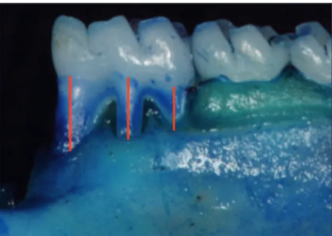
Japan) ixed on a tripod to keep the camera paral-lel to the ground at the minimal focal distance. The specimens were ixed in wax with their occlusal plane kept parallel to the ground and their long axis perpendicular to the camera. Photographs were tak-en of the buccal aspects. To validate measuremtak-ent conversions, a millimetric ruler was photographed together with all specimens.17 Alveolar bone loss
was determined on the buccal surface of the man-dibular irst molars by the distance of the CEJ from the alveolar bone crest (ABC). Measurements were made along the axis of each root in three regions of the irst molar (three roots) (Figure 1). The total al-veolar bone loss was obtained by taking the sum of the linear recordings from the buccal tooth surface of the roots and dividing by three.
The measurements were taken by the same brated masked examiner, after intraexaminer cali-bration, during which 8 non-study images present-ing alveolar bone loss similar to those in the present study were evaluated. The examiner took the linear measurements of all photographs twice within 24 hours. The Intra Class correlation showed 97% re-producibility.
Toxicity assay
OE 1493 single-dose acute toxicity assay
Nine mice weighing 25–30 g, 6 to 9 weeks old, were used in single-dose acute toxicity assays. Mice
were brought to the vivarium at least two days in advance to become acclimatized to the housing con-ditions. A 12-hour light and dark cycle was applied and water and food were offered ad libitum. Five animals were kept in one cage and four in another. At the end of the experiments, the animals were sac-riiced in a CO2 gas chamber.
In order to prospect toxicity and obtain lethal dose tendency, three mice per group were used to as-sess lethality in each dose.18 Doses of 5.0 g/kg, 2.5 g/
kg and 1.25 g/kg of OE 1493 were intraperitoneally administered. The occurrence of massive toxic re-actions and/or death was observed in the irst 15, 30, 60, 120 and 180 minutes after administration of treatments and every 24 hours in the following 14 days. The corresponding volume of vehicle was administered to the control group. Animals were weighed three times during the observation period, at days 1, 7 and 14. At the end of the period of ob-servation, necropsy was performed on each animal to look for macroscopic reactions in the liver, gut, heart and kidneys. Latency for death was assessed.
OE 1493 multi-dose acute toxicity assay
The multi-dose acute toxicity assay was per-formed using the rats included in the induced peri-odontitis. OE 1493 was diluted in 10% Tween 80/ water to a concentration of 100 mg/mL, and 0.3 mL of this solution was administered three times a day, totaling 910 mg of extract a day. Animals placed in glass cages were observed for toxic reactions in gen-eral activity, central nervous system, autonomous nervous system, psychomotricity and in sensorial reactions.
Statistical analyses
To test the null hypothesis that OE 1493 had no inluence on alveolar bone loss, an intergroup analy-sis was performed using Student’s-t test. In addition, Student’s-t test was used for intragroup compari-sons between ligated and unligated teeth. The sig-niicance level established for all analyses was 5% (p < 0.05). Linear regression was used to obtain the tendency of lethal dose in the toxicity assay (Graph-Pad InStat version 3.00 for Windows 95, Graph(Graph-Pad Software, San Diego, USA).
Results
Clinically, at the time of sacriice, signs of gingi-val inlammation were observed in the ligated teeth of all the groups. No signs of gingival inlammation were observed in the unligated teeth.
Morphometric analysis
Intragroup analysis showed that ligatures placed around the teeth were able to promote bone loss when compared with unligated teeth (p < 0.05) (Con-trol Group: 1.32 ± 0.11 mm and 1.73 ± 0.16 mm, for unligated and ligated teeth, respectively; and Test Group: 1.39 ± 0.13 mm and 1.69 ± 0.08 mm, for unligated and ligated teeth, respectively). Intergroup analysis comparing ligated teeth revealed no differ-ences in bone loss between groups (1.73 ± 0.16 mm and 1.69 ± 0.08 mm for Control and Test Group, respectively) (p > 0.05) (Figure 2).
Toxicity assay
Results obtained from the single-dose acute tox-icity assay performed with mice that received OE 1493 intraperitoneally and were under observation for 14 days showed a tendency of LD50 = 2.19g/ kg, after linear regression (correlation coeficient r = 0.7559; r2 = 0.5714; F = 1.333). Latency for
death was obtained for three doses tested to obtain LD50; the 5 g/kg dose showed latency for death of 95.67 ± 15.31 h; the 2.5 g/kg dose showed latency
for death of 300.00 ± 519.62 h and the 1.25 g/kg dose did not cause the mice to die in a period of 14 days. The standard deviation observed in the latency for death after administration of the 2.5 g/kg dose was noted to be extremely high, and this was caused by the death of only one mouse in the group of three mice. Multi-dose acute toxicity tests showed that OE 1493 did not show any tendency towards tox-icity when 0.3 mL was locally administered three times a day for 11 days. No signs of toxicity were observed when compared with the control group.
Discussion
Over the last few years, new natural medici-nal products have been described as an important source of substances for use in the treatment of oral inlammatory conditions.13,19-22 In this context, it
has been evidenced that OB 1493 presented effective anti-bacterial activity in vitro (unpublished data). However, until now, there has been no information available in the literature as regards the effect of OB 1493 on experimental periodontitis. Therefore, this investigation was designed to determine the effect of OB 1493 on attenuating alveolar bone loss in liga-ture-induced periodontitis in rats. The morphomet-rical assays of the present study demonstrated that topically-administered OB 1493 had no inluence on reducing alveolar bone resorption in experimental periodontitis, presenting bone loss comparable with that of the Control group.
Although very few studies have examined the role of natural products in controlling periodonti-tis, Botelho et al.8 recently tested a locally-applied
carvacrol gel (Lippia sidoides derivatives) and deter-mined its eficacy in controlling bone loss in experi-mental periodontitis in rats. The authors showed evidence that this natural chemical agent preserved alveolar bone resorption and showed anti-inlam-matory and antibacterial activities in periodontitis. In contrast, the treatment using a plant extract de-rived from Ipomoea alba, evaluated in the present study, was not able to reduce the alveolar bone loss promoted by ligature-induced periodontitis. To the best of our knowledge, no pre-clinical or clinical in-vestigations have studied the impact of OB 1493 on periodontitis, hampering comparisons with the
sults of the present research.
In the present study, the ligature was placed around the irst molar teeth for 11 days. The ratio-nale for using this experimental period is based on previous investigations.13,15,23 According to Lima et
al.,23 placement of a ligature around the molar teeth
of rats leads to the presence of inlammatory cells, including osteoclasts, beneath the ligature. These authors reported that the substantial alveolar bone loss originated on day 3 of periodontitis induction reached a maximum between days 7 and 11. In line with this data, Kuhr et al.24 revealed that the
appli-cation of this model of periodontitis induction can only be recommended for short periods (less than 15 days). According to these authors, who evaluated the periodontal destruction following experimental-ly induced periodontitis in rats over a 60-day obser-vation period, it was demonstrated that the ligature-induced bone loss increased most from day 1 to day 15, whereas on days 30 and 60 slighter increases in bone loss were observed, supporting the period of periodontitis induction used in the present study.
Previously unpublished data indicated that OE 1493 exerts an important antibacterial activity against Streptococcus mutans, S. sanguinis and En-terococcus faecalis. Recently, Duarte et al.,25 in their
study in rats, reported that high proportions of the human host–compatible species, such as Streptococ-cus-like species, were observed around ligated teeth after periodontitis induction. Previous research in human subjects, observing the sequential develop-ment of oral bioilm, has established that these spe-cies are the early colonizers of teeth, creating an ad-equate microenvironment for the formation of the late colonizers, such as well-recognized periodontal pathogens.26 In addition, other species belonging to
the same genus have also shown antimicrobial ac-tivities, such as I. tyrianthina27 and I. leptophylla.28
Indeed, Locher et al.29 demonstrated in vitro that a
Hawaiian Ipomoea sp. produced anti-fungal activity against Microsporum canis, Trichophyton rubrum
and Epidermophyton loccosum. Although previ-ous studies have indicated the antimicrobial effect of OB 1493 or of similar genes,25,28-30 the possible
anti-inlammatory activities of this natural product in bone metabolism and periodontal disease remain
to be investigated to explain, at least partly, the out-comes of the present study. Indeed, future research using different doses or administration regimes of this plant extract would contribute to a better un-derstanding of the impact of OB 1493 on the pro-gression of ligature-induced periodontitis.
In the present investigation, a toxicity assay of OE 1493 was also performed. A single-dose acute toxicity evaluation was done in mice, and for ethi-cal and legal reasons, three animals were used per group, providing suficient information to obtain the tendency towards toxicity. Our data indicated that OE 1493 did not show any signs of toxicity in the central nervous system, autonomous nervous system, psychomotricity or sensorial systems. For this reason, after being submitted to toxic assays, OE 1493 was selected for evaluation in the present pharmacological study in rats.
The signiicant antibacterial activity of OE 1493, whose minimal inhibitory concentrations and mini-mal bactericidal concentrations were lower than 0.04 mg/mL, indicated a low toxicity (unpublished data). For this reason, the present study used local administration of 0.3 mL of 10% OE 1493 in Tween 80 to evaluate the prevention of bone resorption in experimental periodontal diseases in rats.
In summary, although in vitro studies have shown a signiicant antibacterial activity of OE 1493 and no signs of toxicity were observed when compared with the control group, the topical use of this drug was not able to reduce bone resorption in experimental periodontitis. Further investigations using distinct protocols for OE 1493 use may be necessary to determine its therapeutic effects in con-trolling periodontal breakdown. The biodiversity of the Amazon may be considered a substantial source of new lead compounds to be assessed.
Conclusion
References
1. Kinane DF, Preshaw PM, Loos BG, Working Group 2 of Sev-enth European Workshop on Periodontology. Host-response: understanding the cellular and molecular mechanisms of host-microbial interactions--consensus of the Seventh Euro-pean Workshop on Periodontology. J Clin Periodontol. 2011 Mar;38 Suppl 11:44-8.
2. Apatzidou DA, Kinane DF. Nonsurgical mechanical treat-ment strategies for periodontal disease. Dent Clin North Am. 2010 Jan;54(1):1-12.
3. Bonito AJ, Lux L, Lohr KN. Impact of local adjuncts to scaling and root planing in periodontal disease therapy: a systematic review. J Periodontol. 2005 Aug;76(8):1227-36.
4. Newman DJ, Cragg DM. Natural Products as sources of new drugs over the last 25 years. J Nat Prod. 2007 Mar;70(3):461-77.
5. Botelho MA, Martins JG, Ruela RS, Rachid I, Santos JA, Soares JB, et al. Protective effect of locally applied carvacrol gel on ligature-induced periodontitis in rats: a tapping mode AFM study. Phytother Res. 2009 Oct;23(10):1439-48. 6. DiSilvestro RA, DiSilvestro DJ, DiSilvestro DJ. Pomegranate
extract mouth rinsing effects on saliva measures relevant to gingivitis risk. Phytother Res. 2009 Aug;23(8):1123-7. 7. Leal IC, dos Santos KR, Itabaiana Júnior I, Antunes OA,
Porzel A, Wessjohann L, et al. Ceanothane and lupane type triterpenes from Zizyphus joazeiro - an anti-staphylococcal evaluation. Planta Med. 2010 Jan;76(1):47-52.
8. Botelho MA, Martins JG, Ruela RS, Santos JA, Soares JB, França MC, et al. Protective effect of locally applied carvacrol gel on ligature-induced periodontitis in rats: a tapping mode AFM study. Phytother Res. 2009 Oct;23(10):1439-48. 9. Suffredini IB, Paciencia ML, Varella AD, Younes RN.
Anti-bacterial activity of Brazilian Amazon plant extracts. Braz J Infect Dis. 2006 Dec;10(6):400-2.
10. Suffredini IB, Paciencia ML, Varella AD, Younes RN. In vitro
cytotoxic activity of Brazilian plant extracts against human lung, colon and CNS solid cancers and leukemia. Fitoterapia. 2007 Apr;78(3):223-6. Epub 2007 Feb 6.
11. Cragg GM, Boyd MR, Cardellina JH 2nd, Newman DJ, Snader
KM, McCloud TG. Ethnobotany and drug discovery: the experience of the US National Cancer Institute. Ciba Found Symp.1994; 185:178-90; 190-6.
12. Benatti BB, Nogueira-Filho GR, Diniz MC, Sallum EA, Sallum AW, Nociti Jr FH. Stress may enhance nicotine effects on periodontal tissues. An in vivo study in rats. J Periodontal Res. 2003 Jun;38(3):351-3.
13. Botelho MA, Rao VS, Carvalho CB, Bezerra-Filho JG, Fon-seca SG, Vale ML, et al. Lippia sidoides and Myracrodruon urundeuva gel prevents alveolar bone resorption in experi-mental periodontitis in rats. J Ethnopharmacol. 2007 Sep 25;113(3):471-8.
14. Botelho MA, Rao VS, Montenegro D, Bandeira MA, Fon-seca SG, Nogueira NA, et al. Effects of a herbal gel
con-taining carvacrol and chalcones on alveolar bone resorption in rats on experimental periodontitis. Phytother Res. 2008 Apr;22(4):442-9.
15. Toker H, Ozan F, Ozer H, Ozdemir H, Eren K, Yeler H. A morphometric and histopathologic evaluation of the effects of propolis on alveolar bone loss in experimental periodontitis in rats. J Periodontol. 2008 Jun;79(6):1089-94.
16. Queiroz-Junior CM, Pacheco CM, Fonseca AH, Klein A, Caliari MV, de Francischi JN. Myeloperoxidase content is a marker of systemic inflammation in a chronic condition: the example given by the periodontal disease in rats. Mediators Inflamm. 2009 Dec 31; [Epub ahead of print] Available from: http://www.hindawi.com/journals/mi/2009/760837/. 17. Fernandes MI, Gaio EJ, Oppermann RV, Rados PV, Rosing
CK. Comparison of histometric and morphometric analyses of bone height in ligature-induced periodontitis in rats. Braz Oral Res. 2007 Jul-Sep;21(3):216-21.
18. Botham PA. Acute systemic toxicity – prospects for tiered testing strategies. Toxicol In vitro.2004 Apr;18(2):227-30. 19. Calixto JB, Campos MM, Otuki MF, Santos AR.
Anti-in-flammatory compounds of plant origin. Part II. modulation of pro-inflammatory cytokines, chemokines and adhesion molecules. Planta Med. 2004 Feb;70(2):93-103.
20. Calixto JB, Otuki MF, Santos AR. Anti-inflammatory com-pounds of plant origin. Part I. Action on arachidonic acid pathway, nitric oxide and nuclear factor kappa B (NF-kap-paB). Planta Med. 2003 Nov;69(11):973-83.
21. Cai X, Li C, Du G, Cao Z. Protective effects of baicalin on ligature-induced periodontitis in rats. J Periodontal Res. 2008 Feb;43(1):14-21.
22. Napimoga MH, Benatti BB, Lima FO, Alves PM, Campos AC, Pena-Dos-Santos DR, et al. Cannabidiol decreases bone resorption by inhibiting RANK/RANKL expression and pro-inflammatory cytokines during experimental periodontitis in rats. Int Immunopharmacol. 2009 Feb;9(2):216-22. 23. Lima V, Bezerra MM, Alencar VBM, Vidal FDP, Rocha FAC,
Brito GAC. Effects of chlorpromazine on alveolar bone loss in experimental periodontal disease in rats. Eur J Oral Sci 2000 Apr;108(2):123-9.
24. Kuhr A, Popa-Wagner A, Schmoll H, Schwahn C, Kocher T. Observations on experimental marginal periodontitis in rats. J Periodontal Res. 2004 Apr;39(2):101-6.
25. Duarte PM, Tezolin KR, Figueiredo LC, Feres M, Bastos MF. Microbial profile of ligature-induced periodontitis in rats. Arch Oral Biol. 2010 Feb;55(2):142-7.
26. Socransky SS, Haffajee AD. Periodontal microbial ecology. Periodontology 2000. 2005;38(1):135-87.
27. León-Rivera I, Mirón-Lopez G, Molina-Salinas GM, Herre-ra-Ruiz M, Estrada-Soto S, Del Carmen Gutiérrez M, et al.
28. Barnes CC, Smalley MK, Manfredi KP, Kindscher K, Loring H, Sheeley DM. Characterization of an anti-tuberculosis resin glucoside from the prairie medicinal plant Ipomoea
lepto-phylla. J Nat Prod. 2003 Nov; 66(11):1457-62.
29. Locher CP, Burch MT, Mower HF, Berestecky J, Davis H, Van Poel B, et al. Anti-microbial activity and anti-complement
activity of extracts obtained from selected Hawaiian medicinal plants. J Ethnopharmacol. 1995 Nov 17;49(1):23-32. 30. Goun E, Conningham G, Chu D, Nguyen C, Miles D.