Cariology
Tarcísio Jorge Leitão(a) Livia Maria Andaló Tenuta(a) Guilherme Ishi(a)
Jaime Aparecido Cury(a)
(a)Department of Physiological Sciences,
Piracicaba Dental School, University of Campinas, Piracicaba, SP, Brazil.
Corresponding author:
Livia Maria Andaló Tenuta E-mail: [email protected]
Received for publication on Nov 04, 2011 Accepted for publication on Fev 02, 2012
Calcium binding to
S. mutans
grown in
the presence or absence of sucrose
Abstract: Sucrose is the most cariogenic dietary carbohydrate because it is a substrate for insoluble extracellular polysaccharide (IEPS) produc-tion in dental bioilms, which can proporproduc-tionally decrease bacterial den-sity and, consequently, the number of bioilm calcium (Ca) binding sites. Ca bound to bacterial cell walls can be released into the bioilm luid during a cariogenic challenge, reducing the driving force for mineral dis-solution provoked by the pH drop. Thus, we investigated the effect of an IEPS-rich extracellular matrix on bacterial Ca binding after treatment with Ca solutions. Streptococcus mutans Ingbritt 1600 was cultivated in culture broths supplemented with 1.0% sucrose or 0.5% glucose + 0.5% fructose. The IEPS concentration in bacterial pellets was determined after alkaline extraction. Bacterial pellets were treated with 1 mM or 10 mM Ca++ solutions at 37
°
C for 10 to 60 min. Ca binding to bacterial pellets, determined after acid extraction using the Arsenazo III reagent, was fast and concentration dependent. Although the IEPS concentration was approximately ten times higher in bacterial pellets cultivated in su-crose as compared to its monossaccharides, bound Ca concentration af-ter Ca treatment was similar in both conditions. These results suggest that IEPS may not inluence the amount of Ca bound to reservoirs of dental bioilms.Descriptors: Sucrose; Streptococcus mutans; Polysaccharides; Calcium; Dental Caries.
Introduction
There is considerable evidence demonstrating that sucrose is the most cariogenic carbohydrate from the human diet,1 with enhanced carioge-nicity as compared to its component monosaccharides, glucose and fruc-tose.2 The reason for this relies on the fact that sucrose, besides being fermented to acids, is also used by oral bacteria to synthesize extracellu-lar polysaccharides (EPS).3 These EPS, mainly the insoluble ones (IEPS), play a signiicant role on the adhesion and accumulation of cariogenic streptococci on the tooth surface, especially S. mutans.4 In addition, they change the bioilm structure, resulting in increased porosity,5 which al-lows fermentable substrates to diffuse and be metabolized in the deepest parts of the bioilm.6
Furthermore, since IEPS decrease the bacterial density of bioilms,7,8 they may reduce the amount of bioilm calcium (Ca) binding sites9, which could also inluence bioilm cariogenicity. Ca bound to bacterial cell Declaration of Interests: The authors
walls might function as a source of mineral ions to the bioilm luid during a pH drop,9 acting as a min-eral buffer by helping to maintain the saturation of plaque luid with respect to tooth mineral, a factor that largely governs its dissolution.10
Therefore, we hypothesized that the number of binding sites for Ca would be reduced in dental bioilm formed in the presence of sucrose, and we investigated this effect by evaluating the rate of Ca binding to S. mutans grown in the presence of su-crose or its component monosaccharides in vitro.
Methodology
Experimental designS. mutans Ingbritt 1600 was cultivated in the presence of sucrose (to allow the production of ex-tracellular polysaccharides) or its component mono-saccharides, glucose and fructose (not substrates for EPS synthesis). Bacterial pellets obtained by centrifugation were analyzed for IEPS, bacterial proteins (as an indicator of bacterial density11), and baseline Ca. Additional pellets were treated with 1 or 10 mM Ca++ (CaCl
2), buffered with 0.05 M PIPES (piperazine-N, N’-bis [2-ethanesulphonate]; Sigma Biochemicals), pH 7.0, at 37
°
C for 10, 30, or 60 min. The Ca concentrations used, 1 and 10 mM, represent the resting Ca bioilm luid concentration12 and the high Ca concentration found in bioilm luid after a pH drop, respectively.10 After the speciied equilibrium time, the bacteria were separated from the test solution by centrifugation, bound Ca was extracted from the bacterial pellet with acid treat-ment, and its concentration was determined.Bacterial preparation
S. mutans Ingbritt 1600 was cultivated in Todd-Hewitt broth (THB) (Difco Labs., Detroit, USA) sup-plemented with 1% sucrose or 0.5% glucose + 0.5% fructose for 18 h at 10% pCO2 and 37°C. Bacterial pellets were separated by centrifugation. In order to remove remnants of culture broth and unbound Ca, the pellets were sequentially washed using sonication (Vibra Cell sonicator, Sonics and Materials, Dan-bury, USA) at 7 W for 1 min, irst in 0.05 M PIPES buffer, pH 7.0, followed by 0.01 M EDTA solution,
the pellet was recovered by centrifugation. After this procedure, the pellet was spread on ilter paper to remove excess moisture. Aliquots were transferred to microcentrifuge tubes for IEPS and protein determi-nation and baseline Ca and Ca-binding analyses.
IEPS and protein determination in bacterial pellets
To extract EPS, aliquots (n = 3) of bacterial pel-lets were weighed (± 0.01 mg), suspended in 0.9% NaCl solution (1 mL/mg wet weight), sonicated at 7 W for 60 s, and centrifuged at 10,000 g for 5 min at 4
°
C to remove soluble EPS.13 IEPS was alkaline extracted from the remaining pellet14 and precipi-tated with ethanol. The carbohydrate concentration in the IEPS extract was estimated by the phenol-sul-furic acid method.15To determine bacterial proteins, aliquots of bac-terial pellets (n = 3) were irst treated with a mild alkaline solution16 to remove extracellular proteins, and the supernatant obtained by centrifugation was discarded. The precipitate was treated with a hot al-kaline solution to extract bacterial proteins,13 whose concentration was determined by the Lowry meth-od.17
Ca-binding assessment
Aliquots of S. mutans pellets were exposed to PIPES buffer (1.5 mL/10 mg of bacteria), pH 7.0, containing 1 or 10 mM Ca, for 10, 30, or 60 min (n = 2 for each time point). The high dilution was used to maintain a stable Ca concentration during the experiment. At each time point, the pellets were collected by centrifugation (21,000 g for 5 min), and the supernatant was carefully vacuum-aspirated with a micropipette under a microscope to remove any treatment solution residue. The eficacy of resid-ual Ca removal was validated by a preliminary ex-periment (data not shown) in which the amount of Ca remaining in the bacterial pellet was conirmed to be bound and not simply trapped in the bacterial pellet luid.
lets by treatment with 0.5 M HCl (0.1 mL/10 mg bacterial wet weight) for 3 h.14 The acid extract was collected after centrifugation, and the Ca concen-tration was measured using the Arsenazo III colo-rimetric reagent, after neutralization with 2.5 M NaOH.18 For the analyses, Ca standards contained HCl and NaOH in the same proportion as the sam-ples. The absorbance of the mixtures was read in 96-well microplates, using a Multiskan Spectrum (Thermo Scientiic) microplate reader at 650 nm.
Statistical analyses
Data of IEPS, bacterial proteins, and baseline Ca in untreated pellets on bioilms cultivated in the presence of sucrose or glucose + fructose were compared by the t-test. Ca binding to the bacterial pellets after treatment with Ca solutions at different times were compared by split plot ANOVA, using cultivation conditions (sucrose or glucose + fructose) as plots and Ca concentration in the treatment so-lution (1 or 10 mM) and time (10, 30, or 60 min) as subplots. The normality of error distribution and the homogeneity of variance were checked for each response variable using the SAS/LAB package (SAS software, version 8.01, SAS Institute Inc., Cary, USA), and data were transformed as suggested by the software, according to Box et al.19 The SAS sys-tem (version 9.2) was used in the analyses, and the
signiicance level was set at 5%.
Results
Comparisons of the composition of bacterial pel-lets cultivated in the presence of sucrose or glucose + fructose revealed signiicantly (p < 0.05) higher EPS and lower bacterial protein and baseline Ca concen-trations for the former condition (Table 1).
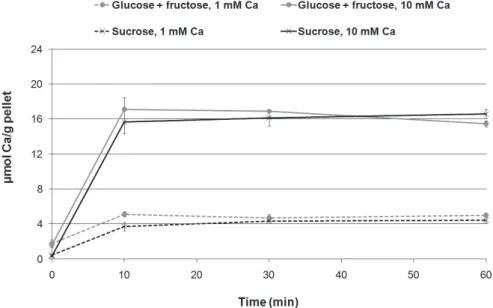
Figure 1 shows that the rate of Ca binding to the bacterial pellets cultivated either in the pres-ence of sucrose or glucose + fructose was fast, with no signiicant difference in bound Ca concentra-tion among the treatment times (p > 0.05). Higher Ca concentrations were found in bacteria treated with 10 mM Ca, as compared to those treated with
Table 1 - Insoluble extracellular polysaccharide (IEPS), pro-tein concentration (mg/g wet weight, mean ± SD; n = 3), and baseline Ca (µmol/g wet weight, mean ± SD, n = 4) in bacterial pellets according to the carbohydrate source used for S. mutans growth.
S. mutans
carbohydrate source IEPS* Bacterial protein Baseline Ca Sucrose 52.5 ± 2.5 39.3 ± 1.8 0.4 ± 0.1 Glucose + fructose 6.0 ± 0.2 75.1 ± 0.5 1.7 ± 0.3
* An inverse transformation of the IEPS data was performed to fit the as-sumptions of the t-test. Significant differences were observed between the treatments for all variables analyzed (p < 0.05).
Figure 1 - Bound Ca
1 mM Ca (p < 0.05). However, the amount of bound Ca was not signiicantly different between the two bacterial growth conditions (presence or absence of sucrose) (p > 0.05).
Discussion
In the present study, the concentration of IEPS was approximately 10 times higher in the bacterial pellets cultivated in the presence of sucrose when compared to its monosaccharides (Table 1). These results conirm the ability of this S. mutans strain to produce EPS in the presence of sucrose,6,8 as a mod-el that simulates what happens in dental bioilm.2 Also, the lower protein concentration in bacterial pellets (Table 1) is in agreement with van Houte
et al.,8 who reported decreased bacterial cell den-sity when this S. mutans strain was grown in THB supplemented with 2% sucrose as compared to the same medium with a ten times lower concentration. Considering that proteins from bacterial cell walls are Ca-binding sites,20 and EPS does not have this property,9 it would be expected that bacterial pellets grown in the presence of sucrose would have a lower ability to bind Ca per weight of bacteria. Although this was observed for the baseline Ca concentration in the bacterial pellets (Table 1), the results of Ca binding after treatment with Ca-containing solu-tions did not support this hypothesis (Figure 1), and the theoretical support to explain these results is presented below.
The concentration of bound Ca found after treatment with Ca solutions is consistent with pre-vious studies of Ca binding to streptococci strains from solutions with varying Ca concentrations,21-23 in which glucose was used as the carbohydrate source for bacterial growth. However, these authors did not evaluate the effect of different sugars used to cultivate bacteria on Ca binding, especially sucrose, which signiicantly affects the bioilm extracellular matrix composition.2,3 This was the aim of the pres-ent study.
Moreover, the results agree with those of previ-ous studies2,12,24 that did not ind a signiicant dif-ference in total Ca concentration in bioilms formed
in situ in the presence of glucose + fructose when
extracellular bioilm matrix is not able to affect whole bioilm Ca binding. In this regard, it is note-worthy that IEPS concentration represents approxi-mately 5% of the total bioilm wet weight, in agree-ment with previous in situ studies.2,13,14,24-26This low percentage, supposedly relevant to induce differ-ences between the bacterial pellets on residual Ca-binding capacity, might not be able to signiicantly affect bound Ca concentration once the bacteria are treated with Ca-containing solutions (i.e., 1 or 10 mM). Furthermore, the results suggest that the EPS concentration is not the reason for the low con-centration of inorganic ions (Ca, inorganic phospho-rus, and luoride) found in dental bioilms formed under exposure to sugars,2,3,12-14,24,26 suggesting that further research should be done to explain this bio-logical phenomenon.
Nevertheless, an alternative hypothesis to ex-plain our indings is based on the effect of sucrose to enhance the number of Ca-binding sites in bacterial plaque formed in its presence. In vivo studies have shown that dental plaque formed in the presence of sucrose has a higher amount of lipoteichoic acid,27 which might enhance Ca-binding capacity because phosphate groups have a higher afinity for Ca ions than carboxyl groups from proteins present in strep-tococcus cell walls.20 Therefore, in the presence of sucrose, a higher concentration of bacterial cell wall components with higher Ca-binding capacity could be expressed and compensate for the lower bacterial density.
Our Ca-binding kinetics results are consistent with those by Tatevossian,28 who studied the kinet-ics of Ca binding in a pool of bacterial plaque us-ing Ca ion-selective electrodes in vitro; this study showed that the binding was rapid and almost reached saturation within 10 min. Although the ex-perimental design used in the present study did not allow for reaction rate determination at times less than 10 min with good precision, the high dilution of the bacterial pellet used ensured that the concen-tration of the treatment solution did not change as binding occurred.
Thus, not only the binding capacity, but also the ki-netics of Ca release as a function of pH should be studied in further detail to provide a better under-standing of these phenomena in dental plaque in vivo.
Conclusion
Our data suggest that Ca binding to bacterial
13. Aires CP, Del Bel Cury AA, Tenuta LM, Klein MI, Koo H, Duarte S, et al. Effect of starch and sucrose on dental biofilm formation and on root dentine demineralization. Caries Res. 2008 Sep;42(5):380-6.
14. Cury JA, Rebello MA, Del Bel Cury AA. In situ relationship between sucrose exposure and the composition of dental pla-que. Caries Res. 1997 Sep-Oct;31(5):356-60.
15. Dubois M, Gilles KA, Hamilton JK, Rebers PA, Smith F. Colorimetric method for determination of sugars and related substances. Anal Chem. 1956 Mar;28(3):350-6.
16. Paes Leme AF, Bellato CM, Bedi G, Cury AA, Koo H, Cury JA. Effects of sucrose on the extracellular matrix of plaque-like biofilm formed in vivo, studied by proteomic analysis. Caries Res. 2008 Nov;42(6):435-43.
17. Lowry OH, Rosebrough NJ, Farr AL, Randall RJ. Protein measurement with the Folin phenol reagent. J Biol Chem. 1951 Nov;193(1):265-75.
18. Vogel GL, Chow LC, Brown WE. A microanalytical procedure for the determination of calcium, phosphate and fluoride in enamel biopsy samples. Caries Res. 1983 Jan-Feb;17(1):23-31. 19. Box GEP, Hunter WG, Hunter JS. Statistics for experimenters:
an introduction to design, data analysis, and model building. New York: John Wiley & Sons Inc.; 1978. 656 p.
20. Rose RK, Matthews SP, Hall RC. Investigation of calcium-binding sites on the surfaces of selected gram-positive oral organisms. Arch Oral Biol. 1997 Sep;42(9):595-9.
21. Rose RK, Turner SJ, Dibdin GH. Effect of pH and calcium concentration on calcium diffusion in streptococcal model-plaque biofilms. Arch Oral Biol. 1997 Dec;42(12):795-800. 22. Rose RK, Turner SJ. Fluoride-induced enhancement of
diffu-sion in streptococcal model plaque biofilms. Caries Res. 1998 May-Jun;32(3):227-32.
23. Rose RK. The role of calcium in oral streptococcal aggrega-tion and the implicaaggrega-tions for biofilm formaaggrega-tion and retenaggrega-tion. Biochim Biophys Acta. 2000 Jun;1475(1):76-82.
24. Vale GC, Tabchoury CP, Arthur RA, Del Bel Cury AA, Paes Leme AF, Cury JA. Temporal relationship between sucrose-associated changes in dental biofilm composition and enamel demineralization. Caries Res. 2007 Aug;41(5):406-12. surfaces is Ca concentration-dependent, but it is not affected by the presence of EPS in the bioilm ma-trix.
Acknowledgments
We are grateful to FAPESP for awarding schol-arships to TJL and GI (Proc. 2009/12907-5 and 2008/00527-0, respectively).
References
1. Zero DT. Sugars - the arch criminal? Caries Res. 2004 May-Jun;38(3):277-85.
2. Cury JA, Rebelo MA, Del Bel Cury AA, Derbyshire MT, Tabchoury CP. Biochemical composition and cariogenicity of dental plaque formed in the presence of sucrose or glucose and fructose. Caries Res. 2000 Nov-Dec;34(6):491-7. 3. Paes Leme AF, Koo H, Bellato CM, Bedi G, Cury JA. The
role of sucrose in cariogenic dental biofilm formation--new insight. J Dent Res. 2006 Oct;85(10):878-87.
4. Bowen WH, Koo H. Biology of Streptococcus mutans-derived glucosyltransferases: role in extracellular matrix formation of cariogenic biofilms. Caries Res. 2011 Apr;45(1):69-86. 5. Dibdin GH, Shellis RP. Physical and biochemical studies of
Streptococcus mutans sediments suggest new factors linking the cariogenicity of plaque with its extracellular polysac-charide content. J Dent Res. 1988 Jun;67(6):890-5. 6. Zero DT, van Houte J, Russo J. The intra-oral effect on
ena-mel demineralization of extracellular matrix material syn-thesized from sucrose by Streptococcus mutans. J Dent Res. 1986 Jun;65(6):918-23.
7. Carlsson J, Sundström B. Variations in composition of early dental plaque following ingestion of sucruse and glucose. Odontol Revy. 1968;19(2):161-9.
8. van Houte J, Russo J, Prostak KS. Increased pH-lowering ability of Streptococcus mutans cell masses associated with extracellular glucan-rich matrix material and the mechanisms involved. J Dent Res. 1989 Mar;68(3):451-9.
9. Rose RK, Dibdin GH, Shellis RP. A quantitative study of calcium binding and aggregation in selected oral bacteria. J Dent Res. 1993 Jan;72(1):78-84.
10. Margolis HC, Moreno EC. Composition of pooled plaque fluid from caries-free and caries-positive individuals following sucrose exposure. J Dent Res. 1992 Nov;71(11):1776-84. 11. Distler W, Petschelt A, Kröncke A. Protein content and wet
weight of plaque microsamples. Caries Res. 1987 May-Jun;21(3):200-3.
25. Tenuta LM, Ricomini Filho AP, Del Bel Cury AA, Cury JA. Effect of sucrose on the selection of mutans streptococci and lactobacilli in dental biofilm formed in situ. Caries Res. 2006 Oct;40(6):546-9.
26. Ccahuana-Vasquez RA, Tabchoury CP, Tenuta LM, Del Bel Cury AA, Vale GC, Cury JA. Effect of frequency of sucrose exposure on dental biofilm composition and enamel demin-eralization in the presence of fluoride. Caries Res. 2007 Dec;41(1):9-15.
27. Rölla G, Oppermann RV, Bowen WH, Ciardi JE, Knox KW. High amounts of lipoteichoic acid in sucrose-induced plaque
in vivo. Caries Res. 1980 Jul-Aug;14(4):235-8.