231
ABSTRACT: Apical sealing is essential for the success of paraendodontic surgery, so any procedure that may favor an adequate sealing of the apical remainder should be performed. The purpose of this study was to evaluate the infl uence of diode laser irradiation on the apical sealing of root-end cavities with MTA retrofi llings. Root canals in twenty extracted human teeth were shaped with K-fi les and fi lled with gutta-percha. The apexes were cut off and root-end preparations were performed. The roots were divided randomly in 2 groups. Group 1 (ten specimens) was retrofi lled with MTA. Group 2 was irradiated with diode laser, with 1 W for 20 seconds, on the apical surface and root end cavity before retrofi lling with MTA. The specimens had their external surfaces impermeabilized with cyanoacrylate, except for the apical surface, and were then immersed in 1% rhodamine B dye for 72 h and placed in plaster stone. After that, the specimens were submitted to longitudinal abrasion until half of the root remained. The linear dye leakage was observed in these mid-roots between the root canal wall and retrofi lling. The linear dye leakage was measured with Image Lab software, and the results were statistically analyzed with Student’s t test. There were no statistically signifi cant differences between the two groups (p > 0.05). The diode laser irradiation did not improve the apical sealing of MTA retrofi llings under the conditions of this in vitro study.
DESCRIPTORS: Lasers; Endodontics; Retrograde obturation.
RESUMO: O selamento apical é fundamental para o sucesso da cirurgia parendodôntica. Assim, procedimentos que melhorem o selamento do remanescente apical devem ser utilizados. O objetivo deste estudo foi verifi car se a irradiação de laser de diodo poderia aumentar o selamento apical em cavidades retrógradas obturadas com MTA. Foram utilizadas 20 raízes de dentes humanos extraídos que, após preparo com lima tipo K, tiveram seus canais obturados com guta-percha. Os ápices foram cortados e sofreram preparo de cavidades retrógradas. As raízes foram divididas aleatoriamente em 2 grupos. O grupo 1 (dez espécimes) foi retrobturado com MTA, e o grupo 2 sofreu irradiação de laser de diodo na potência de 1 W por 20 s na superfície apical e na cavidade retrógrada antes da obturação com MTA. Os espécimes foram impermeabilizados externamente com cianoacrilato, com exceção da superfície apical, imersos em corante rodamina B a 1% por 72 h, incluídos em gesso e posteriormente desgasta-dos no sentido longitudinal até obter-se metade da raiz. Foi feita a leitura da infi ltração linear do corante nessas hemi-raízes, entre a parede do canal radicular e a retroobturação, com auxílio do programa de computação Image Lab. Os resultados foram analisados estatisticamente pelo Teste t de Student. Não houve diferença estatística sig-nifi cante entre os dois grupos (p > 0,05). A irradiação com laser de diodo não proporcionou aumento do selamento apical em retrobturações com MTA sob as condições do presente estudo in vitro.
DESCRITORES: Lasers; Endodontia; Obturação retrógrada.
INTRODUCTION
Apical surgery is a useful procedure when endodontic therapy has failed, and apical seal-ing plays an essential part in its success2,15. This
hermetic seal is intended to prevent the remaining dentinal tubules contamination from reaching the periapical tissues, or to prevent root canal re-infec-tion by the periodontium5.
The apical sealing obtained with an MTA (min-eral trioxide aggregate) retrofilling produces better results than amalgam, Super EBA14, IRM, glass
ionomer and cyanoacrylate1, according to dye
leak-age studies. MTA presents high biocompatibility, equal to that of amalgam16, and induces tissue
reparation with bone regeneration7,11.
* Master of Science Student; **Master of Science; ***Associate Professor – Department of Endodontics, School of Dentistry, Univer-sity of São Paulo.
Braz Oral Res 2006;20(3):231-4
Effect of diode laser irradiation on the apical sealing of MTA
retrofillings
Efeito da irradiação de laser de diodo no selamento apical em
retrobturações com MTA
Eliana Barbosa de Souza*
Crystiane Venditti Gomes de Amorim** José Luiz Lage Marques***
Souza EB, Amorim CVG, Lage-Marques JL. Effect of diode laser irradiation on the apical sealing of MTA retrofi llings. Braz Oral Res 2006;20(3):231-4.
232
Among the present resources in Dentistry, laser irradiation has several applications, and its proper-ties are very interesting in endodontic therapy. It has an effect on dentin permeability3,8,and is used
for cuttinghard and soft tissues, and in coagula-tion, vaporization and disinfection procedures10.It
seems clear that laser has a large amount of study possibilities. Because of its optical properties, laser irradiation has been also used in apical surgery.
The diode laser – GaAlAs – has a wavelength of approximately 809 nm and, like Nd:YAG, has its light in the infrared spectrum. It has been used in the detection of cavities by transillumination, and in dentinal disinfection12. Because its cost is lower
than that of other lasers, diode laser appears as a viable alternative that needs further experimental testing for its use4,9.
The purpose of this study is to evaluate the influence of the diode laser on the apical sealing in root end preparations filled with MTA.
MATERIAL AND METHODS
Twenty single roots were cleansed and kept in saline solution storage. The cervical third was resected to produce specimens with a total length of 15 mm. The specimens had their root canals cleaned and shaped with K-files up to #40 with 0.5% sodium hypochlorite and 10% urea perox-ide – Endo PTC cream (Oficinalis Pharmacy, São Paulo, SP, Brazil), and final irrigation with 17%
EDTA-T (Oficinalis Pharmacy, São Paulo, SP, Bra-zil). After these procedures the specimens were filled with gutta-percha (Dentsply®, Petrópolis, RJ, Brazil) and N-Rickert cement (Oficinalis Pharmacy, São Paulo, SP, Brazil). The apicectomy was done perpendicular to the long axis of the teeth, 3 mm short of the apexes with a diamond bur mounted on a handpiece with water cooling. Three millime-ter-deep, intracanal cavities were made on the root ends with a #2 carbide bur.
The specimens were randomly separated in 2 experimental groups:
• Group 1 (without irradiation) – retrofilled with MTA® (Angelus, Londrina, PR, Brazil).
• Group 2 (with irradiation) – irradiation with di-ode laser (L 808 - Lasering™, Modena, Italy) (Figure 1) with 1 W for 20 seconds, by contact. The apical surface and cavity were scanned in all directions, and were then filled with MTA. The specimens had their external surfaces im-permeabilized with cyanoacrylate (Super Bonder® - Loctite Henkel, Itapevi, SP, Brazil), except for the apical surfaces, before storing in 1% Rhodamine B dye for 72 h.
The specimens were dried in an incubator at 37ºC for 2 h and placed in plaster stone. After that, the samples were submitted to longitudinal wear until half of the root remained. Each specimen provided two linear dye leakage results (one meas-urement in each inner wall).
RESULTS
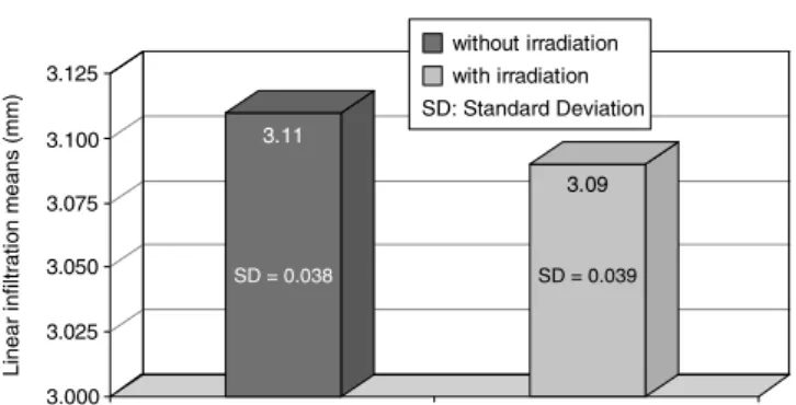
Linear dye leakage was considered from the apex up to the end of the stained area of the inner wall of the canal. The measurements were made with ImageLab® software (Diracom Bio Informática, Vargem Grande do Sul, SP, Brazil). The results were statistically analyzed with Student’s t test. No differences between the two groups were found (Graph 1 and Table 1).
DISCUSSION
Periradicular surgery is a last attempt to save a tooth where re-intervention is not possible or endodontic treatment was not able to produce reso-lution of the disease. The main cause for it is a persisting contamination of the dentinal tubules, despite conventional endodontic treatment. Such microbial permanence in the root canal system is due to iatrogenic procedures, inaccessible infected niches or resistant microorganisms, leading to
233 Souza EB, Amorim CVG, Lage-Marques JL. Effect of diode laser irradiation on the apical sealing of MTA retrofi llings. Braz Oral
Res 2006;20(3):231-4.
ure of the endodontic treatment and retreatment. Surgical intervention aims primarily at obtaining apical sealing in order to stop endodontic-peri-odontal communication2,15.
Among root end filling materials are Super EBA, IRM, silver amalgam, glass ionomer and MTA. Min-eral trioxide aggregate (MTA) is the material which presents the best results in terms of apical sealing when compared to the other materials in dye leakage or endotoxin studies6. It has high
biocompatibil-ity13 and, similarly to amalgam, causes a favorable
inflammatory reaction at 7, 15, 30, 60 and 90 day periods in rats16. In that study the authors observed
dystrophic calcification in the connective tissue ad-jacent to the MTA filling, which they credited to a re-action of calcium oxide (present in MTA) with tissue fluids, forming calcium hydroxide. Those findings are corroborated by total resolution results in root perforations observed in another long term study7.
The diode is a semiconductor chip, which con-sists of two semiconducting materials, one carrying a positive charge, and the other a negative one. They are separated by a non-conductive bandgap layer. Applying positive and negative voltages to conductors leads to a combination that releases energy as a light when the materials are gallium arsenide and aluminium – GaAlAs8.The diode laser
has been studied in photoactivated disinfection of the root canal and external root surface treatment and disinfection10.When applied on external root
surfaces with 1 W of power in vitro without previ-ous scaling or root planning procedures, the diode laser caused dentinal permeability decrease9. SEM
analysis shows few cementum and smear layer changes with diode laser at 1 W of power in ex-tracted teeth with previous scaling and planning procedures. No melting, fusion or carbonization were found12.
In the present study, the diode laser was ap-plied with 1 W on the apical and cavity surfaces
before retrofilling with MTA to avoid interaction between the diode laser and MTA, because this type of laser greatly interacts with dark pigmented surfaces. Using the diode laser after retrofilling with MTA would increase the temperature, which might crack the retrofilling.
The power setting used was based on a previ-ous thermal analysis study12, in which it was found
to be safe. Dye leakage decrease was not observed in this study.This suggests that the laser irradia-tion caused little or no effect on the apical sur-face and on the MTA retrofilling intersur-face. Further studies using a higher power setting or a different scanning dynamics with more intervals should be carried out in order to evaluate a possible improve-ment of the apical sealing.
CONCLUSION
Following the parameters used here and under the conditions of the present study, the specimens submitted to diode laser irradiation did not present any difference in apical sealing when compared with the specimens of the non-irradiated group. TABLE 1 - Linear dye leakage in millimeters.
Group 1: without irradiation
Group 2: with irradiation
3.156 3.073
3.189 3.179
3.122 3.096
3.093 3.065
3.059 3.136
3.091 3.118
3.177 3.048
3.096 3.078
3.089 3.091
3.145 3.065
3.155 3.073
3.171 3.097
3.094 3.129
3.067 3.111
3.126 3.084
3.116 3.059
3.051 3.109
3.158 3.119
3.109 3.088
3.091 3.090
GRAPH 1 - Linear infi ltration means (mm).
3.000 3.025 3.050 3.075 3.100 3.125
SD: Standard Deviation without irradiation with irradiation
L
in
e
a
r
in
fi
lt
ra
ti
o
n
me
a
n
s
(mm) 3.11
SD = 0.038
3.09
Souza EB, Amorim CVG, Lage-Marques JL. Effect of diode laser irradiation on the apical sealing of MTA retrofi llings. Braz Oral Res 2006;20(3):231-4.
234
REFERENCES
1. Dalçóquio C, Schoenau F, Lucena MG, Fedeli Jr A. Sela-mento apical após retrobturações com MTA, IRM, ionô-mero de vidro e cianoacrilato. Rev Assoc Paul Cir Dent 2001;55(3):194-8.
2. Gekelman D. Estudo in vitro dos efeitos da irradiação do laser de Nd:YAG no selamento do terço apical de obturações endodônticas executadas com e sem tampão dentinário [Dissertação de Mestrado]. São Paulo: Faculdade de Odon-tologia da USP; 2000.
3. Gouw-Soares SA, Stabholz A, Lage-Marques JL, Zezell DM, Groth EB, Eduardo CP. Comparative study of dentine permeability after apicectomy and surface treatment with 9.6 μm TEA CO2 and Er:YAG laser irradiation. J Clin Laser
Med Surg 2004;22(2):129-39.
4. Kreisler M, Al Haj H, Daublander M, Gotz H, Duschner H, Willershausen B, et al. Effect of diode laser irradiation on root surfaces in vitro. J Clin Laser Med Surg 2002;20(2):63-9.
5. Lage-Marques JLS, Antoniazzi JH. Versão eletrônica da técnica endodôntica da Faculdade de Odontologia da Uni-versidade de São Paulo [CD ROM]. São Paulo: Ajna Inte-ractive; 2002.
6. Lee SJ, Monsef M, Torabinejad M. Sealing ability of a min-eral trioxide aggregate for repair of latmin-eral root perforations. J Endod 1993;19(11):541-4.
7. Main C, Mirzayan N, Shabahang S, Torabinejad M. Repair of root perforations using mineral trioxide aggregate: a long-term study. J Endod 2004;30(2):80-3.
8. Pearson GJ, Schuckert KH. The role of lasers in dentistry: present and future. Dent Update 2003;30(1):70-6.
9. Souza EB, Amorim CVG, Lage-Marques JLS. Avaliação da permeabilidade dentinária provocada por laser diodo em superfície externa de raiz [resumo Ic23]. Pesqui Odontol Bras 2003;17 Supl 2:82.
10. Stabholz A, Zeltser R, Sela M, Peretz B, Moshonov J, Ziskind D, et al. The use of lasers in dentistry: principles of operation and clinical applications. Compend Contin Educ Dent 2003;24(12):935-48.
11. Tang HM, Torabinejad M, Kettering JD. Leakage evalu-ation of root end filling materials using endotoxin. J Endod 2002;28(1):5-7.
12. Theodoro LH, Haypek P, Bachmann L, Garcia VG, Sampaio JEC, Zezell DM, et al. Effect of Er:YAG and diode laser irradiation on the root surface: morphological and thermal analysis. J Periodontol 2003;74(6):838-43. 13. Torabinejad M, Hong CU, Pitt Ford TR, Kettering JD.
Cytotoxicity of four root end filling materials. J Endod 1995;21(10):489-92.
14. Torabinejad M, Watson TF, Pitt Ford R. Sealing ability of a mineral trioxide aggregate when used as a root end filling material. J Endod 1993;19(12):591-5.
15. von Arx T, Gerber C, Hardt N. Periradicular surgery of molars: a prospective clinical study with a one-year fol-low-up. Inter Endod J 2001;34(7):520-5.
16. Yaltirik M, Ozbas H, Bilgic B, Issever H. Reactions of connective tissue to mineral trioxide aggregate and amal-gam. J Endod 2004;30(2):95-9.