Rafael Morales Vadillo(a) Sonia Julia Sacsaquispe Contreras(b)
Janet Ofelia Guevara Canales(a)
(a)Faculty of Dentistry of the University “San
Martín de Porres”, Lima, Peru.
(b)Department of Medicine, Surgery and
Oral Pathology of the University “Peruana Cayetano Heredia”, Lima, Peru.
Corresponding author:
Janet Ofelia Guevara Canales E-mail: [email protected]
Received for publication on May 23, 2011 Accepted for publication on Aug 26, 2011
Prognostic factors in patients with jaw
sarcomas
Abstract: The aim of this study was to identify the prognostic factors related to the survival of patients with sarcomas of the jaw treated in the Dr. Eduardo Caceres Graziani National Institute for Neoplastic Diseas-es, Lima, Peru. Age, gender, delay in consultation, diagnostic delay, ther-apeutic delay, tumor size, tumor location, facial asymmetry, pain, treat-ment type, and histopathological diagnosis were all evaluated as possible prognostic factors that would inluence survival in those with jaw sarco-mas. In the analysis, the following was used: mortality tables, Kaplan-Meier’s product-limit method, log-rank, and Breslow and Tarone-Ware tests; for the prognostic factors, Cox’s Regression Model was used. The overall survival rate, with the patient being free from disease at two years, was 55%, and that at ive years was 45%. In the independent anal-ysis of the prognostic factors, four variables were statistically signiicant in inluencing survival: gender (p = 0.043), histopathologic diagnosis (p = 0.019), tumor location (p = 0.019), and treatment type (p = 0.030). According to Cox’s Regression Model for the multivariate analysis, sta-tistically signiicant prognostic factors were: gender (p = 0.086), tumor location (p = 0.020), and treatment type (p = 0.092). Thus, the variables of gender, tumor location, and treatment type were determined to be pre-dictive factors for prognosis of survival.
Descriptors: Sarcoma; Jaw; Survival; Prognosis.
Introduction
Sarcomas of the jaws (JS) are infrequent, accounting for about 1% of all the malignant tumors that occur in the oral and maxillofacial region.1
These sarcomas are highly aggressive and as such require an accurate diagnosis and therapy to be treated effectively.2
Prognostic factors for sarcomas are not well-known,3 speciically
re-garding the jawbones. There are some studies that present data on osteo-sarcomas of the jawbones or data on head and neck osteo-sarcomas, but these studies are far from comprehensive.
Studies such as that by Patel et al.4 reviewed the records of 44 patients
with osteogenic head and neck sarcomas and found, at 3 and 5 years, overall survival rates of 81% and 70%, respectively. In this study, only the surgical margins were correlated signiicantly to survival prognosis.
In 2008, Singh et al.5 examined 36 cases of soft-tissue sarcomas of
the head and neck, and Penel et al.6 analyzed 45 similar cases and found
overall survival rates at 5 years of 49% and 52% (± 8%), respectively.
The size of the tumor was the most important prog-nostic factor in the former study, and in the second study, which performed a univariate analysis, the prognostic factors that were statistically signiicant included high levels of malignancy, early iniltration of lymph nodes, absence of surgery, and number of surgical procedures. In the multivariate analysis, the level of malignancy (p = 0.006) and the absence of surgery (p = 0.005) were still signiicant.
The 10-year retrospective research of Ketabchi et al.7 analyzed 25 cases of hard- and soft-tissue
sarco-mas of the head and neck and found a 76% overall survival rate for hard-tissue sarcomas (osteosarco-mas in jawbones) and 80% for soft-tissue sarco(osteosarco-mas within a follow-up time of 12 to 108 months (60 months on average).
The aim of this study was to identify the prog-nostic factors related to the survival of patients with jaw sarcomas, using univariate and multivariate analyses to assess prognostic factors associated with survival, and to determine survival rates at 2 and 5 years for patients with JS treated in the Dr. Eduardo Caceres Graziani National Institute for Neoplastic Diseases (INEN), Lima, Peru, over the period of 1952 to 2007.
Methodology
The present study is a longitudinal, retrospective case series focusing on 155 patients with a diagnosis of JS and registered in the Statistics Department of the INEN, Lima, Peru, from 1952 to 2007. Of these 155 patients, however, 20 had to be excluded due to incomplete case information that could not be ana-lyzed accurately.
Statistical analysis
Descriptive statistics of frequency were devel-oped for each variable. For the percentage of sur-vival at 2 and 5 years, the mortality table was used, and for the univariate survival analysis, Kaplan-Meier’s product-limit method was used. For the bi-variate analysis, the log-rank, Breslow and Tarone-Ware tests were used to compare a variable and other survival variables. The multivariate analysis of the prognostic factors was obtained when Cox’s Regression Model, also called the proportional risks
model, was applied. Values of p ≤ 0.05 (5%) were considered signiicant. The analysis was developed with the SPSS statistical package (15.0 version for Windows, SPSS Inc., Chicago, USA) with Windows XP Operating System (Microsoft, Inc., Seattle, USA).
Results
Distribution of the variables
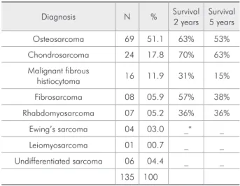
An analysis was performed on 135 clinical re-cords, with appropriate consent having been ob-tained from patients with conirmed diagnosis of JS. The age range was from 1 to 80 years, with the average being 31 years. Most individuals (48.1%) were between 0 and 30 years old. In the analysis re-garding gender, a slight predominance in the female gender (55.6%) (ratio of 1.25 to 1) was observed. Osteosarcoma was the most frequent diagnosis (51.1%), followed by chondrosarcoma (17.8%) (Ta-ble 1). Within a range from 1 to 26 cm, the average tumor size was 5 cm, with most patients (66.7%) having a tumor bigger than 4 cm. The most frequent location was in the upper jaw (maxillary) (53.3%). Facial asymmetry was observed in 87.4%, and the symptom of pain in 62.2%. The two treatment classiications were designated as “combination of treatments” and “surgical treatment” in a total of 114 cases. It was not possible to classify treatments
Table 1 - Survival rates for diagnoses of jaw sarcomas in patients at the Dr. Eduardo Caceres Graziani National Insti-tute for Neoplastic Diseases, Lima, Peru (1952-2007).
Diagnosis N % Survival 2 years
Survival 5 years
Osteosarcoma 69 51.1 63% 53%
Chondrosarcoma 24 17.8 70% 63%
Malignant fibrous
histiocytoma 16 11.9 31% 15%
Fibrosarcoma 08 05.9 57% 38%
Rhabdomyosarcoma 07 05.2 36% 36%
Ewing’s sarcoma 04 03.0 _* _
Leiomyosarcoma 01 00.7 _ _
Undifferentiated sarcoma 06 04.4 _ _
135 100
more speciically, since speciic data on the types of treatment were not described clearly in the clini-cal records. The delay in making an appointment averaged 91 days (3 months), the diagnostic delay averaged 138 days (4.5 months), and the therapeu-tic delay was shown to be 122 days (4 months) on average. The survival time of patients ranged from 12 to 12,205 days (33.42 years), with an average of 355 days.
Survival analysis
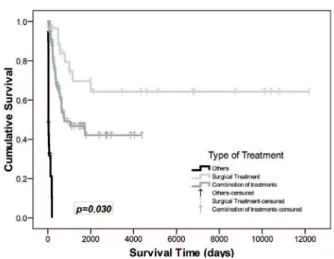
The survival rate at 2 and 5 years, based on mor-tality tables, was 55% and 45%, respectively (Figure 1).
Analysis of relative impact of the prognostic factors of survival
In the univariate analysis, gender (p = 0.043), histopathological diagnosis (p = 0.019), tumor lo-cation (p = 0.019), and treatment type (p = 0.030) were found to be statistically signiicant. Using the Kaplan-Meier survival curve, we observed that fe-male patients had 1.8 times more risk of death than males (Figure 2). By using the log-rank test of sur-vival curves to compare sursur-vival rates between os-teosarcomas and other types of jaw sarcomas, we found that a statistically signiicant difference ex-isted between these two groups (p = 0.019). Patients
with osteosarcomas were found to have a better sur-vival rate than those with diagnoses of other sarco-mas. When analyzing the survival curves regarding the location of the JS, we found a signiicant differ-ence (p = 0.019), demonstrating that patients with sarcomas in the lower jaw (mandibular) had a better survival prognosis than those with maxillary sarco-mas (Figure 3). A statistical difference in survival (p = 0.030) was also found in the type of treatment. It was observed that patients who received surgical treatment (the “surgical treatment” group) had a reduced risk of death by 0.5 times compared with other treatments (the “combined treatment” group) (Figure 4).
Analysis of the influence of the prognostic factors on survival
Univariate and multivariate analyses of all prog-nostic factors were carried out, and three predic-tive variables were seen to inluence survival: male gender (p = 0.086), mandibular tumor location (p = 0.020), and surgical treatment (p = 0.092). Per the results, the female gender presented 1.8 times more risk of death due to this neoplasia than did the male gender. Likewise, it was observed that the maxillary tumor location also had a negative impact on survival, since it caused 2.2 times increased risk of death due to JS compared with the mandibular
Figure 2 - Comparison of survival curves according to gen-der for patients with sarcomas of the jaw, seen at the Dr. Eduardo Caceres Graziani National Institute for Neoplastic Diseases, Lima, Peru (1952-2007) (comparison: Kaplan-Meier’s test) (p ≤ 0.05).
interval, we could not statistically ensure that it be-haves as a protection factor (Table 2).
Risk function of the prognostic factors in survival
The inal model for measurement of the inluence of various variables on survival was the risk func-tion, which is comprised of the multivariate analysis of prognostic factors and Cox’s Regression Model. Overall, 3 of the 4 variables that had a statistical signiicance in the univariate analysis were selected:
Ht = h0(t) Exp (0.563 × Female Gender + 0.8 × Up-per Jaw Location − 0.696 × Surgical Treatment)
Discussion
Regarding the survival risks for patients with JS, Cox’s Regression Model selected 3 of the 4 variables that had a statistical signiicance in the independent analysis: gender, sarcoma location, and treatment type. The fourth variable, histopathological diagno-sis, which was not found to be signiicant in the Cox Regression Model, loses statistical signiicance when analyzed as a whole. It is explained by the joint in-tervention of the other variables, but the speciic rea-son for the occurrence of this phenomenon cannot be determined. Nevertheless, according to the litera-Table 2 - Comparison between the univariate and the
mul-tivariate analyses of the prognostic factors for survival in JS patients at the Dr. Eduardo Caceres Graziani National Insti-tute for Neoplastic Diseases, Lima, Peru (1952-2007).
Risk Factor
Univariate Analysis (p value)
Multivariate Analysis (p value)
Age (years) 0.544 0.729
Gender 0.043* 0.086 (f)
Histopathologic diagnosis 0.019* 0.679
Delay in consulting 0.180 0.377
Diagnostic delay 0.200 0.651
Therapeutic delay 0.143 0.490
Tumor size 0.776 0.993
Location 0.019* 0.020* (f)
Facial asymmetry 0.631 0.436
Pain 0.686 0.790
Treatment type 0.030* 0.092 (f)
The p-value of the univariate analysis was calculated by means of the log-rank analysis (p ≤ 0.05). The p-value of the multivariate analysis was calculated by means of Cox’s Regression Model (p ≤ 0.05). * Values indicating statistical significance; the variable is according to the model. (f)Variable present in the risk function.
Figure 4 - Comparison of survival curves according to type of treatment in patients with sarcomas of the jaw, seen at the Dr. Eduardo Caceres Graziani National Institute for Neoplastic Diseases, Lima, Peru (1952-2007) (Comparison: Kaplan-Meier’s test) (p ≤ 0.05).
Figure 3 - Comparison of survival curves according to tumor location in patients with sarcomas of the jaw, seen at the Dr. Eduardo Caceres Graziani National Institute for Neoplastic Diseases, Lima, Peru (1952-2007) (Comparison: Kaplan-Meier’s test) (p ≤ 0.05).
ture, it is suspected that the delays in consultation and diagnosis, combined with treatment location, lead to similar outcomes for those with a diagnosis of osteosarcoma and those with different types of JS.
In the present study, the age group distribution is from 1 to 80 years, with an average age of 31 years. This value is similar to that described by Brockstein.8
In turn, Yoel9 and co-workers report similar data,
citing greater predominance of JS in the second, third, fourth, and ifth decades of life. The literature describes a bimodal age group distribution for osteo-sarcomas, in which the majority of patients are be-tween 10 and 20 years old, with a secondary group over 50 years old. The present study did not ind this type of distribution and, in fact, found a progressive decrease of cases occurring as age increased. When the diagnoses were compared in the current study, a 51.1% predominance of osteosarcomas was found, which is high compared with 24.6% reported by Yoel et al.9 or 28% reported by Yamaguchi et al.10
Sarcomas are a heterogeneous group of tumors that present, are diagnosed, and are treated at dif-ferent stages and therefore have difdif-ferent overall prognoses. The overall survival at 2 years of head and neck sarcomas in the current study was of 55%, a low value compared with studies by Penel et al.,6
where survival was reported to be 71.7%. Also, Na-gler et al.11 reported a 72% survival rate for
maxil-lofacial sarcomas, Mücke12 and colleagues found a
survival rate of 83.78%, and the cases studied by the Canadian Society of Otorhinolaryngology showed a survival rate of 79%.
At 5 years, the survival rate was 45%, a value close to that found by Singh et al.5 (49%) in the
United Kingdom and by Penel et al.6 (52.3%) in
France, while the value was low compared with that found by Mücke12 and co-workers in Germany
(60.81%) and by Ketabchi et al.7 in the United
King-dom (80%). Overall, much of the literature reports a ive-year survival rate between 57% and 86%.4,6,11,13
For osteogenic sarcomas, Kassir et al.14 report a
survival rate of 37%, and Ketabchi7 and colleagues
note an overall survival rate of 76%. The present study found a survival rate for osteosarcomas of 63% and 53% at 2 and 5 years, respectively. The survival rates for osteosarcomas are better than
those for other sarcomas, since the former exhibit different biological behavior and different dissemi-nation. It is assumed that the differences regarding survival rates are due to the different factors used in the treatment and development of the sarcomas.
In the univariate analysis of risk factors that in-luence survival, the only variables that were found to be statistically signiicant were: gender, histopath-ological diagnosis, tumor location, and treatment type. When all variables were analyzed by Cox’s Re-gression Model (age, gender, delay in consulting, di-agnostic delay, therapeutic delay, tumor size, tumor location, facial asymmetry, pain, treatment type, and histopathological diagnosis), the following prognostic factors emerged: gender, tumor location, and the treatment type received. Patel et al.4 found
that the only prognostic factor was the extension of surgical margins. de Bree15 and colleagues
con-cluded that the prognostic factors for sarcomas that reduce overall survival are: tumors in any anatomi-cal location with a diameter greater than 5 cm, with many abnormalities found on histological examina-tion, and with positive surgical margins. Penel6 and
co-workers found that the variables iniltration level and absence of surgery inluenced survival. August
et al.16 describe the factors of age and surgical
treat-ment as inluencing survival. Ruiz17 and co-workers
refer to tumor size, tumor location, and histological level. Nagler11 and colleagues report type of sarcoma
and young age, and Harb et al.18 mention that tumor
size on presentation generally depends on the loca-tion of the tumor, with the localoca-tion being of greater prognostic impact. Finally, Singh5 and co-workers
found that tumor size was the most important prog-nostic factor.
Fayda et al.19 studied 30 cases and focused on the
roles of surgery and radiotherapy in the treatment of soft-tissue sarcomas of the head and neck during an average follow-up time of 31 months. They found a statistically signiicant difference in the overall sur-vival rate of patients who were treated with surgery and radiotherapy.
osteo-sarcomas differ from those for other types of sar-comas.5 This difference eliminates this factor as an
element in the prognosis of survival. In Peru, there are no data on survival of patients with sarcomas in the jawbones, and therefore prospective studies with more speciic data about this topic are necessary.
Conclusion
In patients with JS, the factors independently
related to survival are: histopathological diagnosis, gender, tumor location, and treatment type. On the whole, the positive prognostic factors were found to be male gender, tumor location in the mandible, and surgical treatment.
Acknowledgements
The authors wish to thank Dr. Matthew J. Pierce and Dr. Mauro Cruz for help with the manuscript.
References
1. Gorsky M, Epstein JB. Head and neck and intra-oral soft tissue sarcomas. Oral Oncol. 1998 Jul;34(4):292-6. 2. Ogunlewe MO, Ajayi OF, Adeyemo WL, Ladeinde AL, James
O. Osteogenic sarcoma of the jaw bones: a single institution experience over a 21-year period. Oral Surg Oral Med Oral Pathol Oral Radiol Endod. 2006 Jan;101(1):76-81.
3. Penel N, Van Haverbeke C, Lartigau E, Vilain MO, Ton Van J, Mallet Y, et al. Head and neck soft tissue sarcomas of adult: prognostic value of surgery in multimodal therapeutic ap-proach. Oral Oncol. 2004 Oct;40(9):890-7.
4. Patel SG, Meyers P, Huvos AG, Wolden S, Singh B, Shaha AR, et al. Improved outcomes in patients with osteogenic sarcoma of the head and neck. Cancer. 2002 Oct 1;95(7):1495-503. 5. Singh RP, Grimer RJ, Bhujel N, Carter SR, Tillman RM,
Abudu A. Adult head and neck soft tissue sarcomas: treatment and outcome. Sarcoma. 2008;2008:654987.
6. Penel N, Mallet Y, Robin YM, Fournier C, Grosjean J, Ceugn-art L, et al. Prognostic factors for adult sarcomas of head and neck. Int J Oral Maxillofac Surg. 2008 May;37(5):428-32. 7. Ketabchi A, Kalavrezos N, Newman L. Sarcomas of the head
and neck: a 10-year retrospective of 25 patients to evaluate treatment modalities, function and survival. Br J Oral Maxil-lofac Surg. 2011 Mar;49(2):116-20.
8. Brockstein B. Management of sarcomas of the head and neck. Curr Oncol Rep. 2004 Jul;6(4):321-7.
9. Yoel J, Gonzáles Aguilar O, Simkin DO, Barg S. Sarcomas of the jaw. Semin Surg Oncol. 1987;3(4):215-27.
10. Yamaguchi S, Nagasawa H, Suzuki T, Fujii E, Iwaki H, Takagi M, et al. Sarcomas of the oral and maxillofacial re-gion: a review of 32 cases in 25 years. Clin Oral Investig. 2004 Jun;8(2):52-5.
11. Nagler RM, Malkin L, Ben-Arieh Y, Laufer D. Sarcoma of the maxillofacial region: follow-up of 25 cases. Anticancer Res. 2000 Sep-Oct;20(5C):3735-41.
12. Mücke T, Mitchell DA, Tannapfel A, Hölzle F, Kesting MR, Wolff KD, et al. Outcome in adult patients with head and neck sarcomas—a 10-year analysis. J Surg Oncol. 2010 Aug 1;102(2):170-4.
13. Fernandes R, Nikitakis NG, Pazoki A, Ord RA. Osteogenic sarcoma of the jaw: a 10-year experience. J Oral Maxillofac Surg. 2007 Jul;65(7):1286-91.
14. Kassir RR, Rassekh CH, Kinsella JB, Segas J, Carrau RL, Ho-kanson JA. Osteosarcoma of the head and neck: meta-analysis of nonrandomized studies. Laryngoscope. 1997 Jan;107(1):56-61.
15. de Bree R, van der Waal I, de Bree E, Leemans CR. Manage-ment of adult soft tissue sarcomas of the head and neck. Oral Oncol. 2010 Nov;46(11):786-90.
16. August M, Magennis P, Dewitt D. Osteogenic sarcoma of the jaws: factors influencing prognosis. Int J Oral Maxillofac Surg. 1997 Jun;26(3):198-204.
17. Ruíz-Godoy RL, Meneses-García A, Mosqueda-Taylor A, De la Garza-Salazar J. Well-differentiated intraosseous os-teosarcoma of the jaws: experience of two cases from the Instituto Nacional de Cancerología, México. Oral Oncol. 1999 Sep;35(5):530-3.
18. Harb WJ, Luna MA, Patel SR, Ballo MT, Roberts DB, Sturgis EM. Survival in patients with synovial sarcoma of the head and neck: association with tumor location, size, and extension. Head Neck. 2007 Aug;29(8):731-40.