Raquel Gonçalves Vieira-Andrade(a) Flávia de Faria Zuquim Guimarães(b)
Charlles da Silva Vieira(b) Sarah Teixeira Carvalho Freire(b) Maria Letícia Ramos-Jorge(a) Anacélia Mendes Fernandes(b)
(a)Departament of Pediatric Dentistry, School of Dentistry, Federal University of Vales of Jequitinhonha and Mucuri, Diamantina, MG, Brazil.
(b) Departament of Stomatology, School of Dentistry, Federal University of Vales of Jequitinhonha and Mucuri, Diamantina, MG, Brazil.
Corresponding Author: Raquel Gonçalves Vieira-Andrade E-mail: [email protected]
Received for publication on May 02, 2011 Accepted for publication on Aug 26, 2011
Oral mucosa alterations in a
socioeconomically deprived region:
prevalence and associated factors
Abstract: This study aimed to evaluate the prevalence and factors as-sociated with oral mucosa alterations in patients from Vale do Jequiti-nhonha, Brazil. The sample consisted of 511 patients of both genders. Questionnaires were used to obtain information about patient gender, age, race, systemic disease state, medication use, cigarette use and alco-hol consumption. Physical examinations were then performed to identify lesions of the oral mucosa. Descriptive analyses, Chi-squared tests and logistic regressions were then used to analyze the results (p < 0.05, 95% CI). In this population, 84.9% (434/511) of patients were found to have alterations in their oral mucosa. The most common alterations were mel-anotic maculae (36.0%), linea alba (33.9%), traumatic ulcers (21.5%), Fordyce’s granules (20.4%), coated tongue (12.5%) and issured tongue (10.0%). Melanotic maculae were more frequently observed in black pa-tients, with an odds ration (OR) of 7.51. Being female was a statistically signiicant predictive factor for having a visible linea alba (OR: 1.90) and a issured tongue (OR: 2.11). No statistically signiicant association was found between the presence of oral lesions and systemic disease, medi-cation use, alcohol use and smoking. The high observed prevalence of melanotic maculae and Fordyce’s granules suggests that these alterations could be considered typical characteristics of the population of the Vale do Jequitinhonha. Coated tongue may be related to the socioeconomic deprivation in the region. Furthermore, the high prevalence of traumatic ulcers may be associated with the traumatic agents that caused patients to seek dental care.
Descriptors: Mouth Mucosa; Mouth Diseases; Prevalence.
Introduction
The diagnosis of oral mucosa alterations depends on the ability of dentists to distinguish between pathological changes and normal varia-tion within the oral structures.1 A knowledge of normal alterations and lesions and their association with systemic changes, deleterious habits and medication use is therefore essential for the diagnosis, treatment and establishment of prevention policies.2
Studies in various parts of the world have reported the prevalence of oral mucosa alterations, as well as their association with systemic changes and with deleterious habits such as smoking and alcohol consumption. The prevalence rates of such alterations usually vary with gender, age, Declaration of Interests: The authors
race and socioeconomic status.3-14
The Vale do Jequitinhonha is a region largely known for its low social indicators and is currently one of the areas of greatest inequality and social ex-clusion in Brazil.15 The microregion of Diamantina is located in this region and is known as the gateway to the Vale do Jequitinhonha.
Despite the socioeconomic deprivation faced by many inhabitants of the Vale do Jequitinhonha, or perhaps because of it, few studies have assessed the oral conditions of the population of this region, especially with regard to the prevalence of altera-tions on the oral mucosa. Once these alteraaltera-tions are studied in different populations, subsequent epide-miological studies will then be able to provide an understanding of the prevalence, extent and severity of oral diseases in order to investigate the factors as-sociated with these diseases and assist in the distri-bution of public health resources.16
Therefore, the purpose of this study was to de-termine the prevalence and associated factors of oral mucosa alterations among 511 patients aged 12 to 78 years attending an oral health service in Vale do Jequitinhonha, Brazil.
Methodology
The present cross-sectional study was carried out on a sample of 511 patients who were treated in the dental clinics of the Federal University of Vales of Jequitinhonha and Mucuri (UFVJM), located in Diamantina, Minas Gerais, Brazil. Patients of both genders and above age 12 who sought care in the clinics between March and August of 2009 were in-cluded in the study.
The research team consisted of ive investiga-tors. The research was divided into two stages. In the irst stage, 20 patients who were not part of the main study were randomly selected and evaluated for the presence or absence of oral mucosa altera-tions. The diagnostic results were compared with a gold standard (minimum kappa value = 0.81, max-imum kappa value = 0.89), and any differences of opinion were discussed among the researchers and resolved through consensus. In the second stage, photographs of oral mucosa alterations in a color at-las were observed. Images of lesions not observed in
the irst clinical stage were selected.
Using questionnaires administered through face-to-face interviews, information such as age, gender, race, medical and pharmacological history and al-cohol and cigarette use was collected. Participants were then subjected to a clinical examination of the oral mucosa through direct inspection of the oral cavity under artiicial light (KAVO, São Paulo, Bra-zil) and with the use of disposable wooden spatulas (Estilo, São Paulo, Brazil). Examinations were per-formed according to World Health Organization criteria,17,18 and biosafety standards were observed. At the time of examination, all participants were in-formed of the state of their oral mucosa and were referred to the appropriate departments depending on their treatment needs.
Twenty-six types of oral mucosa alterations were observed. The cases that required additional tests to conirm the diagnosis were referred to the UFVJM stomatology clinic for biopsy and were included in this study only after a deinitive diagnosis was ob-tained.
The collected data were analyzed by the Statisti-cal Package for Social Science Software (SPSS for Windows, version 17.0, SPSS Inc., Chicago, USA). Initially, we performed a descriptive analysis of the absolute and relative frequencies of all of the vari-ables in the study. Next, we used the Chi-squared test to verify any associations that we found be-tween oral mucosa alterations and each of the inde-pendent variables. A p-value ≤ 0.05 was accepted as signiicant. All independent variables that were sig-niicantly associated (p-values ≤ 0.20) with the more frequent oral mucosa alterations were included in our multivariate logistic regression model.
The study received approval from the Ethics Committee of the UFVJM. The patients were in-formed about the objectives, risks and beneits of the work and signed informed consent forms after agreeing to participate.
Results
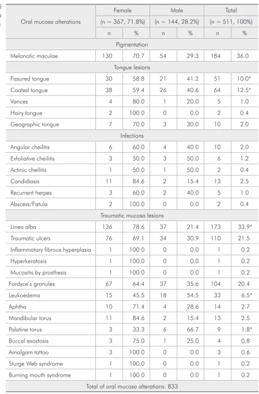
In this study, 833 oral mucosa alterations were found in 434 participants (84.9%), and some indi-viduals had more than one type of alteration (Table 1). A total of twenty-six different mucosal changes were diagnosed, the most common of which were melanotic maculae (36.0%), linea alba (33.9%), traumatic ulcers (21.5%), Fordyce’s granules (20.4%), coated tongue (12.5%) and issured tongue (10.0%) (Table 2).
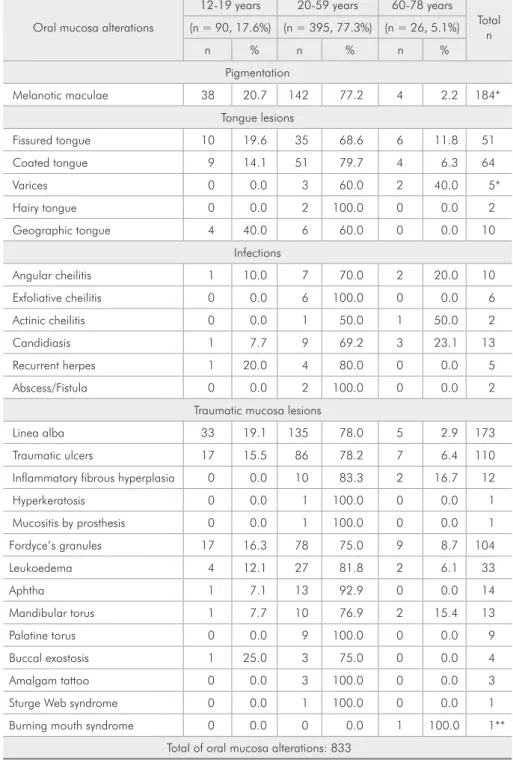
In this study, the prevalence of these alterations was different between the genders. Fissured tongue, coated tongue and linea alba (p < 0.05) were most prevalent in female subjects (p < 0.05; Table 2). Sta-tistically signiicant associations were observed be-tween the group ranging from 20 to 59 years of age and melanotic maculae (p < 0.05; Table 3).
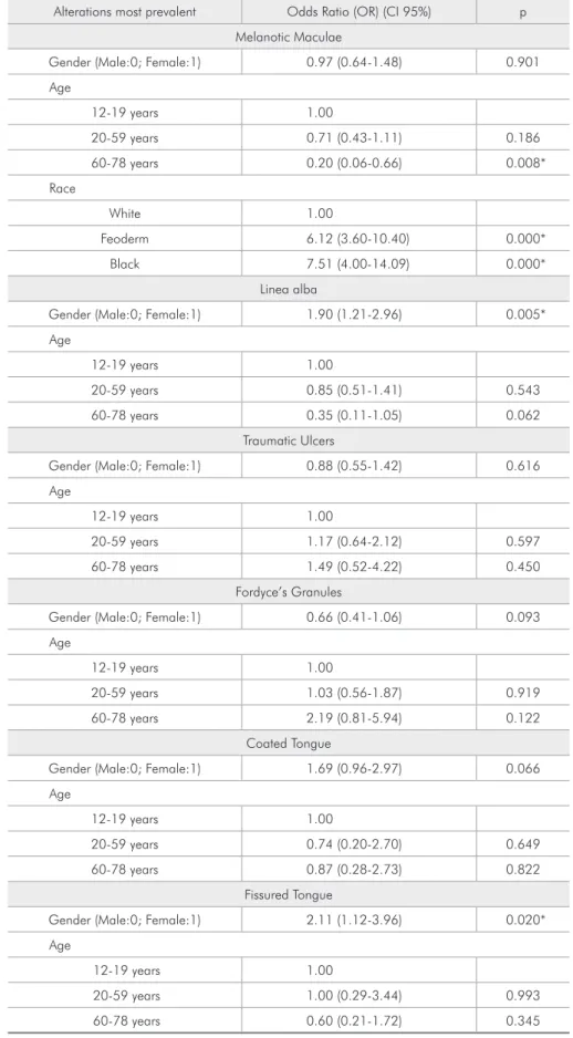
The results of logistic regression analyses on fac-tors most commonly associated with the most prev-alent oral mucosa alterations are presented in Table 4.
Discussion
This study’s indings should be interpreted with caution because we found that the prevalence of certain oral mucosa alterations was relatively low. Furthermore, comparison with other epidemiologi-cal studies is complicated by different experimental methodologies. However, the wide variety of oral mucosa alterations identiied in this study is consis-tent with the indings of other reports.4-7,10,12,14
Of the 511 patients enrolled in this study, 71.8% (n = 367) were female. This overrepresentation of fe-male patients has also been observed in other epide-miological studies of the prevalence of oral mucosa alterations.5,10,13,14 It is possible that this observation relects a greater concern about oral health among women than among men.11,19
The prevalence of oral mucosa alterations found in this study was higher (84.9%) than in similar studies in Spain7 (51.9%) and Turkey10 (41.7%). Such variations in the prevalence rates of oral muco-sa alterations may be the result of geographical dif-ferences, socio-demographic characteristics of the study populations, and a lack of standardized diag-nostic criteria and experimental methodologies.4-12
Lesions of a traumatic origin are those most commonly reported in different parts of the world.11 In this study, traumatic ulcer (21.5%), inlamma-tory ibrous hyperplasia (2.3%), hyperkeratosis (0.2%) and prosthesis-induced mucositis accounted for 58.1% of the total mucosal alterations, which are quite high compared to the previously-reported values of 7.8% and 5.6% reported in two studies of an adult Brazilian population.20,21 This might be due to the fact that the participants in our study were Table 1 - Absolute and relative frequencies of the variables
studied.
Variables Absolute Frequency (relative)
Oral Mucosa Alterations
Yes 434 (84.9%)
No 77 (15.1%)
Gender
Female 367 (71.8%)
Male 144 (28.2%)
Age
12-19 years 90 (17.6%)
20-59 years 395 (77.3%)
60-78 years 26 (5.1%)
Race
White 170 (33.3%)
Phaeoderm 244 (47.7%)
Black 97 (19.0%)
Systemic Diseases
Hypertension 25 (4.9%)
Neurological disorders 17 (3.3%)
Endocrinopathy 16 (3.2%)
Medications
Analgesics 64 (12.5%)
Antihypertensive 41 (8.0%)
Anxiolytic 30 (5.9%)
Anti-inflammatory 8 (1.6%)
Cigarette Use
Yes 80 (15.7%)
No 431 (84.3%)
Alcohol Use
Yes 223 (43.6%)
No 288 (56.4%)
seeking dental care for traumatic agents in the oral cavity, such as maladapted partial or complete re-movable dentures, malocclusion, dental caries or unsatisfactory restorations.
Melanotic maculae were the most prevalent oral mucosa alteration, found in 36.0% of cases and
with a higher frequency in black patients (OR: 7.66). A number of studies also report an association be-tween melanotic maculae and the black race. 22-24 In this study, the lower prevalence of melanotic
maculae among subjects aged 60 to 78 years (OR: 0.20) has no deinite relationship. A knowledge of Oral mucosa alterations
Female Male Total
(n = 367, 71.8%) (n = 144, 28.2%) (n = 511, 100%)
n % n % n %
Pigmentation
Melanotic maculae 130 70.7 54 29.3 184 36.0
Tongue lesions
Fissured tongue 30 58.8 21 41.2 51 10.0*
Coated tongue 38 59.4 26 40.6 64 12.5*
Varices 4 80.0 1 20.0 5 1.0
Hairy tongue 2 100.0 0 0.0 2 0.4
Geographic tongue 7 70.0 3 30.0 10 2.0
Infections
Angular cheilitis 6 60.0 4 40.0 10 2.0
Exfoliative cheilitis 3 50.0 3 50.0 6 1.2
Actinic cheilitis 1 50.0 1 50.0 2 0.4
Candidiasis 11 84.6 2 15.4 13 2.5
Recurrent herpes 3 60.0 2 40.0 5 1.0
Abscess/Fistula 2 100.0 0 0.0 2 0.4
Traumatic mucosa lesions
Linea alba 136 78.6 37 21.4 173 33.9*
Traumatic ulcers 76 69.1 34 30.9 110 21.5
Inflammatory fibrous hyperplasia 1 100.0 0 0.0 1 0.2
Hyperkeratosis 1 100.0 0 0.0 1 0.2
Mucositis by prosthesis 1 100.0 0 0.0 1 0.2
Fordyce’s granules 67 64.4 37 35.6 104 20.4
Leukoedema 15 45.5 18 54.5 33 6.5*
Aphtha 10 71.4 4 28.6 14 2.7
Mandibular torus 11 84.6 2 15.4 13 2.5
Palatine torus 3 33.3 6 66.7 9 1.8*
Buccal exostosis 3 75.0 1 25.0 4 0.8
Amalgam tattoo 3 100.0 0 0.0 3 0.6
Sturge Web syndrome 1 100.0 0 0.0 1 0.2
Burning mouth syndrome 1 100.0 0 0.0 1 0.2
Total of oral mucosa alterations: 833
Chi-square test. *Statistically significant, according to gender (p < 0.05).
this alteration and an ability to properly identify it in patients is important, as melanin pigmentation in the oral mucosa may require a diagnostic biopsy.25,26 Moreover, although this condition was not found to be associated with tobacco use in this study, previ-ous studies have reported a possible association
be-tween smoking and the development of melanotic maculae.22,24
Fordyce’s granules are ectopic sebaceous glands that accounted for 20.4% of the oral mucosa al-terations identiied in our study. This prevalence is higher than that found in studies conducted in Tur-Oral mucosa alterations
12-19 years 20-59 years 60-78 years Total
n (n = 90, 17.6%) (n = 395, 77.3%) (n = 26, 5.1%)
n % n % n %
Pigmentation
Melanotic maculae 38 20.7 142 77.2 4 2.2 184*
Tongue lesions
Fissured tongue 10 19.6 35 68.6 6 11.8 51
Coated tongue 9 14.1 51 79.7 4 6.3 64
Varices 0 0.0 3 60.0 2 40.0 5*
Hairy tongue 0 0.0 2 100.0 0 0.0 2
Geographic tongue 4 40.0 6 60.0 0 0.0 10
Infections
Angular cheilitis 1 10.0 7 70.0 2 20.0 10
Exfoliative cheilitis 0 0.0 6 100.0 0 0.0 6
Actinic cheilitis 0 0.0 1 50.0 1 50.0 2
Candidiasis 1 7.7 9 69.2 3 23.1 13
Recurrent herpes 1 20.0 4 80.0 0 0.0 5
Abscess/Fistula 0 0.0 2 100.0 0 0.0 2
Traumatic mucosa lesions
Linea alba 33 19.1 135 78.0 5 2.9 173
Traumatic ulcers 17 15.5 86 78.2 7 6.4 110
Inflammatory fibrous hyperplasia 0 0.0 10 83.3 2 16.7 12
Hyperkeratosis 0 0.0 1 100.0 0 0.0 1
Mucositis by prosthesis 0 0.0 1 100.0 0 0.0 1
Fordyce’s granules 17 16.3 78 75.0 9 8.7 104
Leukoedema 4 12.1 27 81.8 2 6.1 33
Aphtha 1 7.1 13 92.9 0 0.0 14
Mandibular torus 1 7.7 10 76.9 2 15.4 13
Palatine torus 0 0.0 9 100.0 0 0.0 9
Buccal exostosis 1 25.0 3 75.0 0 0.0 4
Amalgam tattoo 0 0.0 3 100.0 0 0.0 3
Sturge Web syndrome 0 0.0 1 100.0 0 0.0 1
Burning mouth syndrome 0 0.0 0 0.0 1 100.0 1**
Total of oral mucosa alterations: 833
Chi-squared test. *Statistically significant, according to age (P < 0.05). **Statistically significant, according to age (P < 0.001).
Alterations most prevalent Odds Ratio (OR) (CI 95%) p
Melanotic Maculae
Gender (Male:0; Female:1) 0.97 (0.64-1.48) 0.901
Age
12-19 years 1.00
20-59 years 0.71 (0.43-1.11) 0.186
60-78 years 0.20 (0.06-0.66) 0.008*
Race
White 1.00
Feoderm 6.12 (3.60-10.40) 0.000*
Black 7.51 (4.00-14.09) 0.000*
Linea alba
Gender (Male:0; Female:1) 1.90 (1.21-2.96) 0.005*
Age
12-19 years 1.00
20-59 years 0.85 (0.51-1.41) 0.543
60-78 years 0.35 (0.11-1.05) 0.062
Traumatic Ulcers
Gender (Male:0; Female:1) 0.88 (0.55-1.42) 0.616
Age
12-19 years 1.00
20-59 years 1.17 (0.64-2.12) 0.597
60-78 years 1.49 (0.52-4.22) 0.450
Fordyce’s Granules
Gender (Male:0; Female:1) 0.66 (0.41-1.06) 0.093
Age
12-19 years 1.00
20-59 years 1.03 (0.56-1.87) 0.919
60-78 years 2.19 (0.81-5.94) 0.122
Coated Tongue
Gender (Male:0; Female:1) 1.69 (0.96-2.97) 0.066
Age
12-19 years 1.00
20-59 years 0.74 (0.20-2.70) 0.649
60-78 years 0.87 (0.28-2.73) 0.822
Fissured Tongue
Gender (Male:0; Female:1) 2.11 (1.12-3.96) 0.020*
Age
12-19 years 1.00
20-59 years 1.00 (0.29-3.44) 0.993
60-78 years 0.60 (0.21-1.72) 0.345
*Statistically significant (P < 0.05). **Statistically significant (P < 0.001).
key10 (1.3%) and India27 (6.5%), but lower than that reported in studies carried out in Thailand (57.7%), Mexico22 (55.0%) and Malaysia23 (61.8%). This presence of Fordyce’s granules was not signiicantly associated with any independent variables evaluated in the present investigation. However, a number of previous studies have reported that the prevalence of Fordyce’s granules increases with age6,22 and is higher in the male gender.21
Epidemiological studies conducted in different parts of the world have found tongue lesions to be among the most common alterations of the oral mucosa.4,10,22,23 Our study corroborates these ind-ings, as coated tongue (12.5%) and issured tongue (10.0%) were among the seven most prevalent al-terations that we identiied. Coated tongue may be related to a poor oral health status,4,7,28 which has in turn been associated with low socioeconomic status.28 This association between poor overall oral health and low socioeconomic status may explain the high prevalence of this condition in the popula-tion studied. However, a number of studies have re-ported a statistically signiicant association between coated tongue and tobacco smoking,4 which we did not observe in this study.
Linea alba and issured tongue were signiicantly associated with the female gender (p < 0.05), occur-ring approximately twice as frequently in females than in males. This inding was previously reported in a study conducted in Turkey.10 Some studies re-port a higher incidence of issured tongue among men5 and elderly patients.10 Others report a statisti-cally signiicant association between issured tongue
and a history of allergy.9 However, we did not ind these associations in the present study.
The fact that we did not observe a statistically signiicant association between the main oral muco-sa alterations and a patient’s status with respect to systemic disease, alcohol use and tobacco use may be due to the hereditary characteristics of the oral mucosa alterations reported.
The results of this study provide a basis for fu-ture studies involving regions with known socio-economic deprivation, such as the Vale do Jequitin-honha, and may contribute toward public policies directed at oral disease prevention and control pro-grams in this region.
Conclusions
The alterations most commonly identiied in this study were melanotic maculae, linea alba, traumatic ulcers, Fordyce’s granules, coated tongue and is-sured tongue. Furthermore, we found that gender, age and race were all factors that were signiicantly associated with the presence of oral mucosa altera-tions.
The high prevalence of melanotic maculae and Fordyce’s granules indicates that these alterations should be considered normal characteristics of the population of the Vale do Jequitinhonha. On the other hand, the high prevalence of coated tongue may be related to the socioeconomic deprivation in the region and traumatic ulcers may be associated with the traumatic agents that caused patients to seek dental care.
References
1. Canaan TJ, Meehan SC. Variations of structure and appearance of the oral mucosa. Dent Clin North Am. 2005 Jan;49(1):1-14, vii.
2. Rioboo-Crespo M del R, Planells-del Pozo P, Rioboo-García R. Epidemiology of the most common oral mucosal diseas-es in children. Med Oral Patol Oral Cir Bucal. 2005 Nov-Dec;10(5):376-87.
3. Crivelli MR, Águas S, Quarrancino C, Bazerque P. Influence of the socioeconomic status on oral mucosa lesion prevalence in schoolchildren. Community Dent Oral Epidemiol. 1988 Feb;16(1):58-60.
4. Campisi G, Margiotta V. Oral mucosal lesions and risk habits among men in a Italian study population. J Oral Pathol Med. 2001Jan;30(1):22-8.
5. Kovac-Kavcic M, Skaleric U. The prevalence of oral mucosal lesions in a population in Ljubljana, Slovenia. J Oral Pathol Med. 2000 Aug;29(7):331-5.
7. Martínez Díaz-Canel AI, García-Pola Vallejo MJ. Epidemio-logical study of oral mucosa pathology in patients of the Ovie-do School of Stomatology. Med Oral. 2002 Jan-Feb;7(1):4-9. 8. Espinoza I, Rojas R, Aranda W, Gamonal J. Prevalence of oral
mucosal lesions in elderly people in Santiago, Chile. J Oral Pathol Med. 2003 Nov;32(10):571-5.
9. Bessa CF, Santos PJ, Aguiar MC, do Carmo MA. Prevalence of oral mucosal alterations in children from 0 to 12 years old. J Oral Pathol Med. 2004 Jan;33(1):17-22.
10. Mumcu G, Cimilli H, Sur H, Hayran O, Atalay T. Prevalence and distribution of oral lesions: a cross-sectional study in Turkey. Oral Dis. 2005 Mar;11(2):81-7.
11. Castellanos JL, Díaz-Guzmán L. Lesions of the oral mu-cosa: an epidemiological study of 23785 Mexican patients. Oral Surg Oral Med Oral Pathol Oral Radiol Endod. 2008 Jan;105(1):79-85.
12. Mujica V, Riveira H, Carrero M. Prevalence of oral soft tissue lesions in an elderly venezuelan population. Med Oral Patol Oral Cir Bucal. 2008 May;13(5): E270-4.
13. Vasconcelos BC, Novaes M, Sandrini FA, Maranhão Filho AW, Coimbra LS. Prevalence of oral mucosa lesions in diabetic patients: a preliminary study. Braz J Otorrinolaringol. 2008 May-Jun;74(3):423-8.
14. Ferreira RC, Magalhães CS, Moreira AN. Oral mucosal altera-tions among the institutionalized elderly in Brazil. Braz Oral Res. 2010 Jul-Sep;24(3):296-302.
15. Ferreira VA, Silva AE, Rodrigues CA, Nunes NL, Vigato TC, Magalhães R. [Inequality, poverty and obesity]. Cien Saude Colet. 2010 Jun;15 Suppl 1:1423-32. Portuguese.
16. Pack AR. Dental services and needs in developing countries. Int Dent J. 1998 Jun;48(3 Suppl 1):239-47.
17. Kramer IR, Pindborg JJ, Bezroukov V, Infirri JS. Guide to epidemiology and diagnosis of oral mucosal diseases and con-ditions. World Health Organization. Community Dent Oral Epidemiol. 1980 Feb;8(1):1-26.
18. World Health Organization. Application of the international classification of diseases to dentistry and stomatology. Geneva: Word Health Organization; 1995. 246 p.
19. Covington P. Women’s oral health issues: an exploration of the literature. Probe. 1996 Sep-Oct;30(5):173-7.
20. dos Santos PJ, Bessa CF, de Aguiar MC, do Carmo MA. Cross-sectional study of oral mucosal conditions among a central Amazonian indian community, Brazil. J Oral Pathol Med. 2004 Jan;33(1):7-12.
21. Henrique PR, Bazaga Júnior M, Araújo VC, Junqueira JLC, Furuse C. Prevalência de alterações da mucosa bucal em in-divíduos adultos da população de Uberaba, Minas Gerais. RGO. 2009 Jul;57(3):261-7.
22. Cornejo AD, Huerta ERL, Bravo SP, Barrios BA, Riviera DQ, Yañez AB, et al. Distribucuión de condiciones y lesiones de la mucosa bucal en pacientes adultos mexicanos. Rev Cubana Estomatol. 2007 Mar;44(1):181-9.
23. Axell T, Zain RB, Siwamogstham P, Tantiniran D, Thampipit J. Prevalence of oral soft tissue lesions in out-patients at two Malaysian and Thai dental schools. Community Dent Oral Epidemiol. 1990 Apr;18(2):95-9.
24. Hedin CA, Axell T. Oral melanin pigmentation in 467 Thai and Malaysian people with special emphasis on smoker’s mela-nosis. J Oral Pathol Med. 1991 Jan;20(1):8-12.
25. Ciçek Y, Ertas¸ U. The normal and pathological pigmentation of oral mucous membrane: a review. J Contemp Dent Pract. 2003 Aug;4(3):76-86.
26. Meleti M, Vescovi P, Mooi WJ, van der Waal I. Pigmented lesions of the oral mucosa and perioral tissues: a flow-chart for the diagnosis and some recommendations for the manage-ment. Oral Surg Oral Med Oral Pathol Oral Radiol Endod. 2008 May;105(5):606-16.
27. Mathew AL, Pai KM, Sholapurkaar AA, Vengal M. The prevalence of oral mucosa lesions in patients visiting a den-tal school in Southern India. Indian J Dent Res. 2008 Apr-Jun;19(2):99-103.