online | memorias.ioc.fiocruz.br
In vitro activity of the hydroethanolic extract and biflavonoids isolated
from
Selaginella sellowii
on
Leishmania (Leishmania)
amazonensis
Yasmin Silva Rizk1, Alice Fischer2, Marillin de Castro Cunha3, Patrik Oening Rodrigues4,
Maria Carolina Silva Marques5, Maria de Fátima Cepa Matos3, Mônica Cristina Toffoli Kadri6,
Carlos Alexandre Carollo2, Carla Cardozo Pinto de Arruda1/+
1Laboratório de Parasitologia Humana 2Laboratório de Farmacognosia 3Laboratório de Biologia Molecular e Culturas Celulares 4Laboratório de Tecnologia Farmacêutica 5Laboratório de Microbiologia 6Laboratório de Biofisiofarmacologia,
Centro de Ciências Biológicas e da Saúde, Universidade Federal de Mato Grosso do Sul, Campo Grande, MS, Brasil
This study is the first phytochemical investigation of Selaginella sellowii and demonstrates the antileishmanial activity of the hydroethanolic extract from this plant(SSHE), as well as of the biflavonoids amentoflavone and ro-bustaflavone, isolated from this species. The effects of these substances were evaluated on intracellular amastigotes of Leishmania (Leishmania) amazonensis, an aetiological agent of American cutaneous leishmaniasis. SSHE was highly active against intracellular amastigotes [the half maximum inhibitory concentration (IC50)= 20.2 µg/mL]. Fractionation of the extract led to the isolation of the two bioflavonoids with the highest activity: amentoflavone, which was about 200 times more active (IC50 = 0.1 μg/mL) and less cytotoxic than SSHE (IC50 = 2.2 and 3 μg/mL, respectively on NIH/3T3 and J774.A1 cells), with a high selectivity index (SI) (22 and 30), robustaflavone, which was also active against L. amazonensis (IC50 = 2.8 µg/mL), but more cytotoxic, with IC50 = 25.5 µg/mL (SI = 9.1) on NIH/3T3 cells and IC50 = 3.1 µg/mL (SI = 1.1) on J774.A1 cells. The production of nitric oxide (NO) was lower in cells treated with amentoflavone (suggesting that NO does not contribute to the leishmanicidal mechanism in this case), while NO release was higher after treatment with robustaflavone. S. sellowii may be a potential source of biflavonoids that could provide promising compounds for the treatment of cutaneous leishmaniasis.
Key words: cutaneous leishmaniasis - amentoflavone - robustaflavone - antileishmanial activity
Leishmaniases are a group of infectious diseases caused by protozoan parasites of the genus Leishmania
transmitted by the bite of sandflies. American cutaneous leishmaniasis (ACL) is the cutaneous form of the disease in the New World, with clinical manifestations ranging from skin to mucosal lesions, including widespread and diffuse forms (Gontijo & Carvalho 2003, Reithinger et al. 2007). The cutaneous diffuse manifestation is asso-ciated, in Brazil, with Leishmania (Leishmania) ama-zonensis, a form in which anergic individuals develop numerous parasite-rich nodules. A lesion develops at the site of the insect bite and evolves slowly, with multiple non-ulcerated nodules appearing in large extensions of the skin, usually with poor response to treatment (Grimaldi Jr & Tesh 1993).
The first-choice drugs for the treatment of leishma-niasis are the pentavalent antimonials (SbV).
Amphoteri-cin B and pentamidine are used as alternative therapeu-tic options. However, all of these drugs have important limitations as to the safety of use, presenting relevant toxicity and a high frequency of side effects (Croft & Coombs 2003, Sundar & Chatterjee 2006, Oliveira et al.
doi: 10.1590/0074-0276140312 Financial support: CNPq, FUNDECT + Corresponding author: carla.arruda@ufms.br Received 25 August 2014
Accepted 27 November 2014
2011). The antimonials require long-term use and paren-teral administration, resulting in treatment failure and resistant parasites (Sundar et al. 2000), facts which rein-force the urgent need for new therapeutic agents.
Recently, much effort have been applied to the de-velopment of antileishmanial compounds, either natu-rally or synthetically obtained (Monzote 2009). Natural products are a great source of new biologically active compounds that can be used directly or with structural modifications designed to improve their activities and/or reduce their toxicity (Brahmachari 2011).
The genus Selaginella (Selaginellaceae) contains about 750 species, distributed mainly in tropical areas (Jeremy 1990). Many of them are used in traditional medicine in countries like India, China and Brazil (Sah et al. 2005, Zheng et al. 2011, Santos et al. 2012). Several metabolites have been isolated in the genus, such as alkaloids (Wang et al. 2009), lignans and phenylpropanoids (Lin et al. 1994) and mainly biflavonoids (Lin et al. 2000). Studies have explored the biological activities of compounds present in their species, including anticancer, antifungal (Mishra et al. 2011) and antiviral activities (Ma et al. 2001). Kunert et al. (2008) observed antileishmanial activity in bifla-vonoids present in Selaginella bryopteris.
MATERIALS AND METHODS
Plant material - S. sellowii Hieron (Selaginellales: Selaginellaceae) was collected in June 2009 in the state of Mato Grosso do Sul, Brazil. The plant was identified by Dr Arnildo Pott of the Botany Laboratory, Centre for Biological and Health Sciences (CCBS) of the Federal University of Mato Grosso do Sul (UFMS) and voucher material was deposited in the CG/MS Herbarium under registration 27218 (license CGEN/MMA 010273/2013-1).
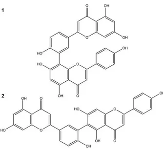
Plant extraction and isolation - The whole dried pulverised plant was submitted to a pressurised fluid extractor (Dionex, model ASE 150) equipped with an extraction cartridge (100 mL). The following parameters were repeated three times: temperature of 130ºC, pres-sure of 1,500 psi, static extraction time of 4 min, 150% volume wash and three cycles of extraction. Plant ma-terial (90.0 g) was first extracted with dichloromethane to remove apolar compounds, followed by a mixture of ethyl acetate:methanol (8:2) and finally ethanol:water (7:3). The latter extraction cycles were concentrated in a rotary evaporator, yielding a hydroethanolic extract de-nominated SSHE. The yield was 10% (w/w). SSHE (2.0 g) was chromatographed on a Sephadex LH-20 column and eluted with methanol (MeOH); 102 fractions of 20 mL were collected. The obtained fractions were grouped after thin-layer chromatography on silica gel 60 plates (Merck), eluted with chloroform:methanol (9:1 and 8.5:1.5) and the revealed with NP/PEG. Fractions 52-63 (14.8 mg) were grouped, obtaining the compound amen-toflavone (1). Fractions 64-101 (1 mg) were identified as robustaflavone (2) (Fig. 1).
Structural elucidation - The compounds were identi-fied by 1H NMR, 13C NMR and DEPT-135º Bruker (300/75
MHz), DPX-300 diluted in deuterated dimethyl sulfoxide (DMSO) (Merck, Germany). Data for mass spectrometry
and tandem mass spectrometry were obtained in high res-olution (ESI-TOF micrOTOF II, Bruker Daltonics) with positive ionisation mode. The presence of amentoflavone and robustaflavone were confirmed by comparison with literature spectra (Agrawal & Bansal 1989).
Liquid chromatography - SSHE was analysed quan-titatively (500 µg/mL) for high-performance liquid chro-matography (HPLC) with diode array detector (DAD) using a liquid chromatograph model LC-20AD coupled with a DAD (model SPD-M20A, Shimadzu), operating at a wavelength of 325 nm and manual injector with 20
μL loop sampling. A reversed phase octadecyl Shimadzu
Shim-pack PREP-ODS (H) kit (250 mm x 4.6 mm, 5 µm) and guard column (4 cm x 3 mm) with the same stationary phase were used for separation. The mobile phase consist-ed of A (H2O) and B (MeOH), both with 1% acetic acid, using a linear gradient system: 0.01-10 min (25% B), 10-25 min (100% B), 25-30 min (100% B) and 30-45 min (25% B). The flow rate was 0.8 mL/min. The chromatographic data were analysed on a computer with the LCsolution operating system (Shimadzu). The solvents were HPLC grade (Vetec) and ultrapure water (Millipore Inc).
Parasites - A standard strain of Leishmania (L.) ama-zonensis (IFLA/BR/1967/PH8) was used for in vitro tests. Amastigote forms were routinely isolated from BALB/c mice’s cutaneous lesions and maintained as promastig-otes at 25ºC in Schneider’s Insect Medium (Sigma-Al-drich), supplemented with 20% foetal calf serum (FCS) (Cultilab) and 140 µg/mL gentamicin (Sigma-Aldrich).
Animals - Female BALB/c mice aged six weeks were used to obtain resident peritoneal macrophages used in the tests. The animals were obtained from the central an-imal facility of the CCBS/UFMS in good health and free of common rodent infections or parasites, maintained in individually ventilated cages equipped with mini-isola-tors, fed a balanced feed (Nuvilab CR-1, Nuvital®) with
free access to water. This study received approval from the local Animal Experimentation Ethical Committee (CEUA/UFMS) under protocol 431/2012.
Antileishmanial activity - Peritoneal macrophages from BALB/c mice were isolated after rinsing with RPMI-1640 medium (Sigma) and placed (1 x 10⁵ cells/
well) in a 24-well plate in RPMI-1640 medium (Sigma), supplemented with 10% FCS (Cultilab) and 140 µg/mL gentamicin (Sigma). After 1 h incubation at 37ºC in 5% CO2, cells were infected with L. amazonensis promas-tigotes (1 x 10⁶ cells/well) and subsequently incubated
at 35ºC for 4 h. SSHE was added at concentrations of 50-12.5 µg/mL. The cells were incubated at 35ºC in 5% CO2, fixed and stained with Giemsa after 24, 48 and 72 h. Amentoflavone and robustaflavone were added at dif-ferent concentrations and the cells stained after 72 h of treatment. Untreated infected cells were used as nega-tive control. Amphotericin B (Sigma) was used as a posi-tive control and analysed after 24 h. The percentage of infected macrophages and the total number of amastig-otes were determined by counting 200 cells in each six replicates. The infection index was determined by mul-tiplying the percentage of macrophages that had at least
one intracellular parasite by the mean of amastigotes per macrophage (Paladi et al. 2012). A non-linear dose-response regression curve was used to calculate the half maximum inhibitory concentration (IC50).
Nitric oxide (NO) evaluation - NO production by L. amazonensis infected cells treated as described in the previous item was evaluated. One hundred microlitres of the supernatants were collected and incubated with an equal volume of Griess reagent (1% sulfanilamide/0.1% naphthalene diamine dihydrochloride/2.5% H3PO4) for 10 min at room temperature for the quantification of the nitrite accumulation (Ding et al. 1988). Absorbance was determined at 540 nm. The conversion of absorbance to µM of NO2- was performed by comparing the samples
to a standard curve obtained with known concentrations (1-10 µM) of sodium nitrite diluted in RPMI medium.
Cytotoxicity assay - Murine macrophages (J774.A1) and fibroblast cells (NIH/3T3) purchased from the Rio de Janeiro Cell Bank (Brazil) were treated with SSHE and isolated compounds at four concentrations from 0.25-250 µg/mL in triplicates for estimating IC50. The plates were incubated with the test substances for 48 h at 37ºC and 5% CO2 amphotericin B (Sigma) was used as reference drug (0.025-25 µg/mL). DMSO (Vetec) was used as a negative control at the concentration used to solubilise higher concentrations of test compounds.
Cell viability was determined using sulforhodamine B (SRB) assay (Skehan et al. 1990). Briefly, the cells
were fixed with 100 μL of ice-cold 20% trichloroacetic
acid (Sigma) and incubated at 4ºC for 30 min. The su-pernatant was then discarded and the plates were washed five times with tap water. The cells were stained for 30
min with 0.1% SRB in 1% acetic acid (50 μL/well) (Sig -ma) and subsequently washed four times with 1% acetic acid to remove the unbound dye. The plates were air-dried and the protein-bound dye was solubilised with
100 μL 10 mM Trizma buffer (Sigma). The plates were
shaken for 10 min on a shaker and the resulting optical density was read in a multiwell plate reader at 540 nm. The growth percentage of each test sample was calcu-lated as described by Monks et al. (1991). A non-linear dose-response regression curve was used to calculate IC50. The selectivity index (SI) was calculated according to Medeiros et al. (2011).
Statistical analysis - NO evaluation and the infection index were expressed as the mean ± standard deviation and the data were analysed using the Student’s t test. Differences were considered significant at p < 0.05 (rep-resented by asterisks).
RESULTS AND DISCUSSION
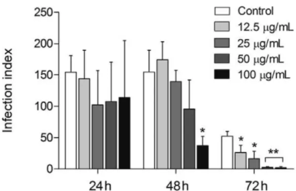
SSHE was tested against intracellular amastigotes by evaluating the kinetics of infection at 24, 48 and 72 h. Our results demonstrated a dose-dependent inhibi-tion of the proliferainhibi-tion of intracellular amastigotes with highest activity registered 72 h after extract addition. At that point, the infection index had decreased from 51.6-96.5%, from the lowest to the highest concentration test-ed (Fig. 2). The IC that rtest-eductest-ed 50% of the intracellular forms of L. amazonensis (IC50) was 20.2 µg/mL with no cytotoxicity to the mammalian cells tested (Table).
SSHE was considered active according to Claudino et al. (2013). Thus their constituents were characterised in order to identify the compounds responsible for the activity. Amphotericin B was used as a reference drug and resulted in IC50 value of 0.03 µg/mL against L. ama-zonensis (Table). Despite its high activity and selectivity, the clinical treatment with amphotericin B is still
high-Fig. 2: antileishmanial activity of the hydroethanolic extract from Se-laginella sellowii (SSHE) on intracellular amastigotes. Peritoneal mac-rophages were infected with Leishmania amazonensis and treated with different concentrations of SSHE. Infection index was calculated 24, 48 and 72 h after treatment. Bars represent the mean ± standard deviation of quadruplicates. p < 0.01 (*) and p < 0.0001 (**) for the different con-centrations compared to untreated cells (control) (Student’s t test).
TABLE
Antileishmanial activity and cytotoxicity of hydroethanolic extract (SSHE) and biflavonoids isolated from Selaginella sellowii
Test sample
Intracellular amastigotes NIH/3T3 J774.A1
IC50 µg/mL(µM)
IC50
µg/mL(µM) SI
IC50
µµg/mL (µM) SI
SSHE 20.2 246.4 12.2 166.3 8.2
ly toxic, costly and associated with cases of resistance (Freitas-Júnior et al. 2012), making the search for a new antileishmanial drug relevant.
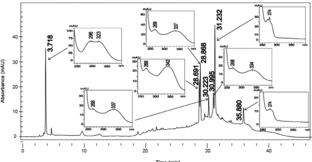
The analysis of ultraviolet spectra of SSHE com-pounds revealed the presence of phenylpropanoids (Sil-verstein et al. 2007) and biflavonoids (Mabry et al. 1970) (Fig. 3). The fractionation led to the isolation of two bi-flavonoids described in the genus Selaginella: amento-flavone (Ma et al. 2001, Zhang et al. 2011) and robusta-flavone (Lin et al. 2000, Zhang et al. 2011).
The 13C NMR profiles of the compounds (1) and
(2) were consistent with those reported previously for amentoflavone and robustaflavone (Agrawal & Bansal 1989). The mass spectrum of (1) exhibited m/z 539,0973 [M+H]+ and fragments which included m/z 497, 403, 347,
335, 283, 153 and 121, characteristic of amentoflavone (Zhang et al. 2011). Compound (2) exhibited a mass spectrum with m/z 539,0956 [M+H]+ and fragments m/z
521, 465, 387, 283, 270, 153 and 121, characteristic of robustaflavone (Zhang et al. 2011).
Amentoflavone shows various biological activities such as antiviral (Ma et al. 2001), antifungal (Jung et al. 2006), antioxidant (Sakthivel & Guruvayoorappan 2013) and anti-inflammatory (Oh et al. 2013). Robusta-flavone is described as a potent inhibitor of hepatitis B virus replication (Zembower et al. 1998). Furthermore, both biflavonoids are suggested for the development of promising anti-dengue drugs (Coulerie et al. 2013) and antielastase agents (Xu et al. 2009).
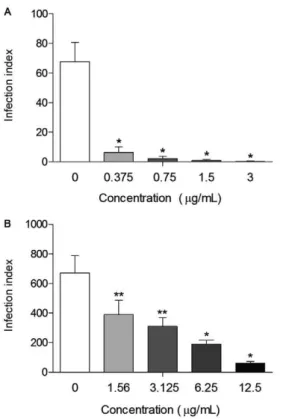
Once the highest activity was found 72 h after the treatment with SSHE, the isolates were evaluated at that point. Purification resulted in compounds with even higher activity. Treatment of intracellular amastigotes at all concentrations tested of amentoflavone caused a significant decrease (p < 0.0001) in the infection index when compared to the control, with a reduction greater
than 90.6%, reaching almost 100% at the highest con-centration (3 µg/mL) (Fig. 4A). Amentoflavone was about 200 times more potent than SSHE (IC50 = 0.1 µg/ mL). The excellent antileishmanial activity of amento-flavone obtained in our study is in agreement with those obtained by Oubada et al. (2014), who observed the ef-fects on L. amazonensis intracellular amastigotes 48 h after the aforementioned treatment. Amentoflavone was not active on axenic amastigotes of Leishmania donovani
(Weniger et al. 2006, Kunert et al. 2008) and showed poor activity on promastigotes of the same species (Ca-macho et al. 2000). Therefore, the antileishmanial action of this compound may be mediated by the host cell. This fact, coupled with the delayed action of SSHE (72 h), leads us to consider the compound as a pro-drug, which needs to be metabolised by the cell to exert its effect.
Robustaflavone also showed antileishmanial activity in a dose-dependent way, with a reduction of the infec-tion index reaching 90.8% (12.5 µg/mL) (Fig. 4B). Ro-bustaflavone was less active than amentoflavone, with an IC50 value of 2.8 µg/mL. Despite being considered as active (IC50 < 10 µg/mL), the compound showed some cytotoxicity on mammalian cells, with an SI below 10 (Lenta et al. 2007), as shown in Table.
Activation of macrophages was investigated by the NO release. NO production was significantly increased (p < 0.05) after treatment with the highest concentra-tion of SSHE (100 µg/mL) (Fig. 5A). NO producconcentra-tion by peritoneal macrophages treated with amentoflavone was significantly lower at the concentrations of 1.5 µg/mL (p < 0.05) and 0.75 µg/mL (p < 0.01), compared to untreated infected cells (Fig. 5B). This may be due to the antioxi-dant properties of amentoflavone. Banerjee et al. (2002) observed the inhibition of inducible NO synthase (iNOS) protein expression by amentoflavone. In addition, Woo et al. (2005) demonstrated the inhibitory effect of
toflavone on NO production induced by
lipopolysaccha-ride, which prevents the activation of NF-κB, the gene
responsible for transcription of iNOS. They suggest, therefore, that the destruction mechanism of intracellu-lar amastigotes by this biflavonoid should not be directly associated with increased release of NO. Amentoflavone could have a direct action on the parasites and/or influ-ence over other cytotoxic mechanisms for intracellular pathogens. Defense mechanisms independent of NO re-lease can be promising in cases of ACL associated with strains of L. amazonensis and Leishmania braziliensis
resistant to NO, as described by Giudice et al. (2007), who demonstrated that the resistance to NO is directly related to lesion size and severity of the disease.
In contrast, robustaflavone had increased the NO release by infected macrophages at the highest concen-tration tested (Fig. 5C). In fact, Yang et al. (2006) dem-onstrated that robustaflavone isolated from Selaginella tamariscina did not affect the expression of iNOS. In the same study, this effect was compared with another iso-lated biflavonoid called sumaflavone, which, as amen-toflavone, has inhibited iNOS and decreased NO levels. The authors also suggest the difference in inhibitory potential of NO due to differences in the location of the C-C bond in the compounds, i.e., IC3’-IIC8” in amento-flavone/sumaflavone and IC3’-IIC6” in robustaflavone.
This is the first phytochemical investigation of S. sellowii. The fractionation of active SSHE allowed the identification of even more active compounds. The an-tileishmanial mechanism of amentoflavone does not seem to involve macrophage activation by increasing the release of NO, unlike the mechanism from robusta-flavone, which induced an increase in its production. S. sellowii may be a potential source of biflavonoids that could provide promising compounds for the treatment of cutaneous leishmaniasis.
REFERENCES
Agrawal PK, Bansal MC 1989. Other Flavonoids. In PK Agrawal, Carbon-13 NMR of flavonoids. Studies in organic chemistry-39, Elsevier, Amsterdam, p. 236-282.
Assis ELM, Labiak PH 2009. Lycophyta da borda oeste do Pantanal, Mato Grosso do Sul, Brasil. Acta Bot Bras 23: 703-712.
Fig. 4: antileishmanial activity of amentoflavone (A) and robustafla-vone (B) on intracellular amastigotes. Peritoneal macrophages were infected with Leishmania amazonensis and treated with different concentrations of the compounds. Infection index was calculated 72 h after treatment. Bars represent the mean ± standard deviation of six replicates. p < 0.01 (**) and p < 0.0001 (*) for the different concentra-tions compared to untreated cells (control) (Student’s t test).
Banerjee T, Van der Vliet A, Ziboh VA 2002. Downregulation of COX-2 and iNOS by amentoflavone and quercetin in A549 hu-man lung adenocarcinoma cell line. Prostaglandins Leukot Es-sent Fatty Acids 66: 485-492.
Brahmachari G 2011. Natural products in drug discovery: impacts and opportunities - an assessment. In G Brahmachari, Bioac-tive natural products: opportunities and challenges in medicinal chemistry, 1st ed., World Scientific Publishing Co. Pte. Ltd., Sin-gapore, p. 1-199.
Camacho MR, Mata R, Castaneda P, Kirby GC, Warhurst DC, Croft SL, Phillipson JD 2000. Bioactive compounds from Celaenoden-dron mexicanum. Planta Med 66: 463-468.
Claudino VD, da Silva KC, Filho VC, Yunes RA, Monache FD, Gimé-nez A, Salamanca E, Gutierrez-Yapu D, Malheiros A 2013. Dri-manes from Drimys brasiliensis with leishmanicidal and antima-larial activity. Mem Inst Oswaldo Cruz 108: 140-144.
Coulerie P, Nour M, Maciuk A, Eydoux C, Guillemot JC, Lebouvier N, Hnawia E, Leblanc K, Lewin G, Canard B, Figadère B 2013. Structure-activity relationship study of biflavonoids on the dengue virus polymerase DENV-NS5 RdRp. Planta Med 79: 1313-1318.
Croft SL, Coombs GH 2003. Leishmaniasis - current chemotherapy and recent advances in the search for novel drugs. Trends Para-sitol 19: 502-508.
Ding AH, Nathan CF, Stuer DJ 1988. Release of reactive nitrogen intermediates and reactive oxygen intermediates from mouse peritoneal macrophages: comparison of activating cytokines and evidence for independent production. J Immunol 141: 2407-2412.
Freitas-Júnior LH, Chatelain E, Kim HA, Siqueira-Neto JL 2012. Vis-ceral leishmaniasis treatment: what do we have, what do we need and how to deliver it? Int J Parasitol Drugs Drug Resist 2: 11-19.
Giudice A, Camada I, Leopoldo PTG, Pereira JMB, Riley LW, Wilson ME, Ho JL, Jesus AR, Carvalho EM, Almeida RP 2007. Resis-tance of Leishmania (Leishmania) amazonensis and Leishmania (Viannia) braziliensis to nitric oxide correlates with disease se-verity in tegumentary leishmaniasis. BMC Infect Dis 7: 7.
Gontijo B, Carvalho MLR 2003. Leishmaniose tegumentar ameri-cana. Rev Soc Bras Med Trop 36: 71-80.
Grimaldi Jr G, Tesh RB 1993. Leishmaniases of the New World: cur-rent concepts and implications for future research. Clin Microbiol Rev 6: 230-250.
Jeremy AC 1990. Selaginellaceae. In KU Kramer, K Kubitzki, PS Green, The families and genera of vascular plants: pteridophytes and gymnosperms, vol. 1, Springer Verlag, Berlin, p. 39-45.
Jung HJ, Sung WS, Yeo SH, Kim HS, Lee IS, Woo ER, Lee DG 2006. Antifungal effect of amentoflavone derived from Selaginella tamariscina. Arch Pharm Res 29: 746-751.
Kunert O, Swamy RC, Kaiser M, Presser A, Buzzi S, Apparao AVN, Schühly W 2008. Antiplasmodial and leishmanicidal activity of biflavonoids from Indian Selaginella bryopteris. Phytochem Lett 1: 171-174.
Lenta B, Vonthron-Sénécheau C, Soh RF, Tantangmo F, Ngouela S, Kaiser M, Tsamod E, Anton R, Weniger B 2007. In vitro antipro-tozoal activities and cytotoxicity of some selected Cameroonian medicinal plants. J Ethnopharmacol 111: 8-12.
Lin LC, Kuo YC, Chou CJ 2000. Cytotoxic biflavonoids from Sela- ginella delicatula. J Nat Prod 63: 627-630.
Lin RC, Skaltsounis AL, Seguin E, Tillequin F, Koch M 1994. Phenolic constituents of Selaginella doederleinii. Planta Med 60: 168-170.
Ma SC, But PPH, Ooi VEC, He YH, Lee SHS, Lee SF, Lin RC 2001. Antiviral amentoflavone from Selaginella sinensis. Biol Pharm Bull 24: 311-312.
Mabry TJ, Markham KR, Thomas MB 1970. The systematic identifica-tion of flavonoids, Springer, New York, 354 pp.
Medeiros MGF, Silva AC, Citó AMGL, Borges AR, Lima SG, Lopes JAD, Figueiredo RCBQ 2011. In vitro antileishmanial activ-ity and cytotoxicactiv-ity of essential oil from Lippia sidoides Cham. Parasitol Int 60: 237-241.
Mishra PK, Raghuram GV, Bhargava A, Ahirwar A, Samarth R, Up-adhyaya R, Jain KS, Pathak N 2011. In vitro and in vivo evaluation of the anticarcinogenic and cancer chemopreventive potential of a flavonoid-rich fraction from a traditional Indian herb Selaginella bryopteris. Br J Nutr 106: 1154-1168.
Monks A, Scudiero D, Skehan P, Shoemaker R, Pau K, Vistica D, Hose C, Langley J, Cronise P 1991. Feasibility of a high-flux anticancer drug screen using a diverse panel of cultured human tumor cell lines. J Natl Cancer Inst 83: 757-766.
Monzote L 2009. Current treatment of leishmaniasis: a review. Int J Antimicrob Agents 1: 9-19.
Oh J, Rho HS, Yang Y, Yoon JY, Lee J, Hong YD, Kim HC, Choi SS, Kim TW, Shin SS, Cho JY 2013. Extracellular signal-regu-lated kinase is a direct target of the anti-inflammatory compound amentoflavone derived from Torreya nucifera. Mediators In-flamm 2013: 1-11.
Oliveira LFO, Schubach AO, Martins MM, Passos SL, Oliveira RO, Marzochi MC, Andrade CA 2011. Systematic review of the ad-verse effects of cutaneous leishmaniasis treatment in the New World. Acta Trop 118: 87-96.
Oubada A, García M, Bello-Alarcó A, Cuesta-Rubio O, Monzote L 2014. Antileishmanial activity of leaf extract from Calophyllum rivulare against Leishmania amazonensis. Emir J Food Agric 26: 807-812.
Paladi CS, Pimentel IAS, Katz S, Cunha RLOR, Judice WAS, Caires ACF, Barbiéri CL 2012. In vitro and in vivo activity of a pallada-cycle complex on Leishmania (Leishmania) amazonensis. PLoS Negl Trop Dis 6: e1626.
Reithinger R, Dujardin JD, Louzir H, Pirmez C, Alexander B, Brooker S 2007. Cutaneous leishmaniasis. Lancet Infect Dis 7: 581-596.
Sah NK, Singh SNP, Sahdev S, Banerji S, Jha V, Khan Z, Hasnain SE 2005. Indian herb ‘Sanjeevani’ (Selaginella bryopteris) can pro-mote growth and protect against heat shock and apoptotic activi-ties of ultra violet and oxidative stress. J Biosci 30: 499-505.
Sakthivel KM, Guruvayoorappan C 2013. Amentoflavone inhibits iNOS, COX-2 expression and modulates cytokine profile, NF-κB signal transduction pathways in rats with ulcerative colitis. Int Immunopharmacol 17: 907-916.
Santos JFL, Pagani E, Ramos J, Rodrigues E 2012. Observations on the therapeutic practices of riverine communities of the Unini River, AM, Brazil. J Ethnopharmacol 142: 503-515.
Silverstein RM, Webster FX, Kiemle DJ 2007. Identificação espec-trométrica de compostos orgânicos, 7th ed., LTC, Rio de Janeiro, 490 pp.
Skehan P, Storeng R, Scudiero D, Monks A, Mcmahon J, Vistica D, Warren JT, Bokesch H, Kenney S, Boyd MR 1990. New colori-metric cytotoxicity assay for anticancer-drug screening. J Natl Cancer Inst 82: 1107-1112.
Sundar S, Chatterjee M 2006. Visceral leishmaniasis - current thera-peutic modalities. Indian J Med Res 123: 345-352.
Sundar S, More DK, Singh MK, Singh VP, Sharma S, Makharia A, Kumar PC, Murray HW 2000. Failure of pentavalent antimony in visceral leishmaniasis in India: report from the center of the Indian epidemic. Clin Infect Dis 31: 1104-1107.
Tang GH 2009. Pyrrolidinoindoline alkaloids from Selaginella moellendorfii. J Nat Prod 72: 1151-1154.
Weniger B, Vonthron-Sénécheau C, Kaiser M, Brun R, Anton R 2006. Comparative antiplasmodial, leishmanicidal and antitrypanosomal activities of several biflavonoids. Phytomedicine 13: 176-180.
Woo ER, Lee JY, Cho IJ, Kim SG, Kang KW 2005. Amentoflavone inhibits the induction of nitric oxide synthase by inhibiting NF-κB activation in macrophages. Pharmacol Res 51: 539-546.
Xu GH, Ryoo IJ, Kim YH, Choo SJ, Yoo ID 2009. Free radical scav-enging and antielastase activities of flavonoids from the fruits of Thuja orientalis. Arch Pharm Res 32: 275-282.
Yang JW, Pokharel YR, Kim MR, Woo ER, Choi HK, Kang KW 2006.
Inhibition of inducible nitric oxide synthase by sumaflavone isolat-ed from Selaginella tamariscina. J Ethnopharmacol 105: 107-113.
Zembower DE, Lin YM, Flavin MT, Chen FC, Korba BE 1998. Ro-bustaflavone, apotential non-nucleoside anti-hepatitis B agent. Antiviral Res 39: 81-88.
Zhang YX, Li QY, Yan LL, Shi Y 2011. Structural characterization and identification of biflavones in Selaginella tamariscina by liquid chromatography-diode-array detection/electrospray ion-ization tandem mass spectrometry. Rapid Commun Mass Spec-trom 25: 2173-2186.