IN METABOLIC DISORDERS
S
TUDIES ON THE MITOCHONDRIAL FLAVOENZYMEETF
Bárbara Joana de Almeida Henriques
Dissertation presented to obtain the PhD degree in Biochemistry at the Instituto de Tecnologia Química e Biológica,
Universidade Nova de Lisboa
Supervisor
Cláudio Emanuel Moreira Gomes Opponents
Ronald J. A. Wanders & Carlos M. S. Farinha
Instituto de Tecnologia Química e Biológica, Universidade Nova de Lisboa
ITQB - Protein Biochemistry Folding and Stability Laboratory Instituto de Tecnologia Química e Biológica, Universidade Nova de Lisboa Av. da República (EAN), 2785-572 Oeiras, PORTUGAL
This dissertation describes the work performed under the supervision of Prof. Cláudio M. Gomes, in the Protein Biochemistry Folding and Stability Laboratory, Instituto de Tecnologia Química e Biológica from October 2006 to June 2010.
The studies here presented aim to contribute to a better understanding of human electron transfer flavoprotein (ETF) folding and stability, towards the elucidation of the molecular rationale of multiple acyl-CoA dehydrogenase deficiency (MADD). First, ETF disease causing missense mutations and the impact of three of those mutations on the protein folding and stability is overviewed. Subsequently, the role of flavinylation on a mutant variant resulting in a mild phenotype was addressed in order to gain a better understanding on the molecular rationale for riboflavin supplementation. Further, two polymorphic ETF variants were analyzed to explore their effects on the protein folding and function and to investigate possible implication in MADD.
This thesis is organised in three parts. The first part is an introduction comprising two chapters: the first describing the state of knowledge on the protein folding problem and protein homeostasis, and the second presenting
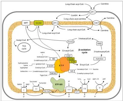
an overview on mitochondrial fatty acid β-oxidation (FAO) enzymes
I would like to express my sincere gratitude to the following people without whom this work would not have been possible:
My supervisor, Cláudio M. Gomes, for all the knowledge and dedication to the work developed in the lab. For his confidence in me and my work. And especially for his passion for science that always keep me motivated.
Peter Bross, for receiving me at Aarhus University Hospital, and for scientific training in molecular biology techniques. I also thank him for all the enthusiastic scientific discussions.
Rikke Olsen, from Aarhus University Hospital for her helpful discussions.
Niels Gregersen and the entire group at Aarhus University Hospital for making my visit there a very pleasant journey.
Mark Fisher, from the University of Kansas Medical Center (US), for sharing immobilized GroEL beads, and for his useful discussions.
My present and past colleagues at the Protein Biochemistry, Folding and Stability Laboratory, for their precious support in the lab, helpful discussions, and amazing work environment.
Hugo Botelho, for being so helpful, and especially for his patience and friendship.
on my work. It is fantastic to share science with you.
Sonia Leal, for the support given during these 6 years, and all the good times we shared.
Ana Paula for always having 5 minutes to listen to me, for her interest in science and for all the laughs in the difficult moments.
All my friends that in someway helped me during these 4 years, especially to Sofia, Patricia, Vera, and Lígia, for always being there and for sharing with me the ups and downs of being a PhD student.
My family for the support and love.
Lipe, for his love, encouragement and for all the support given during this long walk. I could not have done this without you, thanks!
This thesis is dedicated to a wonderful woman, my grandmother Maria, without whom I would not be the person I am today.
Fundação para a Ciência e Tecnologia is acknowledged for financial support, by awarding a PhD Grant SFRH/BD/29200/2006
1. Henriques, B. J., Rodrigues, J. V., Olsen, R. K., Bross, P., Gomes C. M. "Role of flavinylation in a mild variant of multiple acyl-CoA dehydrogenation deficiency: A molecular rationale for the effects of riboflavin supplementation"
J. Biol. Chem.2009, 284 (7):4222-4229
2. Henriques, B. J., Bross, P., Gomes C. M.
"Mutational hotspots in electron transfer flavoprotein underlie defective folding and function in multiple acyl-CoA dehydrogenase deficiency"
BBA - Molecular Basis of Disease 2010,1802 (11):1070-1077
3. Henriques, B. J., Olsen, R. K., Bross, P., Gomes C. M.
"Emerging Roles for Riboflavin in Functional Rescue of Mitochondrial Oxidation Flavoenzymes"
Current Medicinal Chemistry 2010 (in press)
4. Henriques, B. J., Fisher M., Bross P., Gomes, C.M.
"A polymorphic position in electron transfer flavoprotein modulates kinetic stability as evidenced by thermal stress"
FEBS Letters 2010 (in press)
Other publications not included in this thesis
5. Henriques, B. J., Saraiva, L. M., Gomes, C. M.,
"Combined spectroscopic and calorimetric characterisation of rubredoxin reversible thermal transition"
J. Biol. Inorg. Chem. 2006, 11:73-81
6. Henriques, B. J., Saraiva, L. M., Gomes, C. M.,
"Probing the mechanism of rubredoxin thermal unfolding in the absence of salt bridges by temperature jump experiments"
The work presented in this dissertation concerns the study of the electron
transfer flavoprotein (ETF), a protein involved in mitochondrial β-oxidation
whose deficiency is associated to multiple acyl-CoA dehydrogenase deficiency (MADD). The thesis will focus on establishing the functional, cellular and molecular consequences of the genetic variability in ETF, and in particular it aims to clarify the basis for the effect of heat stress on disease progression. Moreover, the beneficial effects of vitamin B2 supplementation will be addressed.
MADD, which is an autosomal recessively inherited disorder of fatty acid, amino acid, and choline metabolisms, results from deficiencies in any of the
following genes: ETFA, ETFB or ETFDH. ETFA and ETFB genes encode
for the α and β subunits of ETF, whereas ETFDH encodes for electron
missense mutations in the above mentioned genes result in proteins with a lower enzymatic activity leading to cellular functional deficiency and thus milder phenotypes. In these cases, the amino acid alterations are likely to cause conformational changes in the expressed gene products which account for disease. This effect is mostly evident in late-onset MADD patients, in which the disease is often intermittent and only becomes evident during periods of illness or catabolic stress. Even so, the molecular genetic basis and functional characterisation of MADD genotypes remains elusive, thus a
detailed in vitro investigation will contribute to a better functional
understanding of the clinical heterogeneity in MADD.
An extensive in silico analysis was carried out on 18 disease associated
missense mutations found in ETF. The analysis revealed that known mutations fall essentially in two groups: 1) mutations affecting protein folding and assembly; 2) mutations impairing catalytic activity and interactions with partner dehydrogenases. We have focused our studies on three of these
mutations, ETFβ Cys42Arg, Asp128Asn and Arg191Cys, which typify
different clinical phenotypes. This part of the investigation involved the study of expressed ETF variants, aiming at evaluating the impact of mutagenesis on ETF intrinsic conformational stability, enzymatic activity, protein assembly and interaction with functionally relevant enzymes.
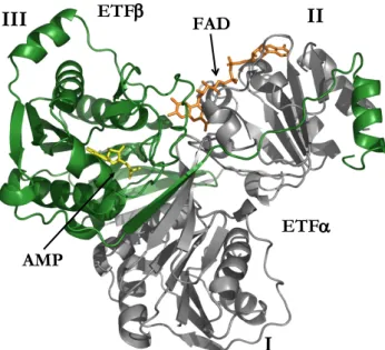
The ETFβ-Cys42Arg mutation, a severe MADD mutation, affects directly
the AMP binding site and the intersubunit contacts impairing protein folding.
In vivo assays following the recombinant expression of the protein in an E. coli
cofactors.
Proteins harboring the other two mutations, ETFβ-Asp128Asn and
ETFβ-Arg191Cys, which are associated to mild MADD, were purified in the
soluble form after heterologous expression. We have used a combination of biophysical and biochemical methods to address the impact of these last two mutations on ETF folding, conformational stability and efficiency in
mediating electron transfer. The two mutant variants had an overall α/β fold
topology identical to native ETF, but both have substantially decreased
enzymatic activity and conformational stability. The ETFβ-Asp128Asn
mutation has a stronger impact on the protein conformational stability, as this mutation is near the dimer interface and the FAD binding site. On the other
hand, the decreased activity observed for the ETFβ-Arg191Cys variant is
likely to result from the impairment of interactions with the electron-donor dehydrogenases, as the mutated residue is within the predicted interface of
the complex formation. Thus, combination of in silico analysis of mutations
with experimental data has allowed to establish structural hotspots within ETF fold, which are useful to provide a rationale for the prediction of effects of mutations in ETF.
Mutations leading to mild clinical phenotypes, such as those selected in this study, are frequently associated to poor catalytic activity of the mutant variant and deficient FAD insertion. Moreover, the molecular consequences of riboflavin supplementation in the functional rescue of defective fatty acid
β-oxidation flavoenzymes are becoming increasingly clear. This has been
made possible with the combination of diverse studies made during the last
decades, ranging from in vitro analysis of purified proteins, to proteomic
availability of FAD, which is the key cofactor in many fatty acid β-oxidation enzymes. It is becoming clear that apart from being an essential chemical component of the active site, FAD may also play a multitude of additional roles such as assisting folding.
This aspect was investigated in detail using the ETFβ-Asp128Asn variant
as a model. Clinical data reports that a homozygous patient for this mutation developed severe disease symptoms in association with a viral infection and fever. In agreement, heat inactivation of this mutant is more significant at
temperatures above 37°C. We have found that cofactor insertion in vitro
substantially improves the folding and stability of this ETF variant. Moreover, the presence of an excess of flavin in a concentration identical to that determined in the mitochondria from muscle of patients that take vitamin B2 orally, prevented proteolytic digestion by avoiding protein destabilization. To mimic a situation of fever in vitro, the flavinylation status was tested at 39°C: FAD improves the protein conformation yielding a more stable and active enzyme, thus acting as a pharmacological chaperone.
The results obtained provide a structural and functional framework for the role of vitamin B2 supplementation in the molecular pathogenesis of MADD and flavoprotein disorders in general.
Apart from disease causing mutations, a number of polymorphic variants have been reported in FAO enzymes. In particular, a polymorphic variation in the alpha-subunit of ETF, which leads to the incorporation of either a threonine or an isoleucine at position 171, has been identified. A previous study has indicated that patients suffering from a mild form of very long chain acyl-CoA dehydrogenase deficiency (VLCADD) displayed an
as yet unknown molecular mechanism. The effect of the variation of the
ETFα-Thr/Ile171 polymorphism on the ETF folding and dynamics under
thermal stress was investigated. The results showed that the two variants have
the identical thermodynamic stabilities (Tm=58°C), although the ETFα
-Thr171 variant has a decreased thermal inactivation midpoint (Tm
inact
=49°C)
in comparison to that of ETFα-Ile171 (Tm
inact
=58°C). Upon thermal stress,
the ETFα-Thr171 variant is prone to faster cofactor (FAD) loss and
conformational destabilization leading to an increase of a heterogeneous population of the variant. Also, this variant has higher conformational
dynamics during thermal stress: upon 2 hours at 39°C, ETFα-Thr171 looses
half of its activity, whereas ETFα-Ile171 activity remains at ~85%. ETFα
-Thr171 can be rescued by the GroEL chaperonin, which captures and refolds the thermally destabilized variant, as observed by the retained enzymatic activity. Therefore, this polymorphic position has an impact on protein conformation and function under thermal stress. The latter is a common metabolic trigger for the manifestation of mitochondrial beta oxidation defects.
O trabalho apresentado nesta dissertação centra-se no estudo da “flavoproteína de transferência electrónica” (ETF), uma proteína
mitocondrial envolvida na β-oxidação dos ácidos gordos, cuja deficiência está
associada à patologia “deficiência múltipla de acil-CoA desidrogenases” (MADD). A tese foca-se na avaliação das consequências funcionais, celulares e moleculares da variabilidade genética na ETF, e em particular, tem como objectivo elucidar a base do efeito de stress térmico na progressão da doença. As consequências moleculares do suplemento com vitamina B2 serão também abordadas.
MADD é uma patologia hereditária autossómica recessiva associada ao metabolismo mitocondrial dos ácidos gordos, aminoácidos e cholina. A patologia resulta de uma deficiência numa das seguintes proteínas: nas subunidades alfa (ETFA) ou beta (ETFB) da (ETF), ou na ETF-ubiquinona oxidoreductase (ETF-QO). Estas enzimas estão envolvidas no metabolismo energético da mitocôndria, actuando concertadamente na matriz mitocondrial catalisando a oxidação dos ácidos gordos de modo a obter energia.
limitado de genótipos foi caracterizado. Os dados disponíveis na literatura sugerem uma relação directa entre o fenótipo clínico e os níveis de ETF com actividade biológica. Claramente, uma mutação nula resulta de um fenótipo severo, e uma mutação pontual no gene da ETF resulta numa menor actividade enzimática conduzindo a uma deficiência funcional ao nível celular. Neste casos, a alteração do aminoácido provavelmente determina alterações conformacionais na proteína que está relacionada com a patologia. Este efeito é mais proeminente nos pacientes com MADD de expressão tardia, nos quais a patologia não é continua, manifestando-se apenas em períodos de doença ou stress catabólico. Assim torna-se necessário um estudo para uma melhor compreensão da variabilidade da expressão clínica da MADD.
Uma ampla análise in silico foi realizada em 18 mutações pontuais,
associadas à MADD, na ETF. A análise revelou que as mutações podem ser agrupadas essencialmente em dois grupos: 1) mutações que afectam a dobragem proteica e a estabilidade conformacional intrínseca; 2) mutações que perturbam actividade enzimática e a interacção com as desidrogenases.
Três destas mutações, ETFβ Cys42Arg, Asp128Asn e Arg191Cys, que
tipificam diferentes cenários no que diz respeito ao fenótipo clínico, foram alvo de um estudo detalhado. Esta parte do trabalho teve como objectivo obter as proteínas variantes da ETF de modo a testar o efeito das mutações na estrutura, dobragem, actividade enzimática, e interacção com co-enzimas funcionalmente relevantes.
A variante ETFα-Cys42Arg, associada a um fenótipo severo da patologia,
afecta directamente o local de ligação do co-factor AMP, e os contactos entre as duas subunidades, logo resulta numa deficiente dobragem proteica. A
Recorrendo à co-expressão com chaperões (GroEL/GroES e dnaK/dnaJ/GrpE)) foi possível expressar a proteína na fracção solúvel, mas não permitiu restaurar a actividade enzimática, provavelmente devido a uma inserção deficiente dos co-factores (AMP e FAD).
Duas variantes associadas com fenótipos menos severos, ETFβ
Asp128Asn e ETF-Arg191Cys, foram heterologamente expressas e purificadas a partir da fracção solúvel. Usou-se uma combinação de métodos bioquímicos e biofísicos de modo a compreender o impacto das mutações na dobragem, estabilidade conformacional e na eficiência da transferência electrónica. Ambas as variantes dobram com uma estrutura semelhante à da proteína selvagem, no entanto, ambas têm a actividade enzimática diminuída assim como a estabilidade conformacional. A mutação Asp128Asn tem um maior efeito na estabilidade conformacional, uma vez que o resíduo 128 está localizado perto da interface do dímero e tem contactos com resíduos que pertencem ao local de ligação do FAD. Por outro lado, o decréscimo da
actividade da variante ETFβ-Arg191Cys parece resultar de ineficiente
interacção com as desidrogenases, pois a mutação está localizada na interface do complexo (ETF-desidrogenase).
A combinação da análise in silico das mutações com os resultados
experimentais permitiu estabelecer “hotspots” estruturais na ETF, que são úteis para estabelecer uma correlação entre a variabilidade genética e o fenótipo.
Mutações que conduzem a fenótipos clínicos menos severos, como as seleccionadas neste estudo, são frequentemente associadas a actividade enzimática deficiente e reduzida inserção de FAD. O uso de suplementos de
vitamina B2 no resgate funcional de flavoenzimas da β-oxidação dos ácidos
décadas, que variam desde a análise de proteínas purificadas, análise proteómica a células de pacientes e aos modelos animais e celulares. A consequência imediata da suplementação com riboflavina é um aumento na
disponibilidade celular de FAD, que é o co-factor de várias enzimas de β
-oxidação dos ácidos gordos.
Este aspecto foi investigado em detalhe usando a variante ETFβ
-Asp128Asn como modelo. Os dados do relatório clínico do paciente homozigótico para esta mutação indicam que ele desenvolveu sintomas severos da doença, em associação com uma infecção viral e febre. Em concordância com os dados clínicos observou-se que a variante é inactivada termicamente para temperaturas acima de 37 ° C. Descobriu-se que a inserção
de FAD in vitro melhora substancialmente a dobragem e estabilidade da
variante. Além disso, a presença de um excesso de flavina, numa concentração idêntica à presente nas mitocôndrias de músculo de pacientes que tomam oralmente vitamina B2, impede a digestão proteolítica da variante
através da estabilização proteica. Para reproduzir uma situação da febre in
vitro, fez-se o estudo da inserção de flavina a 39 °C. A presença de FAD gera uma conformação mais estável e retém a actividade da proteína, actuando deste modo como um chaperão farmacológico.
Estes resultados fornecem uma ferramenta estrutural e funcional que pode ajudar a elucidar a importância do aumento de FAD na célula obtido pelo suplemento com vitamina B2.
Para além das mutações associadas à patologia, uma série de variantes
polimórficas foram identificadas nas enzimas da β-oxidação dos ácidos
cadeia longa (VLCADD) apresentam uma sobre-representação do
polimorfismo ETFα-Thr171, sugerindo que o polimorfismo pode contribuir
para o fenótipo, através de um mecanismo molecular que ainda não está clarificado. Sendo assim, procedemos ao estudo do efeito da variação do
polimorfismo ETFα-171Thr/Ile na dobragem e na dinâmica da ETF durante
stress térmico.
Os resultados mostraram que as duas variantes têm a mesma estabilidade
termodinâmica (Tm= 58 °C) embora a ETFα-Thr171 apresente uma menor
temperatura de inactivação (Tm
inact
= 49 °C) em comparação com a variante
ETFα-Ile171 (Tm
inact
=58 ° C). Sob stress térmico a variante ETFα-Thr171 é
mais propensa a rápida dissociação de FAD e desestabilização conformacional, levando ao aumento de uma população heterogénea da
variante. Além disso, a variante ETFα-Thr171 apresenta uma maior dinâmica
conformacional durante stress térmico e ao fim de 2 horas a 39ºC perde
metade da sua actividade, enquanto a variante ETFα-Ile171 consegue manter
a sua actividade por volta dos 85%. O chaperão GroEL consegue reverter
este efeito na ETFα-Thr171 capturando e re-arranjando
conformacionalmente a variante termicamente desestabilizada, observado pela retenção da actividade enzimática. Deste modo, conclui-se que a posição polimorfica tem um impacto na conformação e função proteica durante stress térmicos, que é um estimulo metabólico comum para a manifestação destas doenças metabólicas.
ACDH Acyl-CoA dehydrogenases
AMP Adenosine monophosphate
ATP Adenosine triphosphate nucleotide
CD Circular dichroism
Cm Midpoint of chemical denaturation curve
DLS Dynamic ligth scattering
ETF Electron transfer flavoprotein
ETF-QO Electron transfer flavoprotein ubiquinone
oxidoreductase
FAD Flavin adenine dinucleotide
FAO Fatty acid oxidation
FMN Flavin mononucleotide
FeS Iron-sulphur
Hsp Heat shock protein
KD Dissociation constant
MCAD Medium-chain acyl-CoA dehydrogenase
MADD Multiple acyl-CoA dehydrogenase deficiency
NAD Nicotinamide adenine dinucleotide
PQC Protein quality control
SCAD Short-chain acyl-CoA dehydrogenase
SPR Surface plasmon resonanse
Tm Midpoint of thermal denaturation curve
VLCAD Very long-chain acyl-CoA dehydrogenase
1. PROTEIN FOLDING AND PROTEIN HOMEOSTASIS...1
1.1. The protein folding problem ... 3
1.2. The native state and protein stability ... 7
1.3. Protein folding in vivo...12
1.4. Regulation of protein homeostasis ...18
1.5. Emerging strategies in proteostasis regulation...21
1.6. References... 24
2. ETF, MITOCHONDRIAL FATTY ACID β-OXIDATION AND RIBOFLAVIN
- AN OVERVIEW...31
2.1. Mitochondrial fatty acid β-oxidation enzymes... 34
2.2. The ETF and ETF-QO hub ... 39
2.3. Fatty acid oxidation disorders ... 44
2.4. Riboflavin, a pharmacological chaperone ... 50
2.5. References... 59
3. MUTATIONAL HOTSPOTS IN ELECTRON TRANSFER FLAVOPROTEIN
UNDERLIE DEFECTIVE FOLDING AND FUNCTION IN MULTIPLE ACYL-COA
DEHYDROGENASE DEFICIENCY... 67
3.1. Summary ... 69
3.3. Material and methods ... 71
3.4. Results and Discussion ... 76
3.5. Conclusion... 90
3.6. Acknowledgments... 91
3.7. References ... 92
4. ROLE OF FLAVINYLATION IN A MILD VARIANT OF MULTIPLE ACYL
-COA DEHYDROGENASE DEFICIENCY: A MOLECULAR RATIONALE FOR THE
EFFECTS OF RIBOFLAVIN SUPPLEMENTATION... 95
4.1. Summary ... 97
4.2. Introduction ... 98
4.3. Material and methods ...100
4.4. Results ...105
4.5. Discussion ...115
4.6. Acknowledgments...119
4.7. References ...119
5. A POLYMORPHIC POSITION IN ELECTRON TRANSFER FLAVOPROTEIN
MODULATES CONFORMATIONAL DYNAMICS AS EVIDENCED BY THERMAL
STRESS...123
5.1. Summary ...125
5.2. Introduction ...126
5.5. Discussion... 138
5.6. Acknowledgments ... 140
5.7. References... 140
6. GENERAL DISCUSSION... 143
6.1. Functional and molecular consequences of ETF genetic
variability ... 145
6.2. Molecular rationale for vitamin B2 effects ... 148
6.3. Role of polymorphisms as modulators of human disease.... 152
P
ROTEIN FOLDING AND PROTEIN HOMEOSTASIS1.1. The protein folding problem...3
1.2. The native state and protein stability...7
1.3. Protein folding in vivo... 12
1.4. Regulation of protein homeostasis ... 18
1.5. Emerging strategies in proteostasis regulation... 21
Chapter 1
1.1. The protein folding problem
Proteins are the most abundant macromolecules in biology, and are one of the key components of the cell, participating virtually in every cellular process. These macromolecules are needed in a wide diversity of process such as: catalysis, structural and mechanical functions, cell signalling and immune response.
In cells, proteins are synthesized in ribosomes as linear chains of amino acids, from information contained within cellular DNA. Following their biosynthesis, and in order to function, they have to acquire a specific three-dimensional structure; this process is called the protein folding.
During the 1960’s, Anfinsen and co-workers, showed that RNase A, a 125
amino acid long protein, could fold spontaneously in vitro, without the
involvement of any other cellular component [1]. From this observation it was postulated that the information contained in the primary sequence of a protein dictates the structure of its native conformation [1-2]. Moreover, Anfinsen proposed that the polypeptide chain folds under the driving force of a free energy gradient, until the thermodynamically most stable conformation is reached [2].
Simultaneously, Levinthal focused on the kinetics and dynamics of the folding process. He rose the question that if the folding mechanism would be a completely random sampling passing all possible conformations, a protein
with 100 amino acids would take 1029 years to fold. Assuming that sampling
of a single conformation would be as fast as single molecular vibration(10-13s),
is clearly that this would be inconsistent with the short time scale (milliseconds to seconds) that a protein takes to fold [3]. This is known as the Levinthal Paradox, and led the author to suggest that proteins must instead fold through specific pathways, during the folding process [4].
understand the folding mechanism, and several models have been proposed, some of which will be briefly addressed here.
The classical nucleation model, proposed by Wetlaufer in 1973, suggested that at the early stages of the folding process, neighbouring residues would form elements of secondary structures that would act as nuclei from which the native structure would propagate, in a stepwise manner [5]. Subsequently, Karplus and Weaver proposed the diffusion–collision model, in which secondary structure elements could form independently of the tertiary structure, and would then diffuse until collide and assemble to form tertiary contacts (Fig. 1.1) [6]. A variation of the latter is the framework model [7]. The hydrophobic collapse model postulated that a protein would rapidly collapse around its hydrophobic residues and rearrange starting from that restricted conformational space (Fig. 1.1) [8-9]. The framework and the hydrophobic collapse models suggested the existence of intermediates, and at this point the presence of these species was taken as essential for the folding process to occur.
Figure 1.1: Pathways of protein folding. Diffusion-collision model: collision of pre-formed elements of secondary structure guides the formation of tertiary structure; hydrophobic collapse model: collapse around hydrophobic residues followed by a rearrange to form secondary structures; nucleation-condensation model: extended nucleus formation precedes secondary structure interactions. From [10].
Chapter 1
without formation of any intermediate at equilibrium, lead to the nucleation-condensation model (Fig. 1.1) [11]. This model combines concepts from the framework and the hydrophobic collapse models, suggesting that secondary structure is guided by native like tertiary interactions.
1.1.1. Energy landscapes
Nowadays, the majority of the authors accept the theoretical formulation of energetic funnel to illustrate the mechanism of protein folding (Fig. 1.2).
Figure 1.2: Three dimensional representation of the folding funnel. Adapted from [12].
conformation, the lowest energy state (Fig. 1.2) [12]. In general native contacts between residues are more stable than non-native interactions, so the number of possible conformations is reduced, and in principle a protein is able to find its lowest energy state [14]. In recent years, reports have described that even small proteins go down hill through the folding funnel, through structural intermediates due to the roughness of the funnel (Fig 1.2) [15-16]. The roughness of the folding funnel, which are local minima, is just a consequence of thousands of mutually supportive weak interactions that a native protein acquires during folding, that can not be satisfied simultaneously [16]. Consequently, the intermediates formed during the process can be productive for folding (on-pathway) or kinetic traps that would need major reorganization to acquire the native conformation (off- pathway) [17].
In the folding funnel model although the polypeptide chain starts to fold from a broad range of conformations at the top of the funnel, it describes the folding process for a single chain at an infinite dilution. Therefore, the model does not take in to account the complex, highly crowed, molecular
environment of the cell. Several in vivo studies on newly synthesized
Chapter 1
Figure 1.3: Energy landscape scheme of protein folding and aggregation. The surface shows the huge number of conformations “funneling” towards the native state via intramolecular contact formation (purple area), or toward aggregates or amyloid formation via intermolecular contacts (pink area). The arrow represents the molecular chaperone rescue. Adapted from [21].
The new areas depicted in the funnel model take into account, not only the off-pathway intermediates, but also illustrate the role of several cellular factors such as molecular chaperones and ribosomes that assist the folding process (see section 1.3) [20]. In this perspective, molecular chaperones would bind to partially folded polypeptide chains in a discrete zone near the junction between productive folding and aggregation (represented by the edge of the two colours, Fig 1.3) [20]. The interaction with molecular chaperones increases the efficiency of the folding reaction, or results in the capture of species that could otherwise go downhill into the aggregation pathway.
1.2. The native state and protein stability
hydrogen bounds, ion pairs, van der Walls attractions and water-mediated hydrophobic interactions (Fig 1.4) [23].
Figure 1.4: The various non-covalent forces that can operate in a protein. From [24].
In the native state the hydrophobic side chains pack in the interior of the protein, and other non-covalent interaction form in order to favour internal organization, thus lowering conformational entropy. Of particular interest are the hydrogen-bond networks formed by the polypeptide backbone that
contribute to the formation of secondary structures, such as α-helix and β
-sheets [25]. Covalent and coordinate chemical bounds such as the ones formed in disulfide bounds, or prosthetic groups, for example heme and iron sulfur centres, also contribute to protein stability.
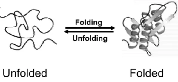
Under physiological conditions the native and unfolded state of a protein
are in equilibrium (Fig. 1.5), and the free energy change (ΔG) of the reaction
is usually referred as conformational stability of a protein [26]. The free energy change of the equilibrium is described by the following thermodynamic equation:
ΔG = ΔH –TΔS
where ΔH and ΔS represent the enthalpic and entropic variations,
~ 3 kcal/mol
2-5 kcal/mol
Chapter 1
respectively, and T represents the temperature. Therefore, the native structure results from a delicate balance between large and opposing forces [22]. The folded conformation is favoured by the negative value of enthalpy due to internal interaction, and the unfolded state is favoured by a positive value of conformational entropy.
Figure 1.5: Schematic representation of a folding-unfolding reaction. The reaction represents a two-state process without the formation of intermediates.
Noteworthy, the native state is known to be only marginally stable and the difference in energy between native and unfolded states is only 5-15
kcal.mol-1[27]. This difference in energy is within the magnitude of only a few
non-covalent interactions.
The determinants of native state stability in aqueous solutions are the primary sequence of the protein as well as the variable conditions of pH, temperature, and concentrations of salt and ligands [28]. Manipulation of environment conditions promoting protein unfolding, such as rise in temperature, variation of pH, or addition of a chemical denaturant, will shift the equilibrium to the unfolded state. (Fig. 1.5 and 1.6) [25]. The unfolding reaction begins with small changes in the folded conformation, such as increase in flexibility and localized conformational alterations, to a point where massive alterations occur, the transition midpoint (Fig. 1.6). At the midpoint of the unfolding curve half of the protein molecules are in the
unfolded state, and the ΔG is zero [25]. The abruptness of the transition is an
evidence of the cooperativity of the unfolding reaction. The cooperative
Unfolded Folded
effect results from the fact that although each interaction has a small stabilization effect, the disruption influences the surrounding interactions, thus lowering the equilibrium constant at each point.
Figure 1.6: Representation of a hypothetical denaturation curve. The cartoons represent the native state (botom) and the unfolded state (Top). Tm or Cm represent temperature or chemical denaturant concentration at the midpoint of the unfolding curve.
The folded and unfolded states have different structural features therefore a diversity of biophysical methods are available to monitor the unfolding reaction. The experimental techniques, the time scale that can be monitored and the information that can be extracted from each technique is summarized in table 1.1 [16]. From the experimental data obtained by the different techniques it is possible to draw curves similar to the one represented in figure 1.6, from which thermodynamic parameters of the reaction can be obtained. The midpoint transition determined from the curves, such as
temperature (Tm) or concentration of chemical denaturant (Cm), is used for
comparing protein stability.
Denaturantcondition
Unf
o
lded Fr
acti
on
0 100
50
Chapter 1
Table 1.1 Experimental techniques that have been applied to the study of protein folding [16].
Technique Timescale Information content Ref
Intrinsic tryptophan
fluorescence > nsª
Environment of tryptophan (through measurement of intensity
and λmax)
[29]
Far UV CD > μsª Secondary-structure content [30] Near UV CD > μsª Packing of aromatic residues [30]
Raman spectroscopy > μsª Solvent accessibility, conformation
of aromatic residues [31] Infrared
spectroscopy > nsª Secondary-structure content [32] ANS > μsª Exposure of aromatic surface area [29]
FRET > psª
Molecular ruler, dependent on the distance between two fluorophores
(r–6 dependence assuming free rotation of the dyes)
[33]
FCS > ps Diffusion time (and hence size and
shape) [34]
Anisotropy > μsª
Correlation time measurements provide information about shape
and size of molecule
[29]
Small-angle X-ray
scattering > μsª Radius of gyration [35] Absorbance > nsª Environment of chromophore [36]
Real-time NMR > min Structural information via chemical
shifts and measurement of NOEs [37] Native-state
hydrogen exchange
h Global stability, detection of
metastable states [38]
Pulsed H/D exchange by
NMR
> ms
Hydrogen exchange protection of folding intermediates on a
per-residue basis
[38]
Pulsed H/D exchange by
ESI-MS
> ms Hydrogen exchange protection of
folding populations [39]
NMR relaxation
methods ~ ms
Nonrandom structure in denatured states and conformational exchange between different species
[37, 40] Protein engineering Depends on probe used
Role of an individual residue in determining the rate of folding and
stability of a species of interest
1.3. Protein folding in vivo
In vivo the protein folding process can begin when the nascent polypeptide
is still attached to the ribosome, in a co-translational manner [42]. Alternatively, part of the folding process may take place in the cytosol, and many proteins fold in specific compartments, such as the endoplasmic reticulum (ER) [43]. Co-translational folding restricts the conformational space, so the polypeptide chain will probably start the folding process from a more defined trajectory, preventing non-productive pathways [20]. In other cases, co-translational folding can have a negative impact, as some proteins have extensive contacts between amino acids in the native structure that are distant in primary sequence. The translational process is relatively slow (~15-75 s for a 300-residues protein) potentially leading to the formation of non-native intramolecular contacts between the N-terminal portion that is being elongated, or intermolecular ones with other molecules in the surroundings [21]. In this context, high macromolecular concentration inside the cell
(300/400 mg.ml-1) play an important role in the process [44]. To overcome
the problem of molecular crowding and increase the efficiency of the folding process, nature has developed specific protein machineries - the protein quality control system (PQC)- that assists the folding process with no effect in the selection of the native structure (Fig. 1.7).
Chapter 1
The PQC system has the ability to supervise folding, disable aggregation, and remove misfolded or damage polypeptide chains before they exert toxic effects (Fig. 1.7) [45]. The system is composed by molecular chaperones, specialized intracellular proteases and accessory factors that regulate the activity of chaperones and proteases or provide communication between the various components [45]. Moreover, the PQC system has the ability to operate during or after protein synthesis, or upon unfolding to facilitate proteins to achieve their correct native structure, or to be degraded. Next, a brief description on different components of the PQC will be presented.
1.3.1. Role of molecular chaperones in de novo folding
Chaperones that assist de novo protein folding in the cytosol can be divided
in two classes: chaperones that stabilize nascent polypeptides on ribosomes and initiate folding, and chaperones that act downstream in completing the folding process [46-47].
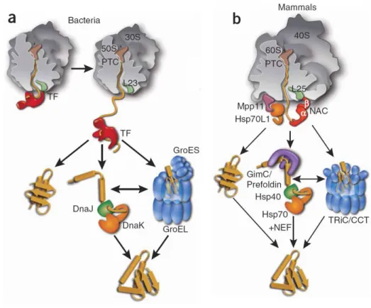
Included in the first class are chaperones that bind directly to the large ribosomal subunit in close proximity to the polypeptide exit site, such as the Trigger factor in bacteria, and a specialized Hsp70 system called RAC (ribosome associated complex) and NAC (nascent chain associated complex), in eukaryotes [48-50] (Fig. 1.8). Trigger factor has an N-terminal domain that binds to the ribosome, a domain with prolyl isomerase activity and a C-terminal chaperone domain. This protein binds not only to the ribosome, but also to the nascent chain forming a complex with the elongation chain, which dissociates when synthesis is complete [51-52]. In mammals, RAC is a heterodimeric complex formed by a J-protein, the Mpp11 that associates with the ribosome, and Hsp70L1 [53-54]. NAC is also a heterodimeric protein
association of the ribosome with the protein translocation machinery of the ER membrane. Therefore, it is believed that it regulates the destiny of the newly synthesized polypeptides, whether they are released into cytosol or are translocated to the ER [57]. This class of chaperones binds linear chain segments enriched in hydrophobic amino acids, and acts by preventing premature chain compaction, and by maintaining the elongation of the polypeptide in a non-aggregated state until the chain is able to fold correctly [21].
Chapter 1
Moreover, Brandt et al have recently provided new insights into the 3D
organization of the bacterial polysome, demonstrating that its three dimensional organization also contributes to prevent interaction of the elongated chain with chains from adjacent ribosomes (Fig. 1.9) [58]. The pseudo helical arrangement of the polysome places the exit channels of each ribosome further apart, thus minimizing unspecific interactions between nascent chains.
Figure 1.9: Model of nascent chain configurations in a representative polysome. From [58].
from these cofactors, a nucleotide exchange factor is also involved, Bag-1/GrpE in mammals and bacteria respectively, to stimulate chaperone activity [63-65]. The Hsp70 family is also very important because it organizes the chaperone network and distribute subsets of proteins to other downstream chaperones, such as Hsp60 (GroEL in bacteria, Hsp60 in mitochondria and TriC/CCT in cytosol in eukarya) and Hsp90 families.
1.3.2. Folding, holding and unfolding chaperones
The chaperonins, Hsp60 family, assemble into double-ring structures, functioning in an ATP dependent way, by enclosing the substrates during the folding event, so they are protected from aggregation [47, 66-67] (Fig. 1.8). These chaperonins are composed of 14 subunits arranged as two stacked rings with a sevenfold rotational symmetry. In general, GroEL and Hsp60 also need a cofactor, respectively GroES and Hsp10. In eukaryotes, the chaperonin interacts directly with Hsp70 and other upstream factors like prefoldin, increasing in this way the folding efficiency by directly recruiting nascent chains that are unable to fold with the chaperone alone [68-69]. Hsp90 acts downstream Hsp70, preventing aggregation of unfolded polypeptides [70-71]. These chaperones are proposed to play an important role in quality control, assembling into large multichaperone complexes that have a broad range of action, including interactions with a wide collection of cell-signalling molecules and transcription factors [72-73].
Chapter 1
Moreover, the Hsp100/Clp family has an unfolding activity but it can also induce structural changes in its substrates, that ultimately change the substrate’s biological activity [75]. This family acts on folded and assembled complexes and on improperly folded and aggregated proteins [75]. These chaperones have the ability to unfold proteins so that they can either be folded in their correct form or be otherwise degraded.
1.3.3. The Proteolytic system
Apart from the molecular chaperones, the PQC also contains the degradation machinery constituted by proteases. These proteins are restricted to specific locations in the cell, that can only be accessed by polypeptides targeted to degradation [45]. The quaternary structure of the different proteases is very similar, and resembles a barrel formed by the proteolytic subunits associated into oligomeric rings that stack up on each other [76]. The proteolytic sites are buried within the central cavity and are only accessible through narrow gates, obstructing folded proteins from entering. One of the most studied members of this family is the proteasome, a central protease in non-lysosomal ubiquitin-dependent protein degradation, which is involved in protein quality control, antigen processing, signal transduction, cell cycle control, cell differentiation and apoptosis [76].
1.3.4. Endoplasmatic reticulum (ER) chaperones
transiently to newly synthesized proteins and in a more permanent way to misfolded, underglycosylated or unassembled proteins that can not be transported from the ER [77]. Other two important ER chaperones, calnexin (CNX) and calreticulin (CRT), recognize monoglucosylated glycan chains on proteins [78]. CNX is a membrane bound protein, and in addition to its chaperone activity, it participates in quality control by delaying the export of incompletely assembled proteins from the ER [79].
1.4. Regulation of protein homeostasis
To ensure protein homeostasis, the cell contains a set of pathways that make up the so called proteostasis network, which regulates protein synthesis, folding, trafficking, aggregation, disaggregation and degradation (Fig. 1.10). This network has the ability to regulate conformation, concentration, binding interactions and location of individual proteins [80]. More than 1000 general and specialized chaperones, folding enzymes, degradation and trafficking components integrate this network [81]. In normal conditions the proteostasis network ensures that each protein reaches its final target in the cell or is eliminated in order to prevent cell damage or dysfunction.
Chapter 1
The control of these pathways is accomplished by signalling pathways that directly regulate the concentration, distribution and activities of the components that make up the proteostasis system. These signalling pathways include: the unfolded protein response (UPR), the heat shock response
(HSR), pathways that regulate the Ca2+ concentration on the ER,
inflammatory response and histone deacetylase (Fig. 1.10).
Several components of the first layer were object of discussion in the section above; here a brief overview on the signalling pathways will be given.
Prolonged exposure to stress can cause proteins to unfold, misfold or aggregate leading to an inefficient function of the cell, demanding an increased synthesis of the proteins from the quality control system. The heat shock response (HSR) controls the proteostasis reaction to these changes at the cytoplasmic level [80]. This signalling pathway is regulated at the transcriptional level by the activity of a family of heat shock transcription factors (HSF) [82]. Transcription of heat shock genes is activated by various acute and chronic conditions such as elevated temperatures, heavy metals, small molecules, infection, and oxidative stress [83]. Several disease states like inflammation, ischemia, tissue wounding and repair, cancer, protein mutations and neurodegenerative diseases are also associated with increase expression of proteins from the heat shock response [83]. Upon activation, an instant induction of genes encoding molecular chaperones, proteases, and other proteins associated with protection and recovery from cellular damage associated to misfolded proteins result in a rapid increase of the cellular levels of these proteins. The heat shock gene superfamily includes Hsp100/Clp, Hsp90, Hsp70, Hsp60, Hsp40, and small heat shock protein (sHsp) families, as referred above.
formed in these types of disorders was identified. It was observed that for example the protein huntingtin aggregates transiently in association with Hsp70, and that the association/dissociation properties identified for these complexes are similar to chaperone interactions with unfolded polypeptides [84]. Also, it is important to mention that neurodegenerative disorders often occur later in life, when the heat shock genes seem to be poorly induced [85-86]. Cancer is another type of disorder where the HSR as been implicated. Tumor cells typically express higher levels of heat shock proteins compared with non-transformed cells, suggesting that the abnormal expression of chaperones is associated with the tumorigenic state [87].
The ER is extremely important to cell regulation as all proteins that enter the secretory pathway first enter in the ER, where they fold and assemble [88]. The “unfolded protein response” (UPR), is the ‘supervising’ pathway for the quality of the folded proteins in this organelle. Unfolded or misfolded proteins identified by the UPR are retained within the ER lumen in complex with chaperones, or are targeted for degradation through the ubiquitin-proteasome, the ER-associated degradation (ERAD) [89].
Cystic fibrosis is related to the UPR response, this is a loss-of-function disease associated to mutations on the cystic fibrosis transmembrane conductance regulator (CFTR), a transmenbranar protein that function as a cAMP-stimulated chloride channel [90] This is the most fatal genetic disease in Caucasians and is caused by mutations on CFTR, a deletion of the
phenylalanine in position 508 (Δ508) is the most prevalent one [91]. This
mutation causes a defect in protein maturation and on its transit to the plasma membrane, due to a change in the protein folding pathway [92]. Due to the UPR actions in the ER misfolded proteins are recognize, retained in ER and
rapidly degraded by the ubiquitin-dependent proteasomal system, thus Δ508
Chapter 1
1.5. Emerging strategies in proteostasis regulation
A decreased ability of the proteostasis network to control the diverse pathways will jeopardize the cell, and can potentially affect in a progressive way, cell, tissue, organ or the organism function. Different factors can compromise protein homeostasis such as genetic and epigenetic pathways, physiological stressors, and metabolites that affect activity of the proteostasis network components (Fig. 1.10). When the cell is no longer able to restore proteostasis, the so call loss- or gain- of function effects emerge. A loss of function disorder is characterized by misfolded proteins that have decreased function and/or are degraded rapidly resulting in a decreased steady-state amount of protein. This type of disorders comprises several inherited autosomal recessive disorders, such as cystic fibrosis, phenylketonuria, fatty acid oxidation defects, and Gaucher’s disease. On the other hand, gain of-toxic function diseases are associated with intra- or extra-cellular accumulation of protein aggregates, like in amyotrophic lateral sclerosis, Alzheimer’s, Parkinson’s and Huntington’s disease. The pathology occurs because the cell is unable to degrade misfolded proteins, leading to the formation of toxic species, in the form of oligomers, aggregates and/or amyloid fibres.
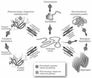
A novel approach to treat protein functional disorders is to regulate at different level the proteostasis network by manipulating the innate biology of the cell with protein replacement, protein stabilization or adaptation of proteostasis network using proteostasis regulators (Fig. 1.11).
recombinant protein reach it destination. The case of lysosomal disorders is one of these examples in which only 5 % of the protein injected enter the lysosome [97]. However, in neuropathic forms of these diseases this therapy is completely useless because recombinant proteins can not cross the blood-brain barrier [98].
Figure 1.11: Proteostasis Regulation. Schematic representation of the different control points of protein homeostasis: red arrows represent the proteostasis pathways, magenta circles represent proteostasis regulators and green squares represent pharmacological chaperones.From [80].
Chapter 1
interaction will prevent an increase in the intrinsic breathing of the protein that could lead to protein misfolding [101]. This is the case of P-glyco-protein, an energy dependent transporter, for which as been shown that several mutants in the presence of substrates (vinblastine and capsaicin) and inhibitors (cyclosporine and verapamil) increase the presence of functional protein in cell surface, probably due to a stabilization of a folding intermediate near native conformation [102-103]. Studies on MNK a copper transporting p-type ATPase showed that the presence of copper can increase stability of the protein [104].
Moreover, cells had already developed their own chemical chaperones to deal with environmental stress; these compounds are the cellular osmolytes and comprise carbohydrates, free amino acids and methylamines. The first two groups include glycerol, sorbitol, arabitol, trehalose, glycine, alanine, proline, taurine that can accumulate to higher concentrations in the cell without affecting it normal function [101]. The methylamines such as betaine, trimethylamine N-oxide (TMAO) and glycero-phosphorycholine are produced to offset the protein denaturing effects of urea. Beside these chemical chaperones other hydrophobic compound can bound to proteins and stabilize their structure in a native like conformation. This is the case of sodium 4-phenylbutyrate (PBA), an orally bioavailable short-chain fatty acid, that can be used as an ammonia-scavenging agent in urea metabolism disorders and it also has beneficial effects in stabilizing CFTR and alpha-1 antitrypsin (AAT) [105-106].
small interfering RNA (siRNA), cDNA, or proteins. It is described in literature that proteostasis regulators can modulate synthesis, folding, trafficking, disaggregation, and aggregation pathways [50, 107-117]. In fact, RNA interference studies have been use with success in cellular models of gain-of function disorders, like Alzheimer and Huntington’s disease. Also, small molecules like diltiazem and verapamil have been used to restore protein homeostasis in lysosomal storage disorders [98]. These two L-type voltage gate calcium channel blockers restore mutant lysosome enzyme function by modulating protein folding in the ER. Unlike the pharmacological chaperones that directly bind to the protein stabilizing the native form for trafficking to the Golgi and to lysosome, the proteostasis regulators diminish
the Ca2+ content in the cytoplasm resulting in the increase of transcription
and translation of several cytoplasmatic and ER chaperones, like BiP and Hsp40 that interact with lysosomal enzymes. One important aspect is the fact that diltiazem can cross the blood-brain barrier, in contrast to the protein replacement therapy [98]. Celastrol, a natural product derived from the Celastraceae family of plants, induced HSF1 protein expression leading to up-regulation of cytoplasmatic chaperone network [117]. Some of the molecules know to act as chemical chaperones as already mentioned can also be called proteostasis regulators once they also can regulate folding and trafficking machinery, one exemple is the use of 4-PBA and taurine-conjugated ursodeoxycholic acid to alleviated ER-stress in cells and whole animals [118].
1.6. References
1. Anfinsen, C. B.; Haber, E., Studies on the reduction and re-formation of protein disulfide
bonds. J Biol Chem 1961,236, 1361-3.
2. Anfinsen, C. B., Principles that govern the folding of protein chains. Science (New York, N.Y
1973,181 (96), 223-30.
3. Levinthal, C., Mossbauer Spectroscopy in Biological Systems. Proceedings of a Meeting
held at Allerton House, Monticello, Illinois
(Eds.: P. Debrunner, J. C. M. Tsibris, E. Münck) 1969, 22.
4. Levinthal, C., J. Chim. Phys. 1968,65, 44-45.
Chapter 1
Natl Acad Sci U S A 1973,70 (3), 697-701.
6. Karplus, M.; Weaver, D. L., Protein-folding dynamics. Nature 1976,260 (5550), 404-6.
7. Kim, P. S.; Baldwin, R. L., Specific intermediates in the folding reactions of small proteins
and the mechanism of protein folding. Annu Rev Biochem 1982,51, 459-89.
8. Baldwin, R. L., How does protein folding get started? Trends Biochem Sci 1989,14 (7),
291-4.
9. Dill, K. A.; Bromberg, S.; Yue, K.; Fiebig, K. M.; Yee, D. P.; Thomas, P. D.; Chan, H. S.,
Principles of protein folding--a perspective from simple exact models. Protein Sci 1995,4 (4), 561-602.
10. Udgaonkar, J. B., Multiple routes and structural heterogeneity in protein folding. Annu Rev
Biophys 2008,37, 489-510.
11. Fersht, A. R., Nucleation mechanisms in protein folding. Curr Opin Struct Biol 1997,7 (1),
3-9.
12. Dill, K. A.; Chan, H. S., From Levinthal to pathways to funnels. Nat Struct Biol 1997,4 (1),
10-9.
13. Bryngelson, J. D.; Onuchic, J. N.; Socci, N. D.; Wolynes, P. G., Funnels, pathways, and the
energy landscape of protein folding: a synthesis. Proteins 1995,21 (3), 167-95.
14. Dinner, A. R.; Sali, A.; Smith, L. J.; Dobson, C. M.; Karplus, M., Understanding protein
folding via free-energy surfaces from theory and experiment. Trends Biochem Sci 2000,25 (7), 331-9.
15. Brockwell, D. J.; Radford, S. E., Intermediates: ubiquitous species on folding energy
landscapes? Curr Opin Struct Biol 2007,17 (1), 30-7.
16. Bartlett, A. I.; Radford, S. E., An expanding arsenal of experimental methods yields an
explosion of insights into protein folding mechanisms. Nat Struct Mol Biol 2009,16 (6), 582-8.
17. Jahn, T. R.; Radford, S. E., The Yin and Yang of protein folding. FEBS J 2005,272 (23),
5962-70.
18. Mitraki, A., King, J., Protein Folding Intermediates and Inclusion Body Formation. Nature
Biotechnology 1989,7, 690-697.
19. Wetzel, R., For protein misassembly, it's the "I" decade. Cell 1996,86 (5), 699-702.
20. Clark, P. L., Protein folding in the cell: reshaping the folding funnel. Trends Biochem Sci
2004,29 (10), 527-34.
21. Hartl, F. U.; Hayer-Hartl, M., Converging concepts of protein folding in vitro and in vivo. Nat
Struct Mol Biol 2009,16 (6), 574-81.
22. Shirley, B. A., Urea and guanidine hydrochloride denaturation curves. Methods Mol Biol
1995,40, 177-90.
23. Dill, K. A.; Ozkan, S. B.; Shell, M. S.; Weikl, T. R., The protein folding problem. Annu Rev
Biophys 2008,37, 289-316.
24. Crowe, J.; Bradshaw, T.; Monk, P., Chemistry for the Biosciences. Oxford University Press:
New York, 2006.
25. Fersht, A., Structure and Mechanism in Protein Science. W. H. freeman and Company: New
York, 1999.
26. Pace, C. N.; Scholtz, J. M., Measuring the conformational stability of a protein. In Protein
structure- A Pratical approach, Creighton, T. E., Ed. Oxford University Press: New York, 1997; pp
299-321.
27. Yang, J. S.; Chen, W. W.; Skolnick, J.; Shakhnovich, E. I., All-atom ab initio folding of a
diverse set of proteins. Structure 2007,15 (1), 53-63.
28. Pace, C. N., Conformational stability of globular proteins. Trends Biochem Sci 1990,15 (1),
14-7.
29. Royer, C. A., Probing protein folding and conformational transitions with fluorescence.
Chem Rev 2006,106 (5), 1769-84.
30. Kelly, S. M.; Jess, T. J.; Price, N. C., How to study proteins by circular dichroism. Biochim
Biophys Acta 2005,1751 (2), 119-39.
31. Balakrishnan, G.; Weeks, C. L.; Ibrahim, M.; Soldatova, A. V.; Spiro, T. G., Protein
dynamics from time resolved UV Raman spectroscopy. Curr Opin Struct Biol 2008,18 (5), 623-9.
32. Fabian, H.; Naumann, D., Methods to study protein folding by stopped-flow FT-IR. Methods
2004,34 (1), 28-40.
33. Schuler, B.; Eaton, W. A., Protein folding studied by single-molecule FRET. Curr Opin Struct
Biol 2008,18 (1), 16-26.
34. Haustein, E.; Schwille, P., Fluorescence correlation spectroscopy: novel variations of an
established technique. Annu Rev Biophys Biomol Struct 2007,36, 151-69.
35. Lipfert, J.; Doniach, S., Small-angle X-ray scattering from RNA, proteins, and protein
complexes. Annu Rev Biophys Biomol Struct 2007,36, 307-27.
37. Dyson, H. J.; Wright, P. E., Elucidation of the protein folding landscape by NMR. Methods
Enzymol 2005,394, 299-321.
38. Krishna, M. M.; Hoang, L.; Lin, Y.; Englander, S. W., Hydrogen exchange methods to study
protein folding. Methods 2004,34 (1), 51-64.
39. Maier, C. S.; Deinzer, M. L., Protein conformations, interactions, and H/D exchange.
Methods Enzymol 2005,402, 312-60.
40. Korzhnev, D. M.; Kay, L. E., Probing invisible, low-populated States of protein molecules by
relaxation dispersion NMR spectroscopy: an application to protein folding. Acc Chem Res 2008,41
(3), 442-51.
41. Zarrine-Afsar, A.; Davidson, A. R., The analysis of protein folding kinetic data produced in
protein engineering experiments. Methods 2004,34 (1), 41-50.
42. Hardesty, B.; Kramer, G., Folding of a nascent peptide on the ribosome. Prog Nucleic Acid
Res Mol Biol 2001,66, 41-66.
43. Hartl, F. U.; Hayer-Hartl, M., Molecular chaperones in the cytosol: from nascent chain to
folded protein. Science (New York, N.Y 2002,295 (5561), 1852-8.
44. Ellis, R. J.; Minton, A. P., Cell biology: join the crowd. Nature 2003,425 (6953), 27-8.
45. Gregersen, N.; Bross, P.; Vang, S.; Christensen, J. H., Protein misfolding and human
disease. Annu Rev Genomics Hum Genet 2006,7, 103-24.
46. Albanese, V.; Yam, A. Y.; Baughman, J.; Parnot, C.; Frydman, J., Systems analyses reveal
two chaperone networks with distinct functions in eukaryotic cells. Cell 2006,124 (1), 75-88.
47. Langer, T.; Lu, C.; Echols, H.; Flanagan, J.; Hayer, M. K.; Hartl, F. U., Successive action of
DnaK, DnaJ and GroEL along the pathway of chaperone-mediated protein folding. Nature 1992,356
(6371), 683-9.
48. Kramer, G.; Boehringer, D.; Ban, N.; Bukau, B., The ribosome as a platform for
co-translational processing, folding and targeting of newly synthesized proteins. Nat Struct Mol Biol 2009,
16 (6), 589-97.
49. Chang, H. C.; Tang, Y. C.; Hayer-Hartl, M.; Hartl, F. U., SnapShot: molecular chaperones,
Part I. Cell 2007,128 (1), 212.
50. Tang, Y. C.; Chang, H. C.; Hayer-Hartl, M.; Hartl, F. U., SnapShot: molecular chaperones,
Part II. Cell 2007,128 (2), 412.
51. Hesterkamp, T.; Hauser, S.; Lutcke, H.; Bukau, B., Escherichia coli trigger factor is a prolyl
isomerase that associates with nascent polypeptide chains. Proc Natl Acad Sci U S A 1996,93 (9),
4437-41.
52. Valent, Q. A.; Kendall, D. A.; High, S.; Kusters, R.; Oudega, B.; Luirink, J., Early events in
preprotein recognition in E. coli: interaction of SRP and trigger factor with nascent polypeptides.
EMBO J 1995,14 (22), 5494-505.
53. Hundley, H. A.; Walter, W.; Bairstow, S.; Craig, E. A., Human Mpp11 J protein:
ribosome-tethered molecular chaperones are ubiquitous. Science (New York, N.Y 2005,308 (5724), 1032-4.
54. Otto, H.; Conz, C.; Maier, P.; Wolfle, T.; Suzuki, C. K.; Jeno, P.; Rucknagel, P.; Stahl, J.;
Rospert, S., The chaperones MPP11 and Hsp70L1 form the mammalian ribosome-associated
complex. Proc Natl Acad Sci U S A 2005,102 (29), 10064-9.
55. Spreter, T.; Pech, M.; Beatrix, B., The crystal structure of archaeal nascent
polypeptide-associated complex (NAC) reveals a unique fold and the presence of a ubiquitin-polypeptide-associated domain. J
Biol Chem 2005,280 (16), 15849-54.
56. Beatrix, B.; Sakai, H.; Wiedmann, M., The alpha and beta subunit of the nascent
polypeptide-associated complex have distinct functions. J Biol Chem 2000,275 (48), 37838-45.
57. Wiedmann, B.; Sakai, H.; Davis, T. A.; Wiedmann, M., A protein complex required for
signal-sequence-specific sorting and translocation. Nature 1994,370 (6489), 434-40.
58. Brandt, F.; Etchells, S. A.; Ortiz, J. O.; Elcock, A. H.; Hartl, F. U.; Baumeister, W., The native
3D organization of bacterial polysomes. Cell 2009,136 (2), 261-71.
59. Bukau, B.; Horwich, A. L., The Hsp70 and Hsp60 chaperone machines. Cell 1998,92 (3),
351-66.
60. Hartl, F. U., Molecular chaperones in cellular protein folding. Nature 1996,381 (6583),
571-9.
61. Kelley, W. L., The J-domain family and the recruitment of chaperone power. Trends
Biochem Sci 1998,23 (6), 222-7.
62. Rassow, J.; Voos, W.; Pfanner, N., Partner proteins determine multiple functions of Hsp70.
Trends Cell Biol 1995,5 (5), 207-12.
63. Bimston, D.; Song, J.; Winchester, D.; Takayama, S.; Reed, J. C.; Morimoto, R. I., BAG-1, a
negative regulator of Hsp70 chaperone activity, uncouples nucleotide hydrolysis from substrate
release. EMBO J 1998,17 (23), 6871-8.

![Figure 1.4: The various non- non-covalent forces that can operate in a protein. From [24]](https://thumb-eu.123doks.com/thumbv2/123dok_br/15769787.641155/34.892.267.554.184.437/figure-various-non-non-covalent-forces-operate-protein.webp)
![Table 1.1 Experimental techniques that have been applied to the study of protein folding [16]](https://thumb-eu.123doks.com/thumbv2/123dok_br/15769787.641155/37.892.130.641.143.948/table-experimental-techniques-applied-study-protein-folding.webp)
![Figure 1.9: Model of nascent chain configurations in a representative polysome. From [58]](https://thumb-eu.123doks.com/thumbv2/123dok_br/15769787.641155/41.892.138.433.341.630/figure-model-nascent-chain-configurations-representative-polysome.webp)