Genotoxicity assessment of a pharmaceutical effluent using four bioassays
Adekunle A. Bakare
1, Alabi A. Okunola
2, Olusanmi A. Adetunji
1and Hafeez B. Jenmi
11
Cell Biology and Genetics Unit, Department of Zoology, University of Ibadan, Oyo State, Nigeria.
2
Department of Basic & Applied Science, Babcock University, Ogun State, Nigeria.
Abstract
Pharmaceutical industries are among the major contributors to industrial waste. Their effluents when wrongly han-dled and disposed of endanger both human and environmental health. In this study, we investigated the potential genotoxicity of a pharmaceutical effluent, by using theAllium cepa, mouse- sperm morphology, bone marrow chro-mosome aberration (CA) and micronucleus (MN) assays. Some of the physico-chemical properties of the effluent were also determined. TheA. cepa and the animal assays were respectively carried out at concentrations of 0.5, 1, 2.5, 5 and 10%; and 1, 5, 10, 25 and 50% of the effluent. There was a statistically different (p < 0.05), concentra-tidependent inhibition of onion root growth and mitotic index, and induction of chromosomal aberrations in the on-ion and mouse CA test. Assessment of sperm shape showed that the fracton-ion of the sperm that was abnormal in shape was significantly (p < 0.05) greater than the negative control value. MN analysis showed a dose-dependent in-duction of micronucleated polychromatic erythrocytes across the treatment groups. These observations were pro-voked by the toxic and genotoxic constituents present in test samples. The tested pharmaceutical effluent is a potentially genotoxic agent and germ cell mutagen, and may induce adverse health effects in exposed individuals.
Key words:genotoxicity, pharmaceutical effluent, mouse,Allium cepa, chromosome, spermatozoa, micronucleus.
Received: September 16, 2008; Accepted: January 22, 2009.
Introduction
Pharmaceuticals are produced and used in increas-ingly larger amounts every year, this having caused the in-dustry to become one of the major contributors to industrial waste. Generally, pharmaceutical industries do not gener-ate uniform waste streams, due to the variety of medicines produced during any given processing period (Houk, 1992). In recent times, a wide range of pharmaceuticals have been found in fresh and marine waters, and it has been shown that even in reduced quantities, some of these com-pounds are potentially capable of causing harm to both aquatic and terrestrial life forms (Jonathan and Nicolaos, 2005). The presence of pharmaceutical chemicals in the en-vironment is a matter of concern due to their lipophilic and non-biodegradability nature, as well as their biological ac-tivities (Velagaleti and Burns, 2006). Currently, there is scarce of measurable evidence of the environmental impact of pharmaceutical chemicals on human health (Christen-sen, 1998).
A review of studies using microbial assays has shown that pharmaceutical waste does not appear to be particu-larly mutagenic, although it may be genotoxic by other
mechanisms (McGeorgeet al., 1985; Molleret al., 1985; Houk and DeMarini, 1988; Sanchez et al., 1988). Houk (1992) suggested that, even though there is no specific evi-dence to suggest this, the chemical composition of pharma-ceutical waste may make it unsuitable for microbial assays, since it could contain antibiotics or bacterial growth inhibi-tors. Thus, to be able to arrive at sufficient conclusions on the potentially genotoxic, mutagenic and masking effects of pharmaceutical waste constituents, it is important that other test systems be utilized, and also chemical character-ization carried out on this group of wastes. In the present study, four eukaryotic mutagenicity assays, namely the Allium cepaassay, the mouse sperm morphology assay, the micronucleus (MN) test and the chromosome aberration (CA) assay in mouse bone marrow cells, were used to eval-uate the genotoxic and mutagenic potential of effluents from a pharmaceutical company. These are the standard bioassays that best reflect the delicate balance between pathways for activation and inactivation of chemicals in human beings.
Materials and Methods
Effluent collection
The raw effluent from a pharmaceutical plant in Lagos State, Nigeria was collected in two 10 L plastic con-tainers, from the point of discharge into the environment.
www.sbg.org.br
Send correspondence to Adekunle. A. Bakare. Cell Biology and Genetics Unit, Department of Zoology, University of Ibadan, Oyo State, Nigeria. E-mail: [email protected], [email protected].
The company produces analgesics, anti-malarias, anesthet-ics, multivitamins, antibiotanesthet-ics, antihistamines, human vac-cines, sulphonamides and antiemetics. The collected material was filtered and the pH taken, to be then kept at 4 °C until use.
Biological materials
Onions (Allium cepa, L., 2n = 16, Family Amaryllidaceae), obtained commercially at the Bodija market, Ibadan, Nigeria, were sun-dried for 2 weeks. The dry bulbs, rotten ones excluded, were later used for the test. Young male Swiss albino mice (Mus musculus) of 6-10 weeks old, which had been inbred for several generations, were obtained from the animal breeding unit of the Depart-ment of Physiology, University of Ibadan, Nigeria. They were kept in a pathogen free, well ventilated animal house at the Department of Zoology, University of Ibadan, for 2 weeks in order to acclimatize. They were maintained in the same room throughout the period of this study. Food (Ladokun pelleted feed®) and drinking water were sup-pliedad libitum. The mice were divided into 3 categories for animal assays: the CA assay and MN test for mice 8 weeks old, and the sperm morphology assay for mice 12-14 weeks old.
Physico-chemical properties and heavy metal analysis
The effluent was analyzed for a number of standard physico-chemical properties, including chemical oxygen demand (COD), total dissolved solids (TDS), alkalinity, biochemical oxygen demand (BOD), chlorides, nitrates, ammonia and phosphates, according to methods described by APHA (1998). Nine metals (including eight heavy met-als) namely aluminum (Al), cadmium (Cd), copper (Cu), chromium (Cr), iron (Fe), mercury (Hg), zinc (Zn), nickel (Ni) and manganese (Mn) were analyzed in the effluent sample according to standard analytical methods (USEPA, 1996; APHA, 1998). Briefly, 100 mL of the effluent was di-gested by heating with concentrated HNO3,and the volume
reduced to 3-5 mL. This volume was made up to 10 mL with 0.1 N HNO3. Concentrations of the metals were
esti-mated by using an Atomic Absorption Spectrophotometer (Perkin Eelmer E. Analyst, 2000, USA).
Allium cepatest
The modifiedA. cepaassay (Fiskesjo, 1997; Bakare and Wale-Adeyemo, 2004; Babatunde and Bakare, 2006) was employed in this study. The outer scales of the onion bulbs and any brownish bottom plate were removed, leav-ing the rleav-ing of primordial root intact. The peeled bulbs were placed into fresh tap water during the cleaning procedure, so as to protect the primordial from drying. Thereafter, the bulbs were placed into 100 mL beakers containing 0.5%, 1.0%, 2.5%, 5% and 10% concentrations (v/v, effluent/dis-tilled water) from the effluent. Twelve onion bulbs were set
up in each concentration, out of which those 10 presenting the best root growth were selected for analysis of root growth inhibition. Distilled water was used as negative control. The experiment was performed in the dark at 27±1 °C. Test liquids were changed daily. On the second day (48 h), root tips of two bulbs in the experimental and control groups were fixed in ethanol:glacial acetic acid (3:1, v/v), to be then squashed on slides for chromosomal analysis, as previously described (Bakareet al., 2000; Ba-kare, 2002). On the third day (72 h), measurements were taken of the length of each root from the bulb, by test sam-ple concentration. The percentage of reduction in root-growth in relation to the control and EC50 was obtained
from the values thus come by. ASTM (1994) minimal sta-tistical guidelines for conducting early seedling growth tests were used in the analysis of measured root length. The effects of the effluent on the morphology of growing roots were also examined.
Sperm morphology assay
Induction of sperm abnormalities was studied accord-ing to Wyrobeket al(1983) and Bakareet al., (2005). Five concentrations of 1%, 5%, 10%, 25% and 50% (v/v, efflu-ent/distilled water) of the effluent were considered together with the positive (cyclophosphamide 20 mg/kg body weight) and negative (distilled water) controls. A single intraperitoneal (IP) injection of 0.5 mL of the different test-sample concentrations was administered to the mice daily for 5 consecutive days. The IP route was favored since it is one of the fastest and most efficient means of delivering test-chemicals into test-animals in a short-term-assay. The exposure period was 35 days and 5 mice were treated for each effluent concentration. Sperm was sampled from the caudal epididymes at 5 weeks from the first injection, since spermatogenesis in mice takes 34.5 days until completion (Bartkeet al., 1974). The mice were sacrificed by cervical dislocation and their epididymes surgically removed. Two sperm suspensions were prepared from the cauda of each testis by mincing the cauda in physiological saline. Smears were prepared on grease-free slides after staining with 1% Eosin Y for 45 min. The slides were air-dried and coded for subsequent microscopic examination under oil immersion at 1000x. For each mouse, 800 sperm cells were assessed for morphological abnormalities according to the criteria of Wyrobek and Bruce (1975).
Chromosome aberration assay
Mice were injected with an aqueous solution of colchicine (2.5 mg/kg bw, IP), 2 h prior to scheduled killing by cervi-cal dislocation. The bone-marrow cells were aspirated into 2.2% (w/v) sodium citrate and centrifuged at 2000 rpm. for 5 min. The pellets obtained were mixed in an aqueous solu-tion of KCl (0.075 M) and left for 30 min at room tempera-ture (29 °C). Cells were re-centrifuged, fixed in cold Carnoy fluid (methanol: glacial acetic acid, 3:1 v/v) and dropped onto clean chilled slides. Finally, the slides were air-dried and stained with 5% Giemsa (v/v, stock Giemsa stain/distilled water). The mitotic index was calculated by counting the metaphase cells from approximately 3000 cells/concentration and expressed in percent. CAs were scored blind to treatment and at least 50 well spread meta-phase cells/mouse were analyzed.
Micronucleus test
Five groups of mice (4 mice per group, 22-30 g each) were utilized in this assay, considering concentrations 1%, 5%, 10%, 25% and 50% (v/v, effluent/distilled water) of the test sample as against negative (distilled water) and pos-itive (cyclophosphamide 20 mg/kg body weight) controls. Each mouse/group was IP exposed to 0.5 mL of each con-centration for 96 h. Bone marrow preparation for micro-nuclei assessment was according to the procedure of Schmid (1975, 1976). Briefly, the animals were sacrificed by cervical dislocation. The femurs were removed from each and bone marrow flushed from the bones with Foetal Bovine Serum (Sigma Aldrich Cheme GmbH, Germany). Cells were centrifuged at 2000 rpm for 5 min and slides stained with May-Grunwald and Giemsa stains. At least 1000 cells/animal were scored for micronuclei in polychro-matic erythrocytes (MNPCE). The differential staining of PCEs (bluish-purple) and normochromatic erythrocytes (NCEs, pinkish-orange), and the relative size of the eryth-rocytes, are indices for differentiating them.
Statistical analysis
The SPSS® 14.0 statistical package was used for data analysis. Data obtained were expressed as percentage fre-quency and mean±standard error (mean±standard devia-tion in the CA test). Significance at the different dose-level of each assay was tested by using the Dunett t- test. Data on root-growth inhibition were expressed with 95% confi-dence limits and ANOVA was used for testing significance. Differences between the negative control-group and indi-vidual dose-groups were analyzed at the 0.05 and 0.001 probability levels.
Results
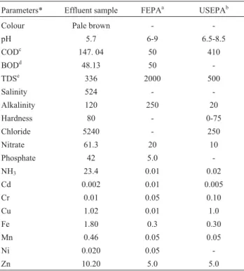
Physical and chemical characteristics of the pharma-ceutical effluent are shown in Table 1. The pH was 5.70, and there was an offensive odour. Chloride and nitrate lev-els were very high, likewise with Zn, Fe and Cu.
Allium cepaassay
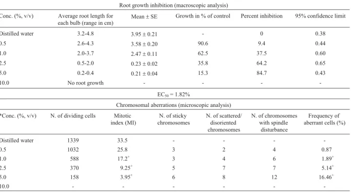
Table 2 shows the results from macroscopic and mi-croscopic analysis of treated Allium cepa roots. Root growth attained a maximum in the control (distilled water). Here, the roots were whitish in color, elongated and straight, with no morphological deformities. At the various concentrations of the test sample, there was a gradual statis-tically significant (p < 0.05) concentration-dependent inhi-bition of root growth. The least mean root growth and the highest mean root growth were obtained at the 5% and 0.5% concentrations, respectively. There was no root growth at a concentration of 10%. Morphological deformi-ties such as very short, bent, spiral and crochet-like roots were also observed at tested concentrations, especially at a concentration of 5%. The EC50value obtained from the %
inhibition value was 1.82%. Under microscopic analysis, there was a concentration-dependent reduction in mitotic index, compared to the negative control value of 33.5%, in all concentrations. Chromosomal aberrations (Figure 1a-c) were induced in all the different concentrations, all (except at the 0.5%) being statistically significant (p < 0.05).
Chromosome aberration assay
Exposure of mice to the effluent sample for 48 h in-hibited MI in bone-marrow cells in a dose-dependent man-ner, but this was only statistically significant (p < 0.05) at
Table 1- Physico-chemical characteristics of the pharmaceutical effluent assessed for genotoxicity.
Parameters* Effluent sample FEPAa USEPAb
Colour Pale brown -
-pH 5.7 6-9 6.5-8.5
CODc 147. 04 50 410
BODd 48.13 50
-TDSe 336 2000 500
Salinity 524 -
-Alkalinity 120 250 20
Hardness 80 - 0-75
Chloride 5240 - 250
Nitrate 61.3 20 10
Phosphate 42 5.0
-NH3 23.4 0.01 0.02
Cd 0.002 0.01 0.005
Cr 0.01 0.05 0.10
Cu 1.02 0.01 1.0
Fe 1.80 0.3 0.30
Mn 0.46 0.05 0.05
Ni 0.020 0.05
-Zn 10.20 5.0 5.0
*All values are in mg/L except pH and salinity (ppt.).aFEPA: Federal En-vironmental Protection Agency (2001).bUSEPA: United States Environ-mental Protection Agency (1989). cCOD: Chemical oxygen demand. d
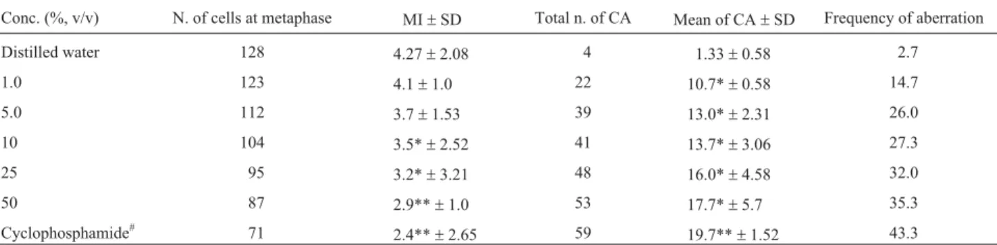
the 10, 25 and 50% concentrations of the test sample (Table 3). Different types of dose-dependent and statistically sig-nificant (p < 0.05) CAs were observed (Figure 2a-d).
Micronucleus test
Figure 3 shows the micronuclei induced in the bone marrow cells after exposure of mice to the test sample. Compared with the negative control, there was a statisti-cally significant (p < 0.001), dose- dependent increase in MN at all concentrations, except 1% and 5% (Figure 4). The dose-response equation for this effect is y = 7.45 + 1.65x (y = 0.99).
The positive control induced a significant induction of CA and MN in positive control groups.
Sperm morphology assay
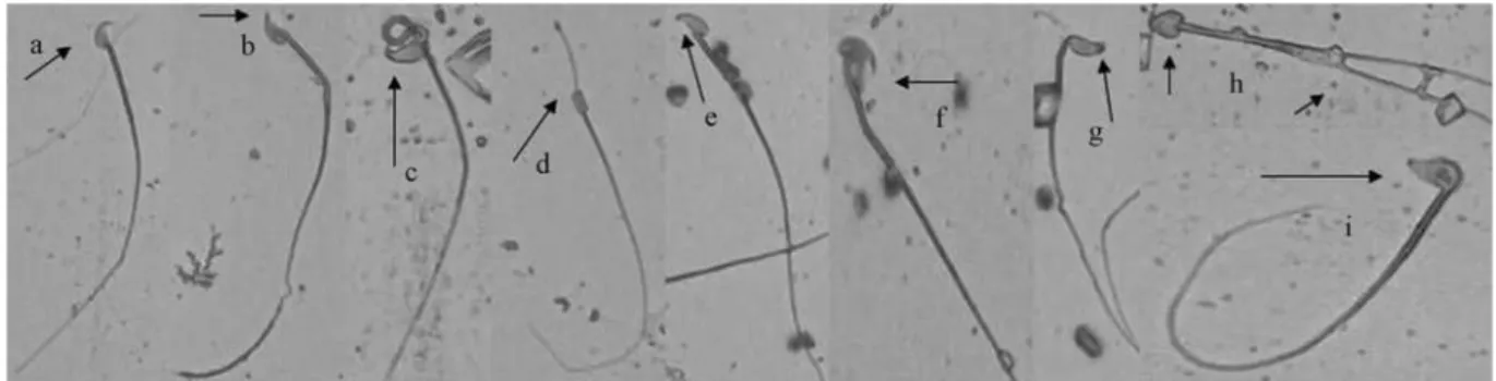
Figure 5 (a-g) shows the different types of abnormal sperm cells observed, 5 weeks from the 1stday of exposure
of male mice to the test pharmaceutical-effluent. The fre-quency of abnormal sperm cells in the negative control was 9.85%. There were 27.8%, 31.0%, 33.3%, 40.6% and 47.2% of abnormal sperm cells at the tested concentrations of 1%, 5%, 10%, 25% and 50%, respectively. This induc-tion of abnormalities was statistically significant (p < 0.05) and concentration-dependent at all concentrations except at 1%. Generally speaking, sperm with wrong-angled hooks (Figure 5b) were the most prominent (26.1%), whereas double-tailed sperm cells (Figure 5h) were very few in number (0.6% occurrence).
Discussion
Industrial discharge is recognized as one of the major sources of toxic chemicals in the environment. In the pres-ent study, the genotoxicity and mutagenicity of a pharma-ceutical effluent was assessed by using a battery ofin vivo assays in plant and animal systems. The results of the
Table 2- Inhibitory and cytological effects of the pharmaceutical effluent onAllium ceparoot.
Root growth inhibition (macroscopic analysis)
Conc. (%, v/v) Average root length for each bulb (range in cm)
Mean±SE Growth in % of control Percent inhibition 95% confidence limit
Distilled water 3.2-4.8 3.95±0.21 - 0 0.38
0.5 2.6-4.3 3.58±0.20 90.6 9.4 0.44
1.0 2.0-3.7 2.47±0.11 62.5 37.5 0.60
2.5 0.5-2.0 0.23±0.02 35.8 64.2 0.65
5.0 0.2-0.4 0.21±0.04 15.3 84.7 0.43
10.0 No root growth - - -
-EC50= 1.82%
Chromosomal aberrations (microscopic analysis)
*Conc. (%, v/v) N. of dividing cells Mitotic index (MI)
N. of sticky chromosomes
N. of scattered/ disoriented chromosomes
N. of chromosomes with spindle
disturbance
Frequency of aberrant cells (%)
Distilled water 1339 33.5 - - -
-0.5 1032 25.8 3 2 4 0.87
1.0 588 17.2+ 3 4 6 1.89+
2.5 370 9.25+ 5 7 7 5.14+
5.0 158 3.95+ 6 8 12 16.46+
10.0 - - -
-* 4000 cells (4 slides) per concentration and the control.
+ Values are significantly different from the control at p < 0.05 (ANOVA).
physico-chemical characteristics of the effluent showed the presence of certain sample-constituents at concentrations beyond the permitted limits set by international regulatory authorities (Table 1). Heavy metal analysis of effluent-samples showed the presence of Cu, Ni, Cr, Cd, Mn, Al, Fe, and Zn at various concentrations. These metals have the po-tential to induce mutation and cancer in living cells. A mix-ture of these may result in synergistic chemical combina-tions that are more harmful than the individual constituent itself. Studies in experimental animals indicate that Ni (Haugenet al., 1994) and Cd (Elinder and Jarup, 1996) are
carcinogenic. Hexavalent Cr was reported to have induced chromosomal aberrations, and micronuclei and single-strand breaks in mammalian cells (Wiseet al., 2002), be-sides gene mutation in bacteria (DeFloraet al., 1990). Tri-valent Fe was also reported as having been responsible for the high level of micronuclei in newt larvae (Godetet al., 1996). The exposure of mice to Zn results in both sin-gle-strand breaks in DNA, as measured by the comet assay (Banuet al., 2001), as well as chromosomal aberrations (Guptaet al., 1991). Ni is known to produce highly selec-tive damage to heterochromatin (Costaet al., 1994). It has
Figure 2- Chromosomal aberrations (arrowed) induced in bone marrow cells of mice exposed to the pharmaceutical effluent. (a) chromatid break, (b) ring chromosome, (c) chromatid exchange, (d) dicentric chromosome. Magnification 1000x.
Table 3- Chromosome aberrations (CAs) induced in bone marrow cells of mice exposed to different concentrations of the pharmaceutical effluent.
Conc. (%, v/v) N. of cells at metaphase MI±SD Total n. of CA Mean of CA±SD Frequency of aberration
Distilled water 128 4.27±2.08 4 1.33±0.58 2.7
1.0 123 4.1±1.0 22 10.7*±0.58 14.7
5.0 112 3.7±1.53 39 13.0*±2.31 26.0
10 104 3.5*±2.52 41 13.7*±3.06 27.3
25 95 3.2*±3.21 48 16.0*±4.58 32.0
50 87 2.9**±1.0 53 17.7*±5.7 35.3
Cyclophosphamide# 71 2.4**±2.65 59 19.7**±1.52 43.3
*p < 0.05, ** p < 0.001: levels of significance of chromosome damage in bone-marrow cells of micevs.distilled water (negative control). MI: Mitotic Index (3000 cells/concentration).#: 20 mg/kg body weight.
also been reported that Cd, Cu and Fe gave rise to reactive oxygen species in eukaryotic systems (Ghioet al., 2002; Radetskiet al., 2004). The constituents analyzed herein do not represent all, or even most, of the chemicals that could have been included in the test-sample. An effluent is a com-plex mixture of organic and inorganic chemicals, and of many unidentified toxicants known as non-conventional pollutants (NCPs), all of which may pose risks of an un-known magnitude to humans.
From data obtained through bioassays, it is shown that the tested effluent is cytotoxic, mutagenic and geno-toxic. The results of theA. cepaassay are indicative of a lin-ear relationship between macroscopical (root-growth inhi-bition) and microscopical (genotoxicity) parameters. The macroscopic effect appears to be the most sensitive param-eter, since any genotoxic effect manifest in a test sample, either directly or indirectly, is likely to result in inhibition of growth (Fiskesjo, 1997). The EC50value indicated that
the tested sample is highly toxic, with the highest tested concentration (10%) totally inhibiting root-growth in the treated bulbs. Microscopic examination allowed for assess-ing chromosome damage and cell-division disturbances, thus providing additional information regarding severity or
mechanism of the toxic effect or of potential mutagenicity. In A. cepa, whenever chromosome aberrations occurred, there were almost always certain growth restrictions (Fis-kesjo, 1997). This became evident in this study, and corrob-orates previous observations on this assay, when done in our laboratory (Bakare and Wale-Adeyemo, 2004; Baba-tunde and Bakare, 2006). Similar observations were also reported on dye wastewater (Somashekar and Gowda, 1983), sugar-cane factory wastewater (Mishra, 1993) and carbonaceous sugar-mill effluents (Kumar, 2000), when using theA. cepatest.
In the sperm morphology assay, the criteria for a posi-tive response were satisfied: there was an increase in abnor-mal sperm morphology to at least double the negative control level (at all treatment levels). There was also evi-dence of a concentration-dependent increase in the number of aberrant sperm cells. Sperm morphology tests provide a direct measure of the quality of sperm production in chemi-cally treated animals. Studies evaluating the genetic conse-quences of chemically induced sperm changes have mainly focused on understanding the genetic basis of chemically shaped abnormalities in mice. A number of lines of evi-dence suggest that an induced change in sperm morphology is reflected by genetic damage in the male germ cell (Topham, 1980). Wyrobek et al. (1983) also noted that when male germ cells are exposedin vivoto a test sample, a positive result demonstrates the sample ability to damage spermatogenesis. Our observations are in accordance with those of Muller and Kasper (2000) who showed that pharmaceuticals are not only capable of inducing abnormal sperm cells, but are also carcinogenic in mice. Similarly, Caldwell (1993) concluded that pharmaceutical com-pounds are potentially genotoxic to mouse sperm cells, and therefore suggested that the waste from such products may be equally dangerous. Thus, the sperm abnormalities ob-served herein are an indication that the effluent chemical constituents, in fact, exerted an effect on sperm from treated spermatogonial cells. This goes to show that the ef-fluent constituents, in this case, were capable of interacting with the genetic processes involved in spermatogenesis in mice.
Figure 4- Frequency of MN induced in bone-marrow cells of mice ex-posed to the pharmaceutical effluent Zero: negative control (distilled wa-ter) Pct. Positive control (cyclophosphamide 20 mg/kg body weight).
Results from the MN and CA assays showed that pharmaceutical effluents caused a decrease in MI, and in-duced high CA and MNPCE frequency in mouse bone-marrow cells. Marked inhibition of MI indicates test-effluent cytotoxic potentiality in mice. In the test-sample, the possible mitotic inhibition mechanism is the most likely action in microtubule functioning and/or formation (Stehrer-Schmid and Wolf, 2005). The induction of various types of structural CA elicits the clastogenic potential of pharmaceutical effluents, which, through long-term expo-sure, can cause somatic mutation. Increased frequency in the occurrence of acentric type aberrations indicates micro-tubule distortion, similar to that observed in theA. cepa as-say in the form of chromosomes with disturbed spindle. A possible mechanism for the induction of anomalies in mouse bone-marrow cells is that the effluent was absorbed into the cells and caused change in pH within and outside cells, which might affect the activities of enzymes and the structure of DNA (Menget al., 2002). Other possible mech-anisms might have been the formation of free radicals, either through auto-oxidation, pharmacodynamics or en-zyme-catalyzed oxidation of electrophilic components of the effluent. Free radicals, endogenously generated on ex-posure to the effluent, could react with the lipid content of the cell membrane, thus resulting in lipid peroxidation in the tissues, thereby causing breakage of the DNA chain by oxidating the base component of the membrane. The free radicals could also have reacted with those protein-enzymes involved in the DNA repair mechanism, the alter-ation of repair enzyme activity resulting in increased fre-quency in DNA damage. It is important to note that the reactions of DNA and the free radicals, especially the reac-tive oxygen species, result in the production of covalently modified bases known as DNA adducts, which are muta-genically potent contributors to the aetiology of genetic dis-eases (Marnett and Burcham, 1993).
The CA and MN assays were primarily devised for evaluating the ability of test-chemicals to induce structural and/or numerical chromosomal damage. There is a correla-tion between the two assays. Generally, micronuclei are forms resulting from the aggregation of whole chromo-somal or chromosome/chromatid fragments, aberrations and disturbances in the mitotic process (Grover and Kaur, 1999). Both types of damage are associated with the ap-pearance and/or progression of tumours, and adverse repro-ductive and developmental outcomes (Krishna and Haya-shi, 2000).
The genotoxicity of industrial effluents has been ex-tensively assessed (Houk and DeMarini, 1988; Houk, 1992; Snyder and Green, 2001; Babatunde and Bakare; 2006; Siddiqueet al., 2008). Our study provides additional and originalin vivoinformation on existingin vitrostudies on the genotoxicity of pharmaceutical effluents. The mech-anism of induction of genetic damage in the four assays may be as previously described for similar complex
mix-tures (Alimbaet al., 2006; Liet al, 2006a; 2006b; Bakareet al., 2007). Our findings are of importance as chemically in-duced genetic damage has been implicated in the aetiology of many genetic diseases. The increased genetic damage caused by industrial effluent/waste mixtures in eukaryotic cells indicates a potential genetic hazard. This is of great importance to public health, seeing that environmental waste management in many developing nations is wholly inadequate.
Acknowledgments
We thank the head of the Department of Zoology, University of Ibadan, Nigeria and Professor Alok Dhawan of the Indian Institute of Toxicology Research, Lucknow, India for assisting with facilities.
References
Alimba CG, Bakare AA and Latunji CA (2006) Municipal landfill leachates induced chromosome aberrations in rat bone mar-row cells. Afr J Biotech 5:2053-2057.
American Public Health Association (1998) Standard Methods for the Examination of Water and Wastewater. 20th ed. American Public Health Association, Washington DC, 1220 pp.
ASTM (1994) Standard practice for conducting early seedling growth tests 1. American Society for Testing and Material Designation E 1598-94:1493-1499.
Bakare AA (2002)In vivomutagenic and acute effects of leachate from three wastes dump sites in South-West Nigeria. PhD Thesis, University of Ibadan, Nigeria.
Babatunde BB and Bakare AA (2006) Genotoxicity screening of wastewaters from Agbara industrial estate, Nigeria evalu-ated with theAlliumtest. Poll Res 25:227-234.
Bakare AA and Wale-Adeyemo AR (2004) The potential muta-genic and cytotoxic effects of leachates from domestic wastes and Aba-Eku landfill, Nigeria onAllium cepa. J Na-ture Environ Poll Technol 3:455-462.
Bakare AA, Mosuro AA and Osibanjo O (2000) Effect of simu-lated leachate on chromosomes and mitosis in roots of Allium cepa(L). J Environ Biol 21:263-271.
Bakare AA, Mosuro AA and Osibanjo O (2005) Anin vivo evalu-ation of induction of abnormal sperm morphology in mice by landfill leachates. Mutat Res 582:28-34.
Bakare AA, Pandey AK, Bajpayee M, Bhargav D, Chowdhuri DK, Singh KP, Murthy RC and Dhawan A (2007) DNA damage induced in human peripheral blood lymphocyte by industrial solid waste and municipal sludge leachates. Envi-ron Mol Mutagen 48:30-37.
Banu BS, Devi KD, Maliboob M and Jamil K (2001)In vivo genotoxic effect of zinc sulfate in mouse peripheral blood leukocytes using comet assay. Drug Chem Toxicol 24:63-73.
Bartke AJ, Weir A, Mathison P, Roberson C and Dalterio S (1974) Testicular function in mouse strains with different age of sexual maturation. J Hered 65:204-208.
as-say but are not genotoxic carcinogens. Terat Carcinogen Mutagen 13:185-190.
Christensen TH (1998) Pharmaceuticals in the environment – A human risk? Regulat Toxicol Pharmacol 28:212-221. Costa M, Salnikow K, Consentino S, Klein CB, Huang X and
Zhaung Z (1994) Molecular mechanism of nickel carci-nogenesis. Environ Health Perspect 102:127-130.
DeFlora S, Begnasco M, Serra D and Zanacchi P (1990) Geno-toxicity of chromium compounds. Mutat Res 238:99-178. Elinder CG and Jarup L (1996) Cadmuim exposure and health
risks: Recent findings. Ambio 25:370-373.
Federal Environmental Protection Agency (2001) Federal Minis-try of Environment National Guidelines and Standards for Water in Nigeria, Lagos, Nigeria; Federal Environmental Protection Agency.
Fiskesjo G (1997) Assessment of a chemical’s genotoxic potential by recording aberration in chromosomes and cell divisions in root tips of Allium cepa. Environ Toxicol Water Qual 9:235-241.
Ghio AJ, Silbajoris R, Carson JL and Samet JM (2002) Biologic effects of oil fly ash. Environ Health Perspect 110:89-94. Godet F, Babut M, Burnal D, Verler AM and Vasscur P (1996)
The genotoxicity of iron and chromium in electroplating effluents. Mutat Res 370:19-28.
Grover IS and Kaur S (1999) Genotoxicity of wastewater samples from sewage and industrial effluent detected by theAllium root anaphase aberration and micronucleus assays. Mutat Res 426:183-188.
Gupta T, Talukder G and Sharma A (1991) Cytotoxicity of zinc chloride in micein vivoBiol Trace Elem Res 30:95-101. Haugen A, Maehle L, Mollerup S, Rivedal E and Ryberg D (1994)
Nickel induced alteration in human renal epithelial cells. En-viron Health Perspect 102:117-118.
Houk VS (1992). The genotoxicity of industrial wastes and effluents: A review. Mutat Res 227:91-138.
Houk VS and DeMarini DM (1988) Use of the microscreen phage-induction assay to assess the genotoxicity of 14 haz-ardous industrial wastes. Environ Mol Mutagen 11:13-29. Jonathan PB and Nicolaos V (2005) Household disposal of
phar-maceuticals as a pattern for aquatic contamination in the United Kingdom. Environ Health Perspect 113:1705-1711. Krishna G and Hayashi M (2000)In vivorodent micronucleus as-say: Protocol, conduct and data interpretation. Mutat Res 455:155-166.
Kumar A (2000) Carbonaceous sugar mill effluent retards growth and yield ofHordeum vulgare1B65. Adv Plant Sci 13:93--96.
Li G, Sang N and Guo D (2006a) Oxidative damage induced in hearts, kidneys and spleens of mice by landfill leachate. Chemosphere 65:1058-1063.
Li G, Sang N and Wang Q (2006b) Oxidative damage induced in brains and livers of mice by landfill leachate. Ecotox Envi-ron Saf 65:134-139.
Marnett LJ and Burcham P (1993) Endogenous DNA adducts: Po-tential and paradox. Chem Res Toxicol 6:771-785. McGeorge LJ, Loius JB, Atherholt TB and McGarity GJ (1985)
Mutagenicity analyses of industrial effluents: Results and considerations for integration into water pollution control programmes. In: Waters MD, Sandhu SS, Lewtas J, Claxton L, Strauss G and Nesnow S (eds) Short-Term Bioassays in
the Analysis of Complex Environmental Mixtures, IV. Ple-num Press, New York, pp 247-268.
Meng ZQ, Sang N and Zhang B (2002) Effects of derivatives of sulfur dioxide on micronuclei formation in mouse bone mar-row cellsin vivoBull Environ Contam Toxicol 69:257-264. Mishra K (1993) Cytotoxic effects of distillary waste onAllium
CepaL. Bull Environ Contam Toxicol 50:199-204. Møller MA, Bjørseth A and Houk VS (1985) Chemical separation
andin situmutagenicity testing. In: Zimmermann FK and Taylor-Mayer RE (eds) Mutagenicity Testing in Environ-mental Pollution Control. J. Wiley and Sons, New York, pp 47-67.
Muller L and Kasper P (2000) Human biological relevance and the use of threshold arguments in regulatory genotoxicity as-sessment: Experience with pharmaceuticals. Mutat Res 464:19-34.
Preston RJ, Kean BJ and Galloway S (1987) Mammalianin vivo cytogenetic assays: Analysis of chromosome aberrations in bone-marrow cells. Mutat Res 198:157-165.
Radetski CM, Ferrari B, Cotelle S, Masfaraud JF and Ferard JF (2004) Evaluation of the genotoxic, mutagenic and oxidant stress potentials of municipal solid waste incinerator bottom ash leachates. Sci Total Environ 333:209-216.
Sanchez PS, Sato MIZ, Paschoal CMRB, Alves MN, Furlan EV and Martins MT (1988) Toxicity assessment of industrial effluents from S. Paulo State, Brazil, using short-term mi-crobial assays. Tox Assess 3:55-80.
Schmid W (1975) The micronucleus test. Mutat Res 31:9-15. Schmid W (1976) The micronucleus test for cytogenetic analysis.
In: Hollander A (ed) Chemical Mutagens, Principles and Methods for Their Detection, v. 4. Plenum Press, New York, pp 31-53.
Siddique HR, Sharma A, Gupta SC, Murthy RC, Dhawan A, Saxena DK and Chowdhuri DK (2008) DNA damage in-duced by industrial solid-waste leachates in Drosophila
melanogaster: A mechanistic approach. Environ Mol
Mutagen 49:206-216.
Snyder RD and Green JW (2001) A review of the genotoxicity of marketed pharmaceuticals. Mutat Res 488:151-169. Somashekar RK and Gowda MTG (1983) Cellular damage
in-duced by dye industry wastewater inAllium cepa. Curric Sci 52:317-319.
Stehrer-Schmid P and Wolf HU (1995) Genotoxic evaluation of three heterocyclic N-methylcarbanate pesticides using the mouse bone marrow micronuleus assay andSaccharomyces cerevisaestrain D and D6 M. Mutat Res 345:111-125. Topham JC (1980) Chemically induced transmissible
abnormali-ties in sperm-head shape. Mutat Res 70:109-114.
United States Environmental Protection (1976) Pharmaceutical industry: Hazardous waste generation, treatment and dis-posal. USEPA, Washington, DC., pp 44.
United State Environmental Protection Agency (1996) Acid di-gestion of sediments, sludges and soil method - 3050B. USEPA, Washington, DC.
Wise JP, Wise SS and Little JE (2002) The cytotoxicity and genotoxicity of particulate and soluble hexavalent chro-mium in human lung cells. Mutat Res 517:221-229. Wyrobek AJ and Bruce WR (1975) Chemical induction of sperm
Wyrobek AJ, Gordon LA, Burkhart JG, Francis MW, Kapp Jr RW, Letz G, Malling HG, Topham JC and Whorton MD (1983) An evaluation of the mouse sperm morphology test and other sperm tests in non-human mammals. A report of the United States Environmental Protection Agency Gene – Tox Programme. Mutat Res 115:1-72.
Internet Resources
United States Environmental Protection Agency (1989) www.epa.gov/safewater/mcl.html (April 22, 2008).
Velagaleti R and Burns PK (2006) The Industrial ecology of phar-maceutical raw materials and finished products with empha-sis on supply chain management activities. http://www.epa.gov/esd/chemistry/ppcp/images/ecol-ogy.pdf (September 1, 2006).
Associate Editor: Catarina S. Takahashi