Article
J. Braz. Chem. Soc., Vol. 22, No. 10, 1880-1887, 2011. Printed in Brazil - ©2011 Sociedade Brasileira de Química 0103 - 5053 $6.00+0.00
A
*e-mail: [email protected]
Molluscicidal Activity of Compounds Isolated from Euphorbia conspicua N. E. Br.
Rosalina C. S. Mata,a Dina I. M. D. de Mendonça,*,b Liliana Vieira,b
Aldenir F. dos Santos,c Luciana A. da Silva,c Jorge F. Gaspar,d Célia Martins,d
José Rueffd and Antônio E. G. Sant’Anac
aChemistry Department, Agostinho Neto University, Luanda, Angola
bTextile and Paper Materials Center, University of Beira Interior, 6200-001 Covilhã, Portugal
cChemistry Department, Federal University of Alagoas, 57092-970 Maceió-AL, Brazil
dDepartmentof Genetics , Faculty of Medical Sciences, New University of Lisbon, Lisbon, Portugal
O latex de Euphorbia conspicua foi fracionado nas frações triterpênica e irritantes I e II. Da fração triterpênica foram isolados 15 compostos já conhecidos e um novo triterpeno denominado 3β-(E)-cinamoileuforbol. A fração irritante II forneceu o 20-O-acetil-3-O-angeloil-ingenol. A atividade moluscicida dos compostos eufol, 3β-acetoxieufa-8,24-dieno, 3β-(E)-cinamoileuforbol e 20-O-acetil-3-O-angeloil-ingenol foi avaliada. O 20-O-acetil-3-O-angeloil-ingenol apresentou uma LC100 de 1 µg mL
-1, a qual foi equivalente ao moluscicida padrão niclosamida. Os compostos
eufol, 3β-acetoxieufa-8,24-dieno e 3β-(E)-cinamoileuforbol apresentaram uma fraca atividade moluscicida. O 3β-(E)-cinamoileuforbol foi submetido a testes de mutagenicidade (teste de Ames com TA 98, 100 e 102) na presença e ausência de ativação metabólica (mistura S9). Foram também realizados os ensaios de citotoxicidade (teste MTT) e genotoxicidade (teste dos micronúcleos, CBMN) com e sem mistura S9, em células V79 de Hamster chinês. O 3β-(E)-cinamoileuforbol revelou-se fracamente citotóxico e sem atividade mutagênica ou genotóxica.
Euphorbia conspicua latex was fractionated into triterpenic and irritant fractions I and II. The triterpenic fraction afforded 15 known compounds and a new triterpene, 3β -(E)-cinnamoyleuphorbol. 20-O-Acetyl-3-O-angeloylingenol was isolated from irritant fraction II. The compounds euphol, 3β-acetoxyeupha-8,24-diene, 3β-(E)-cinnamoyleuphorbol and 20-O-Acetyl-3-O-angeloylingenol were evaluated for molluscicidal activity. 20-O-Acetyl-20-O-Acetyl-3-O-angeloylingenol presented LC100 value of 1 µg mL
-1, equivalent to that of the standard molluscicide niclosamide.
Compounds euphol, 3β-acetoxyeupha-8,24-diene and 3β-(E)-cinnamoyleuphorbol showed low molluscicidal activity. Mutagenic assays (Ames test with strains TA 98, 100 and 102) were performed with 3β-(E)-cinnamoyleuphorbol in the presence and absence of metabolic activation (S9 mix). In V79 cells, the cytotoxicity of 3β-(E)-cinnamoyleuphorbol was evaluated using the MTT assay and the genotoxicity was assessed using the cytokinesis-block micronucleus assay (CBMN) with or without S9 mix. Mutagenic or genotoxic activity was not detected, and no signiicant cytotoxicity was observed for 3β-(E)-cinnamoyleuphorbol at lower doses.
Keywords: Euphorbia conspicua, molluscicidal activity, mutagenic activity, cytotoxic activity, genotoxicity, 3β-(E)-cinnamoyleuphorbol, 20-O-acetyl-3-O-angeloylingenol
Introduction
Euphorbia conspicua N. E. Br.1 (Euphorbiaceae) is a succulent tree endemic to Angola and is traditionally used as a treatment for dermatitis and leprosy wounds.2 The genus Euphorbia is the largest in the spurge family, with more than 1000 species divided into many subgenera and
sections.Many studies have investigated the use of several of these plants in folk medicine to treat cancerous conditions.3-5 Some Euphorbia species have been studied for molluscicidal properties against schistosomiasis or bilharzia, leading to the discovery of milliamines, the most potent molluscicides identiied so far.6
S. japonicum. Because freshwater snails are intermediate hosts for these parasites,7 the use of molluscicides is desirable in the integrated control of schistosomiasis.7 Currently, only niclosamide is widely used in control programs, and it is highly active at all stages of the snail life cycle and on schistosome larvae. Natural molluscicidal compounds isolated from a large number of plants have received much attention in hope that they might provide cheap, biodegradable and effective control agents in rural areas where schistosomiasis is endemic.8,9
As part of our ongoing study of the plants of Angola,2,10,11 we observed that Euphorbia conspicua latex exhibits strong molluscicidal activity2 and may be a potential source of bioactive compounds. This work intended to isolate and describe the constituents of the active fractions and to evaluate their effects. As a result, sixteen compounds were characterized, including a new natural product named 3β -(E)-cinnamoyleuphorbol (3). Compounds 1-3 and 16 were evaluated for their molluscicidal activity. The cytotoxicity, mutagenic and genotoxicity activities of compound 3 were evaluated using the MTT test, Ames test and cytokinesis-block micronucleus assay (CBMN), respectively.
Results and Discussion
Compounds euphol (1),12 3β-acetoxyeupha-8,24-diene (2) ,1 3 c e m b r e n e - A (4) ,1 4 g e r m a c r e n e - B (5) ,1 5 3β-acetoxyeuphorbol (6),16 3β-acetoxycycloart-24-ene (7),17 β-amyrin (8),18,19 3β-(E)-cinnamoyl-β-amyrin (9),19 3β-(E)-cinnamoyloxyeupha-8,24-diene (10),12 cycloart-24-en-3β-ol (11),17 24-methylenecycloartan-3β-ol (12),12 boeticol (13),20 3β-acetoxylophenol (14),21 cholesterol (15)22 and 20-O-acetyl-3-O-angeloylingenol (16)23 (Figure 1) were identiied by comparison of experimental 1H and 13C NMR results with spectral data in the literature.
Compound 3 was obtained as a white amorphous solid, and its molecular formula C40H58O2 was established by HREIMS, showing a molecular ion peak m/z of 570.4417 [M+] (calc. 570.4436) and 12 degrees of unsaturation. The IR spectrum revealed absorption bands for an ester (1720 and 1153 cm-1), a terminal methylene group (890 cm-1), a fully substituted double bond (1640 cm-1) and an aromatic ring (1580, 850, 820 and 679 cm-1). The 1H NMR spectrum (Table 1) exhibited a signal for a terminal methylene group [dH 4.66 and 4.72 (s, 1H, each)], a secondary (E)-cinnamoyloxy group [dH 6.44 and 7.67 (d, 1H, J 16.8 Hz, each)] and eight methyl groups (ive singlets [dH 0.77, 0.88, 0.93, 0.96 and 1.01, (3H each)] as well as three doublets [dH 1.02 and 1.03 (3H, J 6.6 Hz each) and 0.94 (3H, J 6.0 Hz)]). The methyl doublets dH 1.02 and 1.03 (J 6.6 Hz) and the methine septet dH 2.23 (J 6.6 Hz)
suggested the existence of an isopropyl group in the molecule; moreover, the multiplicity of the methine as a septet implied that the isopropyl group was bonded directly to a quaternary carbon. The 13C NMR spectrum (Table 1) showed signals of two sp2 carbons of a tetrasubstituted double bond (dC 133.6 and 134.0) along with signals of a terminal methylene group (dC 156.9 and 106.0) and a cinnamate moiety (dC 166.9, 144.3 and 118.9).
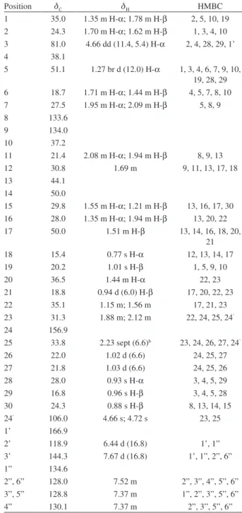
Table 1.1H NMR and 13C NMR data and HMBC correlations of compound 3 in CDCl3a
Position dC dH HMBC
1 35.0 1.35 m H-α; 1.78 m H-β 2, 5, 10, 19 2 24.3 1.70 m H-α; 1.62 m H-β 1, 3, 4, 10 3 81.0 4.66 dd (11.4, 5.4) H-α 2, 4, 28, 29, 1’
4 38.1
5 51.1 1.27 br d (12.0) H-α 1, 3, 4, 6, 7, 9, 10, 19, 28, 29 6 18.7 1.71 m H-α; 1.44 m H-β 4, 5, 7, 8, 10 7 27.5 1.95 m H-α; 2.09 m H-β 5, 8, 9
8 133.6
9 134.0
10 37.2
11 21.4 2.08 m H-α; 1.94 m H-β 8, 9, 13
12 30.8 1.69 m 9, 11, 13, 17, 18
13 44.1
14 50.0
15 29.8 1.55 m H-α; 1.21 m H-β 13, 16, 17, 30 16 28.0 1.35 m H-α; 1.94 m H-β 13, 20, 22 17 50.0 1.51 m H-β 13, 14, 16, 18, 20,
21 18 15.4 0.77 s H-α 12, 13, 14, 17
19 20.2 1.01 s H-β 1, 5, 9, 10
20 36.5 1.44 m H-α 22, 23
21 18.8 0.94 d (6.0) H-β 17, 20, 22, 23 22 35.1 1.15 m; 1.56 m 17, 21, 23 23 31.3 1.88 m; 2.12 m 22, 24, 25, 24’
24 156.9
25 33.8 2.23 sept (6.6)b 23, 24, 26, 27, 24’
26 22.0 1.02 d (6.6) 24, 25, 27
27 21.8 1.03 d (6.6) 24, 25, 26
28 28.0 0.93 s H-α 3, 4, 5, 29
29 16.8 0.96 s H-β 3, 4, 5, 28
30 24.3 0.88 s H-β 8, 13, 14, 15 24’ 106.0 4.66 s; 4.72 s 23, 25
1’ 166.9
2’ 118.9 6.44 d (16.8) 1’, 1”
3’ 144.3 7.67 d (16.8) 1’, 1”, 2”, 6”
1” 134.6
2”, 6” 128.0 7.52 m 2”, 3”, 4”, 5”, 6” 3”, 5” 128.8 7.37 m 1”, 2”, 3”, 5”, 6”
4” 130.1 7.37 m 2”, 3”, 5”, 6”
aSpectra were recorded at 600 MHz for 1H NMR and 150.9 MHz for 13C NMR; 2D NMR experiments recorded in accordance; coupling
Figure 2. Relenvat NOESY and HMBC correlations of 3.
Table 2. Molluscicidal activity of terpenes from Euphorbia conspicua N. E. Br. latex on Biomphalaria glabrata Say
Compound Snail diameter / mm
Concentration / (µg mL-1)
Dead snails / %
After 24 h of exposure After 24 h of recovering After 48 h of recovering
1 13-20 13-20 13-20 15-21 15-21 100 50 20 10 1 0 0 0 0 0 20 20 0 0 0 20 20 0 0 0 2 15-20 13-20 13-20 15-21 15-21 100 50 20 10 1 0 0 0 0 0 40 40 0 0 0 40 40 0 0 0 3 15-20 13-20 13-20 15-21 15-21 100 50 20 10 1 0 0 0 0 0 20 0 0 0 0 20 0 0 0 0 16 15-17 15-20 15-20 13-21 13-21 13-21 100 10 1 0.1 0.01 0.001 100 0 0 0 0 0 − 100 100 60 0 0 − − − 60 20 20
Dechlorinated water 15-20 0 0 0
Dechlorinated water with 1% DMSO
15-21 0 0 0
Cupric sulfate 15-20 50 100 − −
Temperature: 25 ± 1 ºC.
The EI mass spectrum revealed fragment ion peaks at m/z 555 ([M]+ – Me), 407 ([M]+ – Me – HO
2CCH=CHC6H5), 297 (loss of C9H17 and HO2CCH=CHC6H5), 255 (297 – C3H6) and 241 (255 – CH2), which corroborated the structure as a D8-9 tetracyclic triterpene bearing a cinnamoyl group at the C-3 position and a side chain containing nine carbon atoms, including isopropyl and C-24 methylene groups.
Me-30, H-17 and Me-21 on the β face of the structure, H-3 and H-5 with Me-28, H-20 with Me-18 and H-12α with Me-21. These data were consistent with a tirucallane-type structure.16,20,23 Thus, compound 3 was identiied as a new natural product and named 3β-(E)-cinnamoyleuphorbol.
Compounds 1-3 and 16 were evaluated for molluscicidal activity against Biomphalaria glabrata, a vector of S. mansoni (Table 2). The molluscicidal activity of
compound 16, with an LC100 of 1 µg mL-1, was equivalent to that of niclosamide, the synthetic compound used for the control of mollusks (LC100 1.5 µg mL-1).24 Compound 16 presented a dose dependent and continuous effect on adult snails after 24 h of exposure, while it was completely inactive against the egg masses. In contrast, the triterpenic compounds displayed weak activity. Compounds 1 and 3 caused 20% mortality, while compound 2 cause 40% of mortality at a concentration of 100 µg mL-1. Due to a shortage of sample materials, the other triterpenic compounds were not tested.
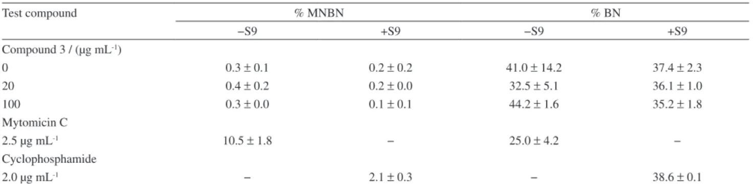
The survival values in V79 cells are given in Table 3, highlighting that only a slight decrease of survival was observed for 250 µg per well. Ames assay outcomes (Table 4) revealed compound 3 as a non-mutagenic agent on the strains tested at doses up to 250 µg per plate. The results obtained for the induction of micronuclei in Chinese hamster cells (V79 cells) at concentrations up to 100 µg mL-1 revealed no signiicant increase compared to the negative control in the absence or presence of S9 mix (Table 5).
Table 3. Effect of compound 3 on cell viability of V79 Chinese hamster cells using the MTT assay
Dose / (µg per well) Viability / %a
25 115.4 ± 5.1
50 123.0 ± 8.1
250 59.7 ± 37.1
aViability is expressed as percentage values relative to control cells;
results are expressed as mean value % viability ± standard deviations (SD) (n = 3); in each independent experiment four replicate cultures were used.
Table 4. Mutagenic activity of compound 3 in the Ames assay, revertants in three strains ofSalmonella typhimurium (TA 98, TA100 and TA102) treated with different concentrations of compound 3 in the presence and absence of metabolic activation (S9)
Dose / (µg per plate)
Revertants per plate
TA 98 TA 100 TA 102
−S9 +S9 −S9 +S9 −S9 +S9
0 17.5 ± 3.5 27.5 ± 9.2 130.5 ± 29.0 123.5 ± 20.5 268.0 ± 56.6 342.0 ± 31.1 5 15.0 ± 1.4 18.5 ± 3.5 127.5 ± 33.2 128.5 ± 23.3 259.5 ± 36.1 296.5 ± 30.4 25 19.5 ± 0.7 18.0 ± 7.1 129.0 ± 14.1 124.0 ± 14.1 231.0 ± 0 309.5 ± 51.6 50 16.5 ± 0.7 28.0 ± 12.7 116.5 ± 14.8 107.5 ± 26.2 288.5 ± 55.9 312.5 ± 88.4 250 14.5 ± 2.1 20.5 ± 13.4 92.5 ± 4.2 87.5 ± 13.4 257.5 ± 96.8 260.5 ± 132.2 Quercetin
10 284.0 ± 77.7 1314.5 ± 102.5 4-NQOa
10 1432 2842
Values are presented as the mean ± standard deviation (SD) (n = 2); dose 0 as negative control; quercetin and 4-NQO as positive controls; a4-NQO:
4-nitroquinoline-1-oxide.
Table 5. Effect of compound 3 on the frequency of micronucleated binucleated cells (% MNBN) in V79 Chinese hamster cells in the presence (+S9) and absence (−S9) of metabolic activation
Test compound % MNBN % BN
−S9 +S9 −S9 +S9
Compound 3 / (µg mL-1)
0 0.3 ± 0.1 0.2 ± 0.2 41.0 ± 14.2 37.4 ± 2.3
20 0.4 ± 0.2 0.2 ± 0.0 32.5 ± 5.1 36.1 ± 1.0
100 0.3 ± 0.0 0.1 ± 0.1 44.2 ± 1.6 35.2 ± 1.8
Mytomicin C
2.5 µg mL-1 10.5 ± 1.8 − 25.0 ± 4.2 −
Cyclophosphamide
2.0 µg mL-1 − 2.1 ± 0.3 − 38.6 ± 0.1
Conclusions
E. conspicua, a succulent tree endemic to Angola, was previously evaluated for its molluscicidal activity,2 but the latex chemical composition was not determined. The present study revealed that E. conspicua latex is composed mainly of triterpenes with euphane and cycloartane skeletons along with other triterpenes and ingenane diterpenes and contains a new compound with a tirucallane skeleton, 3β-(E)-cinnamoyleuphorbol (3). Four of the isolated compounds, 1-3 and 16, were evaluated for their molluscicidal activity. Compound 16
was found to be the most active, with an LC100 value of 1 µg mL-1, which is similar to that for milliamine L, the most powerful molluscicide of plant origin characterized thus far.25 Although compound 16 and milliamine L are structurally differing from each other at the position C-3, where milliamine L bears a dianthraniloyl peptide group and compound 16 an angeloyl group, molluscidal activity remains. At this point compound 16 seems to be the major responsible for E. conspicua molluscicidal activity but further investigation is needed. Compound 16 was found to be totally inactive against the egg masses of B. glabrata. Compound 3 was evaluated for its cytotoxicity, mutagenic activity and genotoxicity using the MTT test, the Ames test and the cytokinesis-block micronucleus assay (CBMN), respectively. No mutagenic or genotoxic activity was detected and little or no cytotoxicity was found concluding that this particular compound as no potential risk regarding their future use as bioactive compound.
The irritant properties of the latex can explain the etnopharmacological use against leprosy wounds and dermatitis in general, but more studies are needed to validate this idea.
Biological and chemical evaluation of other components of E. conspicua latex will require large amounts of latex and will be the subject of a future study.
Experimental
General experimental procedures
O p t i c a l r o t a t i o n s w e r e o b t a i n e d w i t h a Bellingham+Stanley Ltd ADP 220 polarimeter. HREIMS measurements were conducted on a VG Autospec M and recorded at 70 eV. The mass spectrum of 3 was obtained from a GC-MS (Hewlett-Packard 5989 A) spectrometer. The IR spectra were recorded in a Unicam Mattson 5000 FTIR. NMR spectra of 3 were recorded in a Bruker Avance II 600 MHz (1H NMR) and 150.9 MHz (13C NMR) spectrometer in CDCl3. Spectra of compounds 1-2 and 4-16 were
recorded in a Bruker AC 250P 250 MHz (1H NMR) and 62.9 MHz (13C NMR) spectrometer in CDCl
3. Chemical shifts are given in d ppm and are referenced to residual CHCl3, 7.26 ppm for the
1H and 77.0 ppm for 13C.
Two-dimensional experiments were performed with standard Bruker software. Column chromatography was conducted on silica gel 60 (70-230 mesh, Merck, Darmstadt, Germany).
Plant material
Plant material was collected at Cacuaco, Luanda and was identiied by Professor Esperança da Costa from Agostinho Neto University (Biology Department). A voucher specimen (No 4498) has been deposited at the Luanda Herbarium, Luanda, Angola.
Extraction and isolation
(15 mg). Column chromatography of fraction 7 on 10% AgNO3/silica gel 60 (Merck) (m/m) using hexane/EtOAc (95:5, 9:1, 8:2, 7:3, 6:4, 1:1) afforded cycloart-24-en-3β-ol (11) (11 mg) and 24-methylenecycloartan-3β-ol (12) (14 mg). Column chromatography of fraction 8 on silica gel 60 (Merck) using a hexane/EtOAc gradient yielded boeticol (13) (7 mg), 3β-acetoxylofenol (14) (10 mg) and cholesterol (15) (33 mg). Irritant fraction II (2 g) was submitted to column chromatography (CC) over silica gel 60 (Merck) using a gradient elution from 100% hexane to hexane/EtOAc (95:5, 9:1, 8:2 7:3, 1:1). A total of 76 fractions of ca. 50 mL each were collected and pooled into six fractions. Column chromatography of fraction 5 on 10% AgNO3/silica gel 60 (Merck) (m/m) with hexane/diethyl ether (95:5, 9:1, 8:2, 7:3) yielded 20-O-acetyl-3-O-angeloylingenol (16) (22 mg).
3b-(E)-Cinnamoyleuphorbol (3)
White amorphous solid; [α]D20 − 19.0º (c 1.33, CHCl3); IR (KBr) νmax/cm
-1: 3040, 3015, 3021, 1720, 1640, 1580,
1153, 890, 850, 820, 679; 1H NMR (CDCl
3, 600 MHz) and 13C NMR (CDCl
3, 150.9 MHz): see Table 1; HREIMS m/z 570.4417 [M]+ (calcd for C
40H58O2, 570.4436).
Molluscicide activity
The snail colony (B. glabrata) was sustained as described.27,28 Molluscicidal activity for adults and egg masses of B. glabrata was evaluated according to established procedures.27-30
MTT reduction assay.
MTT assay is a cell viability assay based on conversion of MTT dye by mitochondrial enzymes of viable cells into a formazan which can be spectrophotometrically measured. In this assay the absorbance is proportional to the number of viable cells.31
The MTT assay was conducted on V79 Chinese hamster cells. Approximately 104 cells were grown at 37 °C in a 5% CO2 atmosphere for 24 h in 96-well plates in 200 µL of Ham’s F-10 medium supplemented with 10% newborn calf serum and 1% penicillin/streptomycin solution. Different doses of the compound (25, 50 and 250 µg per well) were added, and the cells were incubated for 3 h. The medium was removed, and cells were incubated for 3 h with MTT (0.5 mg mL-1). The cells were washed carefully with PBS, and then 200 µL of DMSO was added to each well. The absorbance of the converted dye was measured at 595 nm in a Zenith 3100 microplate reader. Cell viability was assessed by comparing the absorbance values of treated
cells with that of the control. Absorbance values presented by V79 cell cultures without the addition of compound 3, i.e. control cultures, correspond to 100% of cell viability. Three independent experiments were performed. In each independent experiment four replicate cultures were used.
Ames assay
Mutagenicity testing was conducted by the plate incorporation assay described by Maron and Ames32 using Salmonella typhimurium strains TA 98, TA 100 and TA 102 in the presence or absence of S9 mix.32 At least two independent experiments were performed for each assay. Quercetin and 4-nitroquinoline-1-oxide were used as positive controls and dose 0 as negative control.
Cytokinesis-block micronucleus assay (CBMN)
Approximately 5 × 10⁵ V79 Chinese hamster cells were cultured for 24 h in 25 cm2 culture lasks and then exposed to compound 3 at concentrations of 20 and 100 µg mL-1. Mitomycin C (2.5 µg mL-1) or cyclophosphamide (2.0 µg mL-1) was used as a positive control with and without S9 mix and dose 0 as negative control, respectively. 24 h after the genotoxic treatment, the cells were washed with fresh culture medium, and cytochalasin-B (Cyt-B) was added to a inal concentration of 4.5 µg mL-1. The cells were incubated for additional 16 h, harvested by trypsinization, rinsed and submitted to a mild hypotonic treatment as described elsewhere.33 The centrifuged cells were placed onto dry slides, and smears were made. After air-drying, the slides were ixed with cold methanol for 30 min. One day later, the slides were stained with Giemsa (4% (v/v) in 0.01 mol L-1 sodium phosphate buffer, pH 6.8) for 10 min. For each experimental point, 1000 binucleate V79 cells (BN) with well-preserved cytoplasm were scored. Micronuclei were identiied under a light microscope using a magniication of 1250 × according to the criteria proposed by Caria et al.34 We evaluated MN/BN (data not showed), which represents the average number of micronuclei per binucleated cell, and the frequency of micronucleated binucleated V79 cells (% MNBN), which represents the fraction of cytokinesis blocked (binucleated) cells with micronuclei, regardless of the number of micronuclei per BN cell.35 At least two independent experiments were performed for each assay.
Cell proliferation
of binucleate cells (% BN).35 For this index 1000 cells with well-preserved cytoplasm were analyzed according to number of nuclei at a magniication of 500 ×. Two independent experiments were conducted.
Supplementary Information
1H NMR, 13C NMR, 1H-1H COSY, HSQC, HMBC and
NOESY NMR spectra of compound 3 and physical data of compounds 1, 2 and 16 are available free of charge at http://jbcs.org.br as a PDF ile.
Acknowledgements
This work was supported by Conselho Nacional de Desenvolvimento Cientíico e Tecnológico (CNPq) and Coordenação de Aperfeiçoamento de Pessoal de Nível Superior (CAPES) through scholarships and inancial support. This work was partially funded by projects POCTI/QUI/39380/2001 and FCOMP-01-0124-FEDER-007430 (under COMPETE with FEDER funding) of Fundação para a Ciência e Tecnologia (FCT) and the Textile and Paper Materials Center. Rosalina Mata is thankful to AULP and INABE for inancial support.
References
1. Brown, N. E.; In Fl. Trop. Afr. 1912, 6, 600.
2. dos Santos, A. F.; Lopes, L. A.; Mata, R. C. S.; de Mendonça, D. I. M. D.; Sant’Ana, A. E. G.; Bioresour. Technol.2007, 98, 135.
3. Uemura, B.; Katayama, C.; Luno, E.; Sasaki, K.; Hirata, Y.;
Tetrahedron Lett.1975, 1703.
4. Appendino, G.; Jakupovic, S.; Tron, C. G.; Jakupovic, J.; Milon, V.; Ballero, M.; J. Nat. Prod.1998, 61, 749.
5. Alberto, J. M.; Sanz-Cervera, J. F.; Alberto, Y.; Jakupovic, J.;
J. Nat. Prod.1999, 62, 110.
6. Hecker, E.; Pure Appl. Chem. 1977, 49, 1423.
7. http://www.who.int/topics/schistosomiasis/en/ accessed in June 2011.
8. Hostettman, K.; Marston, A; Ndjoko, K.; Wolfender, J. L.; Curr. Org. Chem.2000, 4, 973.
9. Queiroz, E. F.; Ahua, K. M.; Hostettman, K.; Chimia2005, 59, 299.
10. Borges, C. M. P.; Diakanawma, C.; de Mendonça, D. I. M. D.;
J. Braz. Chem. Soc.2010, 21, 1121.
11. Sebastião, N’S. N.; Cordeiro, I. J. S.; dos Santos, A. F.; Gaspar, J. F.; Martins, C.; Rueff, J.; Diakanamwa, C.; Sant’Ana, A. E. G.; de Mendonça, D. I. M. D.; Phytochemistry2010, 71, 798. 12. Gewali, M. B.; Hattori, M.; Tezuka, Y.; Kikuchi, T.; Namba, T.;
Phytochemistry1990, 29, 1625.
13. Akihisa, T.; Kimura, Y.; Koike, K.; Shibata, T.; Yoshida, Z.; Nikaido, T.; Tamura, T.; J. Nat. Prod.1998, 61, 409.
14. Bai, B.; Jai, M.; Magn. Reson. Chem.2008, 46, 791. 15. Brown, E. D.; Sam, T. W.; Sutherland, J. K.; Torre,
A.; J. Chem. Soc., Perkin Trans. 1 1975, 2326; Adio, A. M.; PhD Thesis, Institute of Organic Chemistry, University of Hamburg, Germany, 2005; http://deposit.ddb. de/cgi-bin/dokserv?idn=974207071&dok_var=d1&dok_ ext=pdf&ilename=974207071.pdf accessed in June 2011. 16. Akihisa, T.; Wijeratne, E. M. K.; Tokuda, H.; Enjo, F.; Toriumi,
M.; Kimura, Y.; Koike, K.; Nikaido, T.; Tezuka, Y.; Nishino, H.;
J. Nat. Prod.2002, 65, 158.
17. Teresa, J. P.; Urones J. G.; Marcos, I. S.; Basabe, P.; Cuadrado, M. J. S.; Moro, R. F.; Phytochemistry1987, 26, 1767. 18. Chen, H.; Jia, Z-J.; Indian J. Chem. B.1996, 35B, 1308. 19. Atta-Ur-Rahman; Pentacyclic Triterpenoids, vol. 2; Elsevier
Science B.V.: Amsterdam, 1994, pp. 21, 443.
20. Ferreira, M. J. U.; Ascenso, J. R.; Tavares, O. S.; J. Nat. Prod
1995, 58, 275.
21. Bonerji, R.; Misra, G.; Nigam, S. K.; Phytochemistry1987, 26, 2644.
22. Kovganko, N. V.; Kashkan, Zh. N.; Borisov, E. V.; Chem. Nat. Compd.2000, 36, 595.
23. Akihisa, T.; Kimura, Y.; Kokke, W. C. M. C.; Takase, S.; Yasukama, K.; Tamura, T.; J. Chem. Soc., Perkin Trans. 11996,
1, 2379.
24. Perret, S.; Whitield, P. J.; Parasitol. Today1996, 12, 156. 25. Zani, C. L.; Marston, A.; Hamburger, M.; Hostettmann, K.;
Phytochemistry1993, 34, 89.
26. Urones, J. G.; Barcala, P. B.; Cuadrado, M. J. S.; Marcos, I. S.;
Phytochemistry 1988, 27, 207.
27. Santos, A. F.; Sant’Ana, A. E. G.; Phytother. Res.1999, 13, 660. 28. Santos, A. F.; Sant’Ana, A. E. G.; Phytomedicine2000, 6, 431. 29. Thiilborg, S. T.; Christensen, S. B.; Cornet, C.; Olsen, C. E.;
Lemmich, E.; Phytochemistry 1994, 36, 753.
30. World Health Organization (WHO); Bull. WHO1965, 33, 567. 31. Carmichael, J.; DeGraff, W. G.; Gazdar, A. F.; Minna, J. D.;
Mitchell, J. B.; Cancer Res.1987, 47, 936.
32. Maron, D. M.; Ames, B. N.; Mutat. Res.1983, 113, 173. 33. Van Hummelen, P.; Kirsch-Volders, M.; Mutagenesis1990, 5,
203.
34. Caria, H.; Chaveca, T.; Laires, A.; Rueff, J.; Mutat. Res.1995,
343, 85.
35. Oliveira, N. G.; Castro, M.; Rodrigues, A. S.; Gonçalves, I. C.; Cassapo, R.; Fernandes, A. P.; Chaveca, T.; Toscano-Rico, J. M.; Rueff, J.; Mutagenesis2001, 16, 369.
Supplementary Information
S
I
J. Braz. Chem. Soc., Vol. 22, No. 10, S1-S4, 2011. Printed in Brazil - ©2011 Sociedade Brasileira de Química 0103 - 5053 $6.00+0.00
*e-mail: [email protected]
Molluscicidal Activity of Compounds Isolated from Euphorbia conspicua N. E. Br.
Rosalina C. S. Mata,a Dina I. M. D. de Mendonça,*,b Liliana Vieira,b
Aldenir F. dos Santos,c Luciana A. da Silva,c Jorge F. Gaspar,d Célia Martins,d
José Rueff,d and Antônio E. G. Sant’Anac
aChemistry Department, Agostinho Neto University, Luanda, Angola
bTextile and Paper Materials Center, University of Beira Interior, 6200-001 Covilhã, Portugal
cChemistry Department, Federal University of Alagoas, 57092-970 Maceió-AL, Brazil
dDepartmentof Genetics , Faculty of Medical Sciences, New University of Lisbon, Lisbon, Portugal
Physical data of compounds 1, 2 and 16
Euphol (1)
White amorphous solid; [α]
D
20 + 35.0º (c 0.70, CHCl 3);
IR (KBr) ν
max/cm
-1: 3580, 3089, 2980, 1640, 1450, 1375,
1151, 1131; 1H NMR (CDCl
3, 250 MHz) d 5.08 (t, 1H,
J 7.0 Hz, H-24), 3.23 (dd, 1H, J 12.1 and 4.8 Hz, H-3), 1.68 (s, 3H, Me-27), 1.60 (s, 3H, Me-26), 0.99 (s, 3H, Me-19), 0.98 (s, 3H, Me-29), 0.87 (s, 3H, Me-28), 0.85 (d, 3H,
J 6.6 Hz, Me-21), 0.79 (s, 3H, Me-30), 0.75 (s, 3H, Me-18).
3b-Acetoxyeupha-8,24-diene (2)
White amorphous solid; [α]
D
20 + 38.3º (c 0.28, CHCl 3);
IR (KBr) ν
max/cm
-1: 3060, 1735, 1640, 1385, 1380, 1250; 1H NMR (CDCl
3, 250 MHz) d 5.09 (br t, 1H, J 6.9 Hz,
H-24), 4.50 (dd, 1H, J 11.1 and 4.9 Hz, H-3), 2.04 (s,
3H, -OOCCH3) 1.68 (s, 3H, Me-27), 1.60 (s, 3H, Me-26) 0.98 (s, 3H, Me-19), 0.97 (s, 3H, Me-29), 0.88 (d, 3H,
J 7.5 Hz, Me-21), 0.87 (s, 6H, Me-28 and Me-30), 0.74 (s, 3H, Me-18).
20-O-Acetyl-3-O-angeloylingenol (16)
Oil; IR (ilm) ν
max/cm
-1: 3460, 3051, 1725, 1705, 1695
1665, 1245, 1160; 1H NMR (CDCl
3, 250 MHz) d 6.16
(m, 1H, H-3’), 6.12 (d, 1H, J 4.1 Hz, H-7), 6.04 (d, 1H,
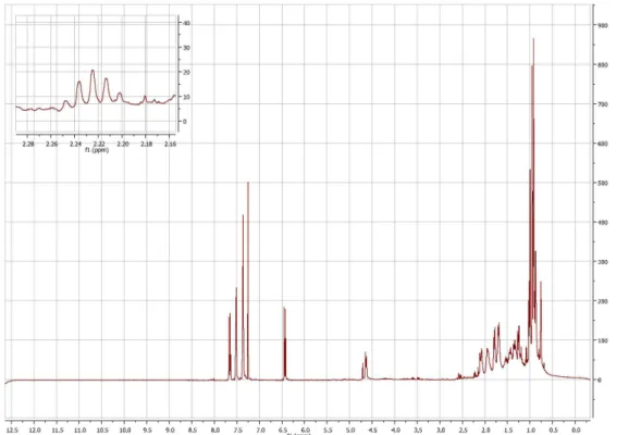
Figure S1.1H NMR spectrum (600 MHz, CDCl
3) of compound 3.
Figure S2.13C NMR spectrum (150.9 MHz, CDCl
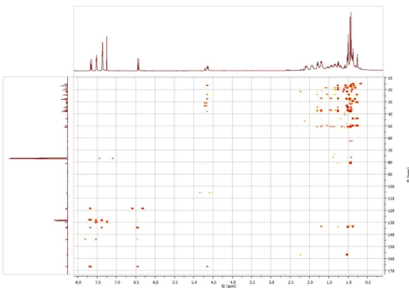
Figure S4. HSQC spectrum (600 MHz, CDCl3) of compound 3.
Figure S3. 1H -1H COSY spectrum (600 MHz, CDCl
Figure S5. HMBC spectrum (600 MHz, CDCl3) of compound 3.