Genotoxicity and mutagenicity of water contaminated with tannery effluents,
as evaluated by the micronucleus test and comet assay using the fish
Oreochromis niloticus
and chromosome aberrations in onion root-tips
Silvia Tamie Matsumoto
1, Mário Sérgio Mantovani
2, Mirtis Irene Ariza Malaguttii
3, Ana Lúcia Dias
2,
Inês Cristina Fonseca
4and Maria Aparecida Marin-Morales
51
Universidade Federal do Espírito Santo, Departamento de Ciências Biológicas, Maruípe, Vitória, ES,
Brazil.
2
Universidade Estadual de Londrina, Departamento de Biologia Geral, Londrina, PR, Brazil.
3
Universidade Estadual Paulista ‘Júlio de Mesquita Filho’, Instituto de Geociências e Ciências Exatas,
Departamento de Petrologia e Metalogenia, Rio Claro, SP, Brazil.
4
Universidade Estadual de Londrina, Departamento de Agronomia, Londrina, PR, Brazil.
5
Universidade Estadual Paulista ‘Júlio de Mesquita Filho’, Instituto de Biociências,
Departamento de Biologia, Rio Claro, SP, Brazil.
Abstract
Cytotoxicity of metals is important because some metals are potential mutagens able to induce tumors in humans and experimental animals. Chromium can damage DNA in several ways, including DNA double strand breaks (DSBs) which generate chromosomal aberrations, micronucleus formation, sister chromatid exchange, formation of DNA adducts and alterations in DNA replication and transcription. In our study, water samples from three sites in the Córrego dos Bagres stream in the Franca municipality of the Brazilian state of São Paulo were subjected to the comet assay and micronucleus test using erythrocytes from the fishOreochromis niloticus. Nuclear abnormalities of
the erythrocytes included blebbed, notched and lobed nuclei, probably due to genotoxic chromium compounds. The greatest comet assay damage occurred with water from a chromium-containing tannery effluent discharge site, sup-porting the hypothesis that chromium residues can be genotoxic. The mutagenicity of the water samples was as-sessed using the onion root-tip cell assay, the most frequent chromosomal abnormalities observed being: c-metaphases, stick chromosome, chromosome breaks and losses, bridged anaphases, multipolar anaphases, and micronucleated and binucleated cells. Onion root-tip cell mutagenicity was highest for water samples containing the highest levels of chromium.
Key words: Allium cepa, chromium, chromosomes aberrations, comet assay, micronucleus,Oreochromis niloticus.
Received: May 2, 2005; Accepted: June 8, 2005.
Introduction
Pollution of water resources is a serious and growing problem but despite the existence of relevant legislation the pollution of the aquatic environment by toxic chemical pol-lutants continues to occur, with domestic and industrial effluents being the main sources responsible for the con-tamination of aquatic environments (Claxtonet al., 1998; White and Rasmussen, 1998).
Chromium compounds are known to have toxic, genotoxic, mutagenic and carcinogenic effects on man and
animals (Von Burg and Liu, 1993; Stohs and Bagchi, 1995; Mount and Hockett, 2000), with both trivalent chromium III (Cr III) and hexavalent chromium VI (Cr VI) being bio-logically active but differing in their ability to cross biolog-ical membranes. Octahedral chromium III is potentially mutagenic but does not pose an immediate danger to cells because it is unable to cross cell membranes, although it may be environmentally transformed into tetrahedral hex-avalent chromium VI (chromate) to which cell membranes are highly permeable (Leonard and Lauwerys, 1980; Beyersmannet al., 1994; Stohs and Bagchi, 1995; Singhet al., 1998; Matsumoto, 2003). According to Sugiyama (1992), hexavalent chromate enters cells via the surface transport system and inside the cell it is reduced to trivalent
Genetics and Molecular Biology, 29, 1, 148-158 (2006) Copyright by the Brazilian Society of Genetics. Printed in Brazil www.sbg.org.br
Send correspondence to Maria Aparecida Marin-Morales. Univer-sidade Estadual de São Paulo, Instituto de Biologia, Departamento de Biologia, Av. 24-A 1515, 13506-900 Rio Claro, SP, Brazil. E-mail: mamm@rc.unesp.br.
chromium which induces genotoxic effects in the cell (Bianchiet al., 1983). However, if trivalent chromium has access to the intracellular medium through processes such as pinocytosis and endocytosis or by the reduction of hex-avalent chromium inside the cell it acts directly on DNA and causes more damage than when it continues in the chromate form (Matsumoto, 2003).
Severalin vivoandin vitrostudies have shown that chromium compounds damage DNA in a variety of ways, including DNA single and double-strand breaks (SDSBs) generating chromosomal aberrations, micronucleus forma-tion, sister chromatid exchanges, formation of DNA ad-ducts, and alteration in DNA replication and transcription (Zhitkovichet al., 1996; O’Brienet al., 2001; Matsumoto, 2003; Matsumoto and Marin-Morales, 2004).
Organisms used in mutagenesis testing should be se-lected using criteria that permit a realistic evaluation of the potential of a suspected mutagen to induce changes in ge-netic material such as structural and/or numerical modifica-tion of chromosomes resulting in chromosome aberramodifica-tions. Aquatic organisms such as fish accumulate pollutants di-rectly from contaminated water or indidi-rectly through the in-gestion of contaminated aquatic organisms. Thus, genotoxic pollutants may lead to the contamination not only of the aquatic organisms themselves but of the entire ecosystem and, finally, of humans through the food chain. A variety of teleost fish, including larvae, have been used for the study of the mutagenic, clastogenic and terato-genic effects of environmental contaminants during the early stages of life. Metcalfe (1988) used the fish
Oncorthychus mykissandOryzias latipesas test organisms for the study of carcinogenesis and model systems based on fish have become important for determining the distribu-tion and toxic effects of aquatic contaminants, within vivo
techniques such as the micronucleus test performed in fish test systems having been shown to be efficient not only for assessing genotoxic potential but also for water quality monitoring (Al-Sabti and Metcalfe, 1995; and Dashwood and Bariley, 1998).
The analysis of the frequency of micronuclei during interphase is a rapid and easy to conduct technique. Teleost erythrocytes possess a nucleus and are a good tool for the detection of clastogenic substances in water, the fish eryth-rocyte micronucleus test having been used as an initial step in evaluating clastogenic potential. Several studies have shown a high incidence of micronuclei in fish peripheral erythrocytes after exposure to different pollutants under both field and laboratory conditions (Al-Sabti, 1986; Metcalfe, 1988; Hoseet al., 1987; Minissiet al., 1996).
Al-Sabti (1994) used the micronucleus test to deter-mined the cytological effects of hexavalent and trivalent chromium in erythrocytes ofCarassus auratus gibeliofish exposed to sub-lethal chromium concentrations in the labo-ratory and fish collected from chromium-contaminated rivers showing a significant frequency of micronuclei when
compared to negative controls and thus demonstrating the true genotoxic effect of chromium.
Nuclear abnormalities are frequently observed in fish erythrocytes, with Carrascoet al. (1990) having described such abnormal nuclei as being blebbed, notched or lobed. Other investigators have also reported nuclear abnormali-ties but simply classified them as ‘genotoxic damage’ (Bombailet al., 2001; Pachecco and Santos, 1998; Ayylon and Garcia-Vazquez, 2000). Eiras (1990) identified nuclear abnormalities in fish with folic acid deficiency or which had been affected by viral necrosis, as well as in specimens exposed to chlorine and cadmium.
The single cell gel electrophoresis (SCGE) assay, commonly called the comet assay, is a genotoxicity test able to detect DNA damage induced by alkylating, interca-lating and oxidizing agents (Tice et al., 2000; Kosz-Vnenchak and Rokosz, 1997). The comet assay has been used as an important tool for monitoring genotoxicity in aquatic environments. For this purpose, fish are used as a test organism in which it is possible to detect DNA damage induced by direct mutagens and pro-mutagens in both fresh and salt water (Mitchelmore and Chipman, 1998; Lemoset al., 2005). This technique has also been employed in the de-termination of the genotoxic potential of water resources such as rivers and lakes.
According to Matsumoto et al. (2003, 2005) the comet test is sensitive enough to be used for the environ-mental monitoring of waters and is suitable for assessing the quality of water contaminated with effluents containing chromium residues, these authors having used the comet assay to detect genotoxicity effects caused by chromium concentrations as low as 0.01 mgL-1which is significantly less than the 0.05 mgL-1currently accepted as the interna-tional standard for chromium (CETESB, 1995).
An efficient test organism for the assessment of chro-mosomal aberrations should have chromosomes which are easy to analyze in terms of size, morphology and number. The higher plants Allium cepa (onion), Tradescantia paludosaandVicia fabahave relatively large monocentric chromosomes in reduced numbers and are accepted as suit-able test organisms for the study of environmental muta-genesis (Rank and Nielsen, 1998; Grover and Kaur, 1999; Kong and Ma, 1999; Moraes and Jordão, 2001; Patra and Sharma, 2002).
The clastogenic and aneugenic effect of atmospheric, water and soil pollutants have been demonstrated by sev-eral authors using the micronucleus assay inA. cepaandV. fabaroot tip cells (Grover and Kaur, 1999; Sudhakaret al., 2001; and Patra and Sharma, 2002), these authors having reported that the chromosomal changes most frequently ob-served were c-metaphases, stick chromosomes and breaks, bridges, laggards, binucleate cells and micronucleated cells.
analysis of the cytotoxic effects of such metals has received special attention due to the fact that they are potentially mutagenic and induce the formation of tumors in experi-mental organisms and humans exposed to them (Garcia-Rodríguezet al., 2001).
Sahiet al. (1998) usedA. cepatest systems to assess the effects of chromium contamination on the waters of an Indian river and showed that at sites where chromium con-centrations were high there was a reduction in mitotic index and an increase in the rate of mitotic abnormalities, thus confirming the cytotoxic and genotoxic effect of chro-mium.
In the municipality of Franca in the Brazilian state of São Paulo there are several leather tanneries which use tri-valent chromium salts for tanning and which discharge effluents into the Córrego dos Bagres stream. The study de-scribed in this paper used the erythrocyte comet assay and micronucleus test in the fishOreochromis niloticusand the cellular cycle of onion root tip cells to investigate three sites along the Córrego dos Bagres stream for the genotoxic and mutagenic potential of tannery effluents which may be con-taminated with chromium.
Material and Methods
Sampling sites and chemical analyses
The Corrego dos Bagres stream in the Franca munici-pality in the Brazilian state of São Paulo is a small stream which receives tannery effluents. We collected water sam-ples from this stream during spring, summer, autumn, and winter of 2001 and 2002 at three locations, one being 200 meters upstream of the tannery effluent discharge site, one at the tannery effluent discharge site and another 500 me-ters downstream of the effluent discharge site (Figure 1). In this context ‘upstream’ means in the higher part of the stream nearer the source of the stream and against the flow of the current while ‘downstream’ means nearer the mouth of the river and in the direction of the water flow.
Chemical analysis of the waters was carried out using standard methods (Franson, 1995). For cation determina-tion, the samples were first acidified with HNO3(pH 1) and
the cations sequentially analyzed by inductively coupled plasma atomic emission spectrometry (ICP-AES) with ul-trasonic nebulization. The following elements were deter-mined: calcium, magnesium, strontium, silicon, iron, manganese, aluminum, zinc, chromium, cobalt, nickel, lead, cadmium, phosphorus, copper, and barium. The stan-dard solutions (1 g L-1) used to construct the calibration curves for the elements were made up in 0.1% HNO3and
appropriate dilutions made in titrisol (Merck).
Micronucleus test and comet assay using the fish Oreochromis niloticus
The water samples colleted in summer of 2001 from the three sites were placed in individual aquaria and diluted 1:1 (v/v) with well-water from State University of Lon-drina, and then aerated continuously for three days, after which 6 adult ofOreochromis niloticuswith 10 cm long health were added to each aquarium and left for 72 h. The fish were kindly provided by the Fish Farm of the State University of Londrina, Paraná, Brazil. Control fish were placed in aquarium containing the same volume of well-water.
For the micronucleus testO. niloticusblood samples were obtained by tail puncture using heparinized syringes. Smears were prepared on slides, with 6 slides being pre-pared (one from each fish) for each water sample. After 24 h, the material was fixed in absolute methanol for 10 min and stained with 5% Giemsa for 20 min. The number of normal erythrocytes without micronuclei and the number of damaged cells with micronuclei were determined by analysis of 2000 cells per fish (Huber 1992), the Kruskal-Wallis test being used to compare the results for fish posed to collection site water with those for control fish ex-posed to well-water only.
For the comet assay blood was obtained from the fish as described above and 10-mL aliquots diluted in 1000mL of fetal bovine serum8. Microscope slides were coated with 120mL of 0.5% (w/v) low melting point agarose at 37 °C containing 10mL of the diluted blood and the slides placed in lysis buffer (10 mM Tris, ~8 g of NaOH and 10 mL of 1% (w/v) sodium lauryl sarcosinate solution plus 1 mL of Tri-ton X-100, 10 mL of DMSO and 89 mL of pH 10 lysis solu-tion containing 2.5 M NaCl, 100 mM EDTA, 10 mM Tris and ~8 g of NaOH) in a refrigerator for 1 h. After lysis, the slides were incubated in 300 mM NaOH + 1 mM EDTA buffer (pH > 13) for 20 min to denature the DNA and then submitted to electrophoresis at 25 V and 300 mA for 20 min. The slides were then neutralized with 0.4 M Tris for 15 min and fixed in ethanol for 10 min. For each fish 100 nuclei were analyzed per blood sample. The slides were stained with ethidium bromide (0.02 mg mL-1) and ana-lyzed under a Nikon fluorescence microscope equipped
150 Genotoxicity and mutagenicity of the tannery effluents
with a B-3A filter (excitation:l= 420-490 nm, emission barrier:l= 520 nm) and a 40x objective lens. The nuclei were visually classified according to fragment migration as undamaged (class 0), slightly damaged (class 1), more damaged (class 2) and highly damaged (class 3). Thec2test was used to compare the total number of altered nuclei from fish exposed to site water with control fish exposed to well-water only.
Onion root-tip mutagenicity test
For this test onion (A. cepa variety Periform Baia) seeds were germinated in Petri plates containing water from the collection sites, pollutant-free Milli-Q water being the negative control (NC) and an aqueous solution of 0.089 mg L-1trivalent chromium the positive control (PC). Two types of treatments were applied, a continuous treat-ment in which the seeds were soaked and germination in the water from the collection sites until the radicals reached 2 cm in lenght and a discontinuous treatment in which the seeds were first soaked in Milli-Q water until the radicals reached 2 cm in length and then transferred to a Petri plates containing water from the collection sites and were left for 20 h (acute treatment) after which some of the rooted seeds were collected at random and assessed, the remaining roots being left under the same conditions until 72 h (chronic treatment) before being collected. For the samples taken in 2002, after chronic treatment some roots were transferred to plates containing Milli-Q water and left to recover for 48 h before being assessed. After treatment roots were fixed in 3:1 (v/v) ethanol/glacial acetic acid (Carnoy solution) for 24 h, carefully squashed and hydrolyzed with 1 N HCl at 60 °C for 8 min, washed with distilled water and Schiff stained for 2 h in the dark. All cells with alterations were counted and the most representative ones for each abnor-mality were photographed. For the mutagenicity assess-ment dividing cells with irregular anaphases (i.e.
disorganized structure, lag chromosomes or multipolar anaphases), cells with stick chromosomes, micronuclei, and binucleate and/or multinucleate cells were recorded. These data were analyzed using the Kruskal-Wallis test.
Results and Discussion
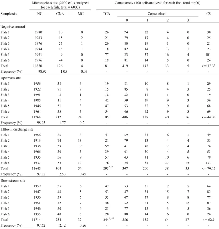
TheO. niloticusmicronucleus test and the comet as-say showed that the water collected at the three sites on the Córrego dos Brages stream was significantly genotoxic as compared to the well-water used as the negative control.
Water from the effluent discharge site produced the highest nuclear abnormality frequency (2.53%) and micro-nuclei frequency (0.45%), with the data showing that, as expected, water from the effluent discharge site was more genotoxic due to the presence of tannery effluent which re-sulted in a higher rate of abnormalities (Table 1, Figures 2 and 3). The nuclear abnormality and micronuclei data sug-gest that concentrations lower than 0,05 mgL-1induce
ef-fects genotoxic in the exposed organisms, supported by the chemical analysis of the water collected from this site (Ta-ble 3). Our results agree with those reported by Von Burg and Liu (1993), Blasiak and Kowalik (2000) and Matsu-motoet al. (2003), who proposed that chromium exerts a genotoxic effect on animals due to its potential to cause var-ious forms of DNA damage
The micronuclei and nuclear abnormality data showed that water from the upstream and downstream sites were less genotoxic than water from the effluent discharge site. At the upstream site, the nuclear abnormality fre-quency was 1.77% and the micronuclei frefre-quency 0.20%, although this micronuclei frequency was not significantly different to that found for the well-water used as the nega-tive control. At the downstream site, the nuclear abnormal-ity frequency was 2.12% and micronuclei frequency 0.26% (Table 1 and Figures 2 and 3). Compared to water from the upstream site and the negative control water, the micro-nuclei frequency was significantly higher for water from the effluent discharge and downstream sites, demonstrating that upstream of the discharge site the genotoxic effect was smaller than at the effluent discharge site and the down-stream site. These data support the view of Matsumotoet al. (2003) that chromium-containing tannery residues in the Franca region represents a genotoxic pollution hazard.
The erythrocyte nuclear abnormalities (Figure 3) were classified as blebbed, notched and lobed (Carrascoet al. 1990; Ayylon and Garcia-Vazquez, 2000; Çavas and Ergene-Gözükara, 2003). Water from the effluent dischar-ge and downstream sites produced a significantly higher frequency of nuclear abnormalities compared to the nega-tive control well-water, indicating that these abnormalities are the consequence of the genotoxic effect of chromium
residues present in the tannery waste water discharged into the stream.
The comet assay has been shown to be efficient in the detection of DNA damage provoked by genotoxic agents present in aquatic environments, Mitchelmore and
Chipman (1998) having stated that the comet assay using fish as the test organism is an effective technique for moni-toring the genotoxic potential of aquatic environments.
The damage detected by the comet assay for water from the upstream site was not significantly different to that
152 Genotoxicity and mutagenicity of the tannery effluents
Table 1- Changes observed in erythrocytes of sixOreochromis niloticusfish placed for 72 h in a 1:1 (v/v) mixture of well-water and water collected at several sites on the Córrego dos Bagres stream. The stream water was collected in summer of 2001 from a tannery industry chromium-containing effluent discharge site and at sites upstream and downstream. Well-water was also the negative control.
Micronucleus test (2000 cells analyzed for each fish, total = 6000)
Comet assay (100 cells analyzed for each fish, total = 600)
Sample site NC CNA MC TCA Comet class1 CS
0 1 2 3
Negative control
Fish 1 1980 20 0 26 74 22 4 0 30
Fish 2 1983 15 2 21 79 17 4 0 25
Fish 3 1976 23 1 20 80 19 1 0 21
Fish 4 1984 15 1 18 82 14 3 1 23
Fish 5 1991 9 0 77 23 57 16 4 101
Fish 6 1956 44 0 19 81 14 5 0 24
Total 11870 126 4 181 419 143 33 5 x = 37.33
Frequency (%) 98.92 1.05 0.03 - - -
-Upstream site
Fish 1 1956 38 6 19 81 10 8 1 29
Fish 2 1922 71 7 15 85 8 4 3 25
Fish 3 1991 8 1 18 82 17 1 0 19
Fish 4 1985 11 4 42 59 29 9 3 56
Fish 5 1946 51 3 47 53 32 9 6 68
Fish 6 1964 33 3 54 46 42 9 3 69
Total 11764 212 24 195 406 138 40 16 x = 44.33
Frequency (%) 98.03 1.77 0.2 - - -
-Effluent discharge site
Fish 1 1956 36 8 41 59 34 6 1 49
Fish 2 1913 74 13 21 79 13 4 4 33
Fish 3 1938 53 9 59 41 48 7 4 74
Fish 4 1966 30 3 39 61 30 4 5 53
Fish 5 1935 56 9 57 43 41 10 6 79
Fish 6 1937 55 12 76 24 34 27 15 133
Total 11645 304 54 293*,** 307 200 58 35 x = 70.17
Frequency (%) 97.02 2.53 0.45 - - -
-Downstream site
Fish 1 1959 35 6 47 53 35 7 5 64
Fish 2 1947 48 5 53 47 31 15 7 82
Fish 3 1956 39 5 53 47 37 8 8 77
Fish 4 1951 42 7 48 52 21 15 12 87
Fish 5 1946 50 4 23 77 15 3 5 36
Fish 6 1955 40 5 20 80 14 6 0 26
Total 11714 254 32 244*,*** 356 152 54 37 x = 62.0
Frequency (%) 97.62 2.12 0.26 - - -
-1Class 0 = undamaged, 1 = slightly damaged, 2 = more damaged and 3 = highly damaged. *significantly different in relation to the negative control site by
detected for the negative control (Table 1 and Figure 4), a finding explainable by the fact that the upstream site re-ceives little or no tannery effluent. The comet assay results revealed significant genotoxic effects for water from the ef-fluent discharge and downstream sites compared to the negative control well-water (Table 1, Figure 4), with the highest damage frequency occurring with water from efflu-ent discharge site, confirming the genotoxic effect of the chromium residues present at the tannery effluent disposal site. These results support those of Matsumotoet al. (2003) who used the comet assay and CHO-K1 cell cultures to
de-tect genotoxicity caused by water contaminated with tan-nery effluent containing a chromium concentration lower than that permitted by the legislation of the State of São Paulo (0.01 mg L-1).
To estimate the mutagenic effect of the water sam-ples, we calculated the ratio of aberrant to dividing onion cells and found that, with one exception, for all the sam-ples collected during the dry and rainy seasons of 2001 and 2002 the aberrant onion cell rate was higher than that recorded for the negative control (Tables 2), the exception being water from the upstream site collected during the dry period of 2002 and used in the continuous exposure experiments.
For water samples collected during the dry period (Auttumn and Winter) of 2001, mutagenic and clastogenic effects were recorded for water from the effluent discharge site continuous exposure experiments and the upstream and effluent discharge sites 20 h discontinuous exposure exper-iments, while for water collected during the rainy season (Spring and Summer) of the same year such effects were observed only for the effluent discharge site 72 h chronic discontinuous exposure experiments. Table 3 shows that during 2001 the chromium concentration at the effluent dis-charge site was higher than the maximum concentration (0.05 mg/L) permitted by the 1995 legislation of the State of São Paulo. These results support the hypothesis that chromium is the contaminant present in the water samples of the Bagres Stream and is responsible for the clastogenic action on onion root-tip cells.
For the water samples collected in 2002 both during the dry and rainy seasons, the frequency of aberrant cells was high for water collected from the effluent discharge and downstream sites (Table 2), again supporting the hy-pothesis that the observed mutagenicity was due to the tan-nery effluent chromium-content (Table 3). The most frequent aberrations observed were c-metaphases, stick chromosomes, anaphase irregularities (dots and multi-polarity), chromosome breaks and cells with micronuclei or binucleate (Table 2, Figure 5, 6 and 7). Some cells showed the loss of whole chromosomes, which persisted up to telophase (Figure 6), such losses probably resulting in micronucleated interphase cells. These results agree with those obtained by Sudhakar et al. (1998), who reported micronucleus induction involving the mitotic spindle and consequent production of laggard chromosomes during anaphase and the loss of a complete chromosome. In our
Figure 3- Erytrocytes (Oreochromis niloticus) with a micronucleus and nuclear abnormalities: A and B-Notched nuclei; C-Lobed nuclei; D-Lobed nuclei (large arrow) and blebbed nuclei (small arrow); E-Broken eggs (large arrow) and blebbed nuclei (small arrow); F-Micronucleus.
154
Genotoxicity
and
mutagenicity
of
the
tannery
effluents
Table 2- Onion root-tip mutagenicity test based on changes inAllium ceparoot tip cells treated with water collected at several sites on the Córrego dos Bagres stream during under different weather conditions in 2001 and 2002. Water was collected from a site at which a chromium-containing effluent was discharged into the stream and at sites upstream and downstream. For continuous exposure seeds were germinated in sample water until the radicals reached 2 cm in length. In the other treatments the seeds were first soaked in pollutant-free Milli-Q water until the radicals reached 2 cm in length and then placed in water from the collection sites for 20 h (acute treatment) or 72 h (chronic treatment). For the 2002 sample, after chronic treatment some seedlings were transferred to Milli-Q water to recover for 48. Milli-Q water was the negative control and an aqueous solution of 0.089 mgL-1trivalent chromium the positive control.
Weather, type of exposure and sample collection site
Micronucleus Anaphase with bridge
Multipolar anaphase
Delayed anaphase Chromosome breaks
Chromosome losses
C-metaphase Adherence Binucleate cell RAC1
2001 2002 2001 2002 2001 2002 2001 2002 2001 2002 2001 2002 2001 2002 2001 2002 2001 2002 2001 2002
Dry weather2
continuous exposure
Negative control 0.6 2.2 0.1 1.6 - - - 0.7 - - - 1.2 - 0.52 c 2.04b
Positive control 1.2 4.4 1.8 2.2 2.2 0.8 0.1 0.9 1 0.5 2 0.5 5.2 1.2 2.7 3.9 - 0.1 18.07 a 14.42 a
Upstream site 1.1 0.2 0.4 0.8 0.3 0.6 0.4 - - - 0.6 0.2 - 0.2 - 0.9 1.7 - 2.61 bc 1.88 c
Effluent discharge site 0.7 1.4 0.4 0.1 0.7 0.2 0.4 0.2 - - 0.5 1 0.1 1 0.2 2.4 2.3 0.6 3.23 b 5.72 ab
Downstream site 0.8 0.5 0.3 1.0 0.5 1.2 0.4 0.4 - - 0.3 0.1 - 0.3 0.1 1.5 1.6 - 2.06 bc 3.51 bc
20 h acute exposure
Negative control 0.6 2.2 0.1 1.6 - - - 0.7 - - - 1.2 - 0.52 c 2.04b
Positive control 1.2 4.4 1.8 2.2 2.2 0.8 0.1 0.9 1 0.5 2 0.5 5.2 1.2 2.7 3.9 - 0.1 18.07 a 14.42 a
Upstream site 1.5 1 0.6 0.6 0.8 0.8 0.2 0.2 - 1.6 0.9 0.9 0.1 1 0.1 1.8 2.7 - 3.88 b 5.49 bc
Effluent discharge site 1 0.2 0.3 2.2 1 1.3 0.3 0.2 - 0.6 1.1 1 - 2.3 0.1 2.3 2.3 - 3.85 b 7.58 b
Downstream site 1.5 0.2 0.3 0.7 0.4 0.9 0.3 - - 0.2 0.2 0.8 - 0.9 - 3.2 0.3 - 1.98 bc 4.86 bc
72 h chronic exposure
Negative control 0.6 2.2 0.1 1.6 - - - 0.7 - - - 1.2 - 0.52 b 2.04b
Positive control 1.2 4.4 1.8 2.2 2.2 0.8 0.1 0.9 1 0.5 2 0.5 5.2 1.2 2.7 3.9 - 0.1 18.07 a 14.42 a
Upstream site 0.7 2.2 0.1 0.9 0.1 0.2 0.4 - - 0.9 - 1 - 1.6 - 1.8 0.6 0.2 1.16 ab 6.57 bc
Effluent discharge site 2.1 0.6 0.1 - 0.3 0.2 0.4 - - 0.4 0.1 0.2 0.1 2.8 - 5.9 1 0.1 1.98 ab 11.93 abc
Downstream site 1.3 2.3 0.1 0.5 - 0.2 - - - 0.1 0.1 0.1 - 0.5 - 4.7 0.2 - 0.78 ab 7.62 bc
48 h recovery
Negative control - 2.2 - 1.6 - - - 2.04b
Positive control - 4.4 - 2.2 - 0.8 - 0.9 - 0.5 - 0.5 - 1.2 - 3.9 - 0.1 - 14.42 a
Upstream site - 1.0 - 0.3 - 1.0 - - - 0.6 - 0.6 - - - 0.3 - 3.35 b
Effluent discharge site - 1.0 - 1.0 - 0.4 - 1.0 - 1.4 - 5.2 - 6.8 - 2.20 - 0.2 - 23.67 a
Downstream site - 1.9 - - - 0.75 - 1.0 - 0.82 - 1.15 - 1.45 - 0.4 - 9.78 a
Rainy weather3
Continuous exposure
Negative control - 2 - - - 0.8 - 0.1 - 0.1 - - - - 0.0 b 0.18 c
Positive control 0.3 1.8 1.2 1.6 0.7 2.0 0.2 - 0.2 1.8 0.4 0.8 1.1 4.4 1.7 1.0 - - 10.95 a 15.81 a
Upstream site 0.8 0.3 0.4 0.7 0.4 0.6 - - - 1.1 0.2 0.5 - 0.4 - 0.3 1 0.1 0.82 ab 5.07 b
Effluent discharge site 0.4 - 0.7 0.7 0.4 0.2 0.2 0.1 - 0.1 0.1 0.2 - 0.6 - 0.2 0.9 - 1.47 ab 3.79 a
Downstream site 1.6 0.2 0.5 0.8 0.5 0.4 - 0.1 - 0.1 - 0.4 - 1.3 - 1 1 1 1.10 ab 7.69 ab
20 h acute exposure
Negative control - 2 - - - 0.8 - 0.1 - 0.1 - - - - 0.0 b 0.18 a
Positive control 0.3 1.8 1.2 1.6 0.7 2.0 0.2 - 0.2 1.8 0.4 0.8 1.1 4.4 1.7 1.0 - - 10.95 a 15.81 b
Upstream site 0.9 0.6 0.3 0.5 - 0.6 - - - 0.4 - 0.5 - 0.9 - 0.5 0.3 0.3 0.87 ab 5.22 a
Effluent discharge site 2.9 0.4 0.1 0.6 - 0.4 - 0.1 - 0.2 - 0.2 - 1.7 0.2 1.5 0.3 0.1 0.96 ab 7.36 a
study, evidence of the action of chromium on the mitotic spindle was confirmed by the presence of c-metaphase cells. We also observed that clastogenic and aneugenic fre -quencies were higher for water from the collection sites where total chromium concentration was highest (Table 3) and this supports the view of Sahi et al . (1998) and Matsu -et al. 155
Weather, type of exposure and sample collection site
Micronucleus Anaphase with bridge
Multipolar anaphase
Delayed anaphase Chromosome breaks
Chromosome losses
C-metaphase Adherence Binucleate cell RAC1
2001 2002 2001 2002 2001 2002 2001 2002 2001 2002 2001 2002 2001 2002 2001 2002 2001 2002 2001 2002
72 h chronic exposure
Negative control - 2 - - - 0.8 - 0.1 0.1 - - - - 0.0 c 0.18 c
Positive control 0.3 1.8 1.2 1.6 0.7 2.0 0.2 - 0.2 1.8 0.4 0.8 1.1 4.4 1.7 1.0 - - 10.95 a 15.81 a
Upstream site 0.5 - 0.4 0.8 0.3 0.8 - - - 0.3 - 0.40 - 0.8 - 0.8 0.7 - 0.79 ab 9.35 b
Effluent discharge site 1.6 0.2 1 0.2 0.1 0.2 0.4 - 0.1 - 0.4 - - 0.5 0.1 - 1.6 - 11.81 a 6.13 ab
Downstream site 0.3 0.3 0.5 0.6 0.2 1.20 - - - 1.40 - 1.0 - 3.0 - 1.0 0.7 - 2.21 cb 13.27 b
48 h recovery
Negative control - 0.6 - 0.6 - - - 0.1 - 0.3 - 0.1 - 0.3 - 0.7 - - - 6.60 b
Positive control - 1.8 - 1.6 - 2.0 - - - 1.8 - 0.8 - 4.4 - 1.0 - - - 15.81 a
Upstream site - 1.2 - 0.5 - 0.2 - - - 0.4 - 0.4 - 0.3 - 0.7 - - - 5.81 b
Effluent discharge site - 0.9 - 0.6 - - - 0.6 - 0.1 - 0.5 - 0.4 - - - 6.76 b
Downstream site - 2 - - - 0.8 - 0.1 - 0.1 - - - 0.18 c
1
Means in the same column followed by the same letters do not differ by the mean rank Kruskal-Wallis test at p = 0.05.2Autumn and Winter in the Southern Hemisphere.3Spring and Summer in the Southern Hemi-sphere. RAC: Ratio of aberrant cells to dividing cells.
motoet al. (2004) who state that chromium has the poten-tial to induce chromosome breaks (clastogenic effect) and losses (aneugenic effect) resulting in chromosome aberra-tions, micronucleus formation and binucleate cells.
The strongest mutagenic effect detected by us were recorded during the dry period from autumn and winter when the water flow is usually low, which results in a higher concentration of chromium. It is known that tannery effluents have the potential to damage the DNA of test or-ganisms (Zhitkovich et al., 1996; O’Brien et al., 2001;
Matsumotoet al., 2003 and Matsumotoet al., 2004) and as such may compromise the quality of the water in the Cor-rego dos Bagres stream. Our data agree with that of Matsu-motoet al. (2003) who used the comet assay and CHO-K1 cells to detect genotoxicicity in water from the Corrego dos Bagres stream collected during the winter dry period.
The legislation of the State of São Paulo establishes a maximum chromium concentration of 0.05 mgL-1for the emission of industrial effluents into rivers. In our study we found genotoxic effects on onion root-tip cells and
erythro-156 Genotoxicity and mutagenicity of the tannery effluents
Table 3- Chemical analysis of water collected at various sites on the Córrego dos Bagres stream during different seasons in 2001 and 2002.
Element (mgL-1
)
Pb Fe Cd Cr P Al
Season and site (site code) 2001 2002 2001 2002 2001 2002 2001 2002 2001 2002 2001 2002
Summer (wet weather)
Upstream site < 0.05 < 0.025 0.21 0.36 < 0.005 < 0.005 0.05 < 0.01 1.5 0.32 0.33 < 0.05 Effluent discharge site < 0.025 < 0.025 0.8 0.28 < 0.005 < 0.005 0.38 < 0.01 2.20 0.24 0.43 0.07 Downstream site < 0.025 < 0.025 0.3 0.27 < 0.005 < 0.005 0.23 < 0.01 0.11 0.21 0.14 0.09
Autumn (dry weather)
Upstream site < 0.025 < 0.012 0.3 0.23 < 0.005 < 0.003 < 0.01 < 0.005 1.5 < 0.1 0.33 0.05 Effluent discharge site < 0.025 < 0.012 0.5 0.22 < 0.005 < 0.003 0.11 < 0.005 1.2 0.11 0.12 0.03 Downstream site < 0.025 < 0.012 0.3 0.27 < 0.005 < 0.003 0.05 < 0.005 1.1 < 0.1 0.07 0.1
Winter (dry weather)
Upstream site < 0.025 < 0.02 0.04 0.76 < 0.005 < 0.003 < 0.01 0.005 1.6 < 0.1 < 0.05 0.3 Effluent discharge site < 0.025 < 0.02 0.18 0.46 < 0.005 < 0.003 0.02 0.01 1.82 0.14 0.05 0.16 Downstream site < 0.025 < 0.02 0.15 0.19 < 0.005 < 0.003 0.02 < 0.005 1.22 < 0.1 < 0.05 0.25
Spring (wet weather)
cytes from fish exposed to water taken from sites where the total chromium concentration was 0.01 mgL-1, significantly less than the value established by the legislation currently in force in the state of São Paulo.
Acknowledgment
We are grateful to Cristiane Márcia Milleo for the as-sistance in preparation of the schematic figure.
References
Al-Sabti K (1986) Clastogenic effects of five carcinogenic chemi-cals on the cells of the commonCyprinus carpio L.Comp. Biochem Physiol 85:5-9.
Al-Sabti K (1994) Chromium-induced micronuclei in fish. J Appl Toxicol 14:333-336.
Al-Sabti K and Metcalfe C (1995) Fish micronuclei for assessing genotoxicity in water. Mutat Res 343:121-135.
Ayylon F and Garcia-Vazquez G (2000) Induction of micronuclei and other nuclear abnormalities in European minnow
Phoxinus phoxinusand molliePaecilia latipinna: An assess-ment of the fish micronucleus test. Mutat Res 467:177-186. Beyersmann D, Block C and Malviya A (1994) Effects of
cad-mium on nuclear protein kinase C. Environ Health Perspect. Envirronmental Health Perspectives 102:177-180. Bianchi V, Celotti L, Lanfreanchi G, Majone F, Marin G,
Montal-di A, Sponza G, Tamino G, Venier P, Zantedeschi A and Le-vis AG (1983) Genetic effects of chromium compounds. Mutat Res 117:279-300.
Blasiak J and Kowalik J (2000) A comparison of the in vitro genotoxicity of tri- and hexavalent chromium. Mutat Res 469:135-145.
Bombail V, Gordon E and Batty J (2001) Application of the comet and micronucleus assays to butterfish (Pholis gunnelus) erythrocytes from the Firth of Forth, Scotland. Chemosphere 44:283-392.
Carrasco KR, Tilbury KL and Mayers MS (1990) Assessment of the piscine micronuclei test as anin situbiological indicator
of chemical contaminants effects. Can J Fish Aquat Sci 47:2123-2136.
Çavas T and Ergene-Gözükara S (2003) Micronuclei, nuclear le-sions and interphase silver-stained nucleolar organizer re-gions (AgNORs) as cyto-genotoxicity indicators in
Oreochromis niloticus exposed to textile mill effluent. Mutat Res 534:93-99.
CETESB - Companhia de Tecnologia de Saneamento Ambiental (1995) Controle de Poluição Ambiental. Legislação Esta-dual, São Paulo.
Claxton LD, Houk VS and Hugles TJ (1998) Genotoxicity of in-dustrial wastes and effluens. Mutat Res 410:237-243. Eiras JC (1990) Observations on erythrocytes abnormalities in
fish. Bull Eur Ass Fish Pathol 10:64-67.
Franson MAH (1995) Standard Methods for the Examination of Water and Wastewater. Publication of American Public Health Association. 19th ed. Washington. 3120- Inductively Coupled Plasma Atomic, 3:33-39.
Garcia-Rodríguez MC, López-Santiago V and Altamirano-Lo-zano M (2001) Effect of chlorophyllin on chromium triox-ide-induced micronuclei in polychromatic erythrocytes in mouse peripheral blood. Mutat Res 496:145-151.
Grover IS and Kaur S (1999) Genotoxicity of wastewater samples from sewage and industrial effluent detected by theAllium cepa root anaphase aberration and micronucleus assays. Mutat Res 426:183-188.
Hose JE, Cross JN, Smith SG and Diehl D (1987) Elevated circu-lating erythrocyte micronuclei in fishes from contaminated sites of Southern California. Mar Environ Res 22:167-176. Huber VS (1992) The genotoxicity of industrial wastes and
effluents. Mutat Res 277:91-138.
Kong MS and MA TH (1999) Genotoxicity of contaminated soil and shallow well water detected by plant bioassays. Mutat Res 426:221-228.
Kosz-Vnenchak M and Rokosz K (1997) The “Comet” assay for detection of potential genotoxicity of polluted water. Folia boil 45:153-239.
Table 3 (cont.)
Element (mgL-1
)
Zn Cu Ba Co Ni
Season and site (site code) 2001 2002 2001 2002 2001 2002 2001 2002 2001 2002
Summer (wet weather)
Upstream site 0.11 0.01 0.01 < 0.01 0.04 0.05 < 0.01 < 0.01 < 0.01 < 0.01 Effluent discharge site 0.04 0.01 0.01 < 0.01 0.05 0.05 < 0.01 < 0.01 < 0.01 < 0.01 Downstream site 0.05 0.01 < 0.01 < 0.01 0.03 0.05 < 0.01 < 0.01 < 0.01 < 0.01
Autumn (dry weather)
Upstream site 0.11 < 0.005 0.01 < 0.005 0.04 0.05 < 0.01 < 0.005 < 0.01 < 0.005 Effluent discharge site 0.05 0.04 < 0.01 < 0.005 0.04 0.05 < 0.01 < 0.005 < 0.01 < 0.005 Downstream site 0.01 < 0.005 < 0.01 < 0.005 0.04 0.05 < 0.01 < 0.005 < 0.01 < 0.005
Winter (dry weather)
Upstream site 0.01 < 0.005 < 0.01 < 0.005 0.04 0.08 < 0.01 < 0.005 0.01 < 0.005 Effluent discharge site 0.02 < 0.005 < 0.01 < 0.005 0.04 0.05 < 0.01 < 0.005 0.01 < 0.005 Downstream site 0.01 < 0.005 < 0.01 < 0.005 0.05 0.01 < 0.01 < 0.005 < 0.01 < 0.005
Spring (wet weather)
Lemos NG, Dias AL, Silva-Souza AT and Mantovani MS (2005) Evaluation of environmental waters using the comet assay in
Tilapia rendalli. Environ Toxicol Pharmacol 19:197-201. Leonard A and Lauwerys RR (1980) Carcinogenicity and
muta-genicity of chromium. Mutat Res 76:227-239.
Majer BJ, Tscherko D, Paschke A, Wennrich R, Kundi M, Kandeler E and Knasmüller S (2002) Effects of heavy metal contamination of soils on micronucleus induction in Trades-cantiaand on microbial enzyme activities: A comparative investigation. Mutat Res 515:111-124.
Matsumoto ST (2003) Efeitos tóxicos e genotóxicos de metais pesados, especificamente do cromo trivalente e hexavalente. Tese de Doutorado, Universidade Estadual Paulista ‘Júlio de Mesquita Filho’, São José do Rio Preto.
Matsumoto ST and Marin-Morales MA (2004) Mutagenic poten-cial of the water of a river that receives tannery effluent us-ing theAlliumcepa test system. Cytologia 69:399-408. Matsumoto ST, Mantovani MS, Mallaguti MI and Marin-Morales
MA (2003) Investigation of the genotoxic potential of the waters of a river receiving tannery effluents by means of the
in vitrocomet assay. Cytologia 68:395-401.
Matsumoto ST, Mantovani MS, Rigonato J and Marin-Morales MA (2005) Evaluation of the genotoxic potential due to the action of an effluent contaminated with chromium, by the comet assay in cho-k1 cultures. Caryologia 58:40-46. Metcalfe CD (1988) Induction of micronuclei and nuclear
abnor-malities in the erythrocytes of mudminnows and brow bull-heads. Bull Environ Contam Toxicol 40:489-495.
Minissi S, Ciccotti E and Rizzoni M (1996) Micronucleus test in erythrocytes ofBarbus plebejus(Teleostei, Pisces) from two natural environments: A bioassay for thein situdetection of mutagens in freshwater. Mutat Res 367:245-251.
Mitchelmore CL and Chipman JK (1998) DNA strand breakage in aquatic organisms and the potential value of the comet assay in environmental monitoring. Mutat Res 399:135-147. Moraes DSL and Jordão BQ (2001) Evaluation of the genotoxic
potential of municipal waste water discharged into the Para-guay river during periods of flood and drought. Environ toxicol 16:113-116.
Mount DR and Hockett JR (2000) Use of toxicity identification evaluation methods to characterize, identify, and confirm hexavalent chromium toxicity in an industrial effluent. Wa-ter Res 34:1379-1385.
O’Brien T, Xu J and Patierno SR (2001) Effects of glutathione on chromium-induced DNA crosslinking and DNA polymerase arrest. Mol Cell Biochem 222:173-182.
Pachecco M and Santos MA (1998) Induction of liver EIROD and erythrocytic nuclear abnormalities by cyclophosphamide and PAHs inAnguilla anguillaL. Ecotoxicol Environ Saf 40:71-76.
Patra M and Sharma A (2002) Relative efficacy ofAllium cepa
and Allium sativum in anaphase-telophase test screening metal genotoxicity. Biologia 57:409-414.
Rank J and Nielsen MH (1998) Genotoxicity testing of waste-water sludge using the Allium cepa anaphase-telophase chromosome aberration assay. Mutat Res 418:113-119. Sahi AN, Singh SK, Sem PK and Singh RN (1998) Gytogenetic
response of hexavalent chromium-induced somatic cell ab-normalities inAllium cepaCytobios 96:71-79.
Singh J, Bridgewater LC and Patierno SR (1998) Differential sen-sitivity of chromium-mediated DNA interstrand crosslinks and DNA-protein crosslinks to disruption by alkali and EDTA. Toxicol Sci 45:72-76.
Stohs SJ and Bagchi D (1995) Oxidative mechanisms in the toxic-ity of metal ions. Free Radic Biol Med 18:321-336. Sudhakar D, Panda KK and Panda BB (1998) Biomonitoring of
low levels of mercurial derivatives in water and soil by
Alliummicronucleus assay. Mutat Res 203:11-23.
Sudhakar R, Ninge-Gowda KN and Venu G (2001) Mitotic abnor-malities induced by silk dyeing industry effluents in the cells ofAllium cepa.Cytologia 66:235-239.
Sugiyama M (1992) Role of physiological antioxidants in chro-mium (VI)-induced cellular injury. Free Radic Biol Med 12:397-407.
Tice RR, Agurell E, Anderson D, Burlinson B, Hartmann A, Kobayashi H, Miyamae Y, Rojas E, Ryu JC and Sasaki YF (2000) Single cell gel/Comet assay: Guidelines forin vitro
andin vivogenetic toxicology testing. Environ Mol Mutag 35:206-221.
Von Burg R and Liu D (1993) Chromium and hexavalent chro-mium. J Appl Toxicol 13:225-230.
White PA and Rasmussen (1998) The genotoxic hazards of do-mestic wastes in surface waters. Mutat Res 410:223-236. Zhitkovich A, Voitkun V and Costa M (1996) Formation of the
amino acid-DNA complexes by hexavalent and trivalent chromiumin vitro: Importance of trivalent chromium and the phosphate group. Biochemistry 35:7275-7282.
Associate Editor: Catarina S. Takahashi