Morphological development of
Corydoras
aff
. paleatus
(Siluriformes,
Callichthyidae) and correlation with the emergence of motor and
social behaviors
Daniel Rodríguez-Ithurralde, Gabriela del Puerto & Fernando Fernández-Bornia
Laboratory of Molecular Neuroscience, Pharmacology and Developmental Toxicology
Instituto de Investigaciones Biológicas Clemente Estable (IIBCE), Av. Italia, 3318, 11600 Montevideo, Uruguay. ([email protected]; [email protected])
ABSTRACT. Here we examine major anatomical characteristics of Corydoras aff. paleatus (Jenyns, 1842) post-hatching development, in parallel with its neurobehavioral evolution. Eleutheroembryonic phase, 4.3-8.8 days post-fertilization (dpf); 4.3-6.4 mm standard length (SL) encompasses from hatching to transition to exogenous feeding. Protopterygiolarval phase (8.9-10.9 dpf; 6.5-6.7 mm SL) goes from feeding transition to the
commencement of unpaired fin differentiation, which marks the start of pterygiolarval phase (11-33 dpf; 6.8-10.7 mm SL) defined by appearance of lepidotrichia in the dorsal part of the median finfold. This phase ends with the full detachment and differentiation of unpaired fins, events signaling
the commencement of the juvenile period (34-60 dpf; 10.8-18.0 mm SL). Eleutheroembryonic phase focuses on hiding and differentiation of
mechanosensory, chemosensory and central neural systems, crucial for supplying the larval period with efficient escape and nutrient detection-capture neurocircuits. Protopterygiolarval priorities include visual development and respiratory, digestive and hydrodynamic efficiencies. Pterygiolarval priorities change towards higher swimming efficacy, including carangiform and vertical swimming, necessary for the high social interaction typical
of this species. At the end of the protopterygiolarval phase, simple resting and foraging aggregations are seen. Resting and foraging shoals grow in complexity and participant number during pterygiolarval phase, but particularly during juvenile period.
KEYWORDS. Catfish, developmental neuroethology, embryo, larvae, teleost.
RESUMEN. Desarrollo morfológico de Corydoras aff. paleatus (Siluriformes, Callichthyidae) y su correlación con la emergencia de sus comportamientos motores y sociales.Examinamos aquí las características anatómicas del desarrollo post-eclosión de Corydoras aff. paleatus
(Jenyns, 1842), en paralelo con su evolución neuroetológica. La fase eleutheroembrionaria, 4.3-8.8 días post-fecundación (dpf), 4.3-6.4 mm de longitud estándar (SL), comprende desde la eclosión hasta la transición a alimentación exógena. La fase protopterygiolarval (8.9-10.9 dpf; 6.5-6.7 mm SL) va de aquella transición al comienzo de diferenciación de aletas impares, inicio de la fase pterygiolarval (15-33 dpf; 6.8-10.7 mm SL),
definido por aparición de lepidotriquia en pliegue medial. Esta fase finaliza con la separación y diferenciación completa de aletas impares, que indican
el inicio del período juvenil (34-60 dpf; 10.8-18.0 mm SL). La fase eleutheroembionaria prioriza el ocultamiento y la diferenciación de sistemas
mecanosensoriales, quimiosensoriales y neurales cruciales para dotar a la larva de neurocircuitos eficientes de huída y detección-captura de nutrientes. La fase protopterygiolarval prioriza el desarrollo visual y la eficiencia respiratoria, digestiva e hidrodinámica. Las prioridades de la pterygiolarva cambian al aumento de la eficacia natatoria, adquiriendo el nado carangiforme y el ascenso vertical, imprescindibles para la alta interacción social característica de esta especie. Al fin de fase protopterigyolarval aparecen agrupamientos simples de reposo y forrajeo. Los shoalings de reposo y forrajeo crecen en complejidad y número de participantes durante la fase pterygiolarval y especialmente en el período juvenil.
PALABRAS-CLAVE. Bagre, embrión, larva, neuroetología del desarrollo, teleósteo.
Callichthyidae family harbors 197 valid species (Britto, 2003), grouped in eight genera; from that total, 156 species are included in Corydoras Lacépède, 1803, the Siluriformes genus comprising the highest number of species (reis, 2003; Ferraris Jr., 2007; tencattet al., 2014). Corydoras representatives are widely distributed throughout all major river basins of South America
(shimaBukuro-Dias et al., 2004) and have relative
economic importance among ornamental fish keepers
(reis, 2003). Corydoras aff. paleatus (Jenyns, 1842) occupies slow moving, shallow streams along a wide geographical extension of Neotropical South America. Being a classical inhabitant of Río de la Plata (River Plate), Uruguay and Paraná river basins (Burgess, 1989), it also reaches colder climates, including, for instance Northern Patagonian rivers (Baigúnet al., 2002). They are usually observed in nature in various sizes of groups, from pairs
to hundreds of fishes (Burgess, 1989). They are benthic, though individuals occasionally surface to exchange a
bubble of air, a behavior known as aeration, part of an
adaptation that allows callichthyids to inhabit stagnant, anoxic waters (kramer & mcclure, 1980).
Formally described long time ago, Corydoras aff. paleatus is found in almost every freshwater aquaria around the world, and despite of being of considerable developmental and ecotoxicological interest (sarikaya
et al., 2004; cazenaveet al., 2006; Pesceet al., 2008; De castilhos & cestari, 2013; guiloskiet al., 2013),
scientific research on this species has been mainly devoted
to molecular phylogeny and systematics (shimaBukuro
-Diaset al., 2004), sound production (Pruzsinszky & laDich, 1998) and reproductive behavior (khoDaet al., 1995), but
little is known on its morphological and neuroanatomical
development. Moreover, although adult “peppered corys”
are well known by amateur fish keepers for displaying a
wide variety of ethological repertories, development of its individual and social behavior has received very little
scientific interest.
The aims of the present study were to establish the major phases of the morphological ontogenesis of
Corydoras aff. paleatus, and to correlate them with the appearance of its main behavioral and socio-behavioral capacities.
MATERIAL AND METHODS
Breeding. Adult Corydoras aff.paleatus (46 males, 16 females.) of very good condition were obtained from a professional pet shop located in Montevideo, Uruguay.
According to their records, the age of this first (parent)
generation (on 15th-May-2011) was of 9-12 months. The age of the second generation, born at our laboratory, was determined with precision, since spawning occurred usually in the presence of laboratory personnel. Parent generation
was distributed in five rectangular 85-liter aquaria (70
cm x 32 cm x 38 cm), at an approximate male:female
ratio of 3:1, i.e., 9-10 males plus 3-4 females per tank. They were kept under a 14h light-10h darkness cycle in a temperature-controlled (23±0.5°C) room. Each tank was provided with a filtration system, water pH kept between 7.8–8.5 and conductivity lower than 300 μS/cm. Animals
were fed daily with Tetramin tablets and Artemia salina
nauplii. To induce spawning behavior, dry-season followed by rain season conditions were imitated as described by Fuller (2001). Parents were moved from the reproduction
tank immediately after ending the spawning cycle, and fertilized eggs kept in the spawning aquaria (at 23±0.5°C) until 30 days upon hatching, having therefore each tank
specimens of the same age.
Characterization of developmental periods and phases. For embryo-larval staging, period and phase
definitions were used as stated in Balon (1975, 1999)
and Belangeret al. (2010). Morphological terminology
and criteria previously published by huysentruytet al. (2009) for the related species Corydoras aeneus (Gill, 1858) were also used. Start of exogenous feeding was determined for each individual. Once two external feeding events
were observed, individual fish was separated to a different tank and start of protopterygiolarval phase diagnosed.
Initiation of the pterygiolarval phase was determined upon stereomicroscopic examination of unstained specimens (Duránet al., 2011), by the first appearance of actinotrichia and mesenchymatic buds of lepidotrichia within the median
finfold (kaPoor & khanna, 2004).
In vivo stereo-photomicroscopy and microscopic biometry. Individuals aged from 5-23 days post fertilization (dpf) and 27 dpf were measured alive (and in some cases photographed and filmed) under a stereomicroscope equipped with a high resolution camera, calibrated in micrometers (µm). Specimens from 30-160 dpf (measured
every five days), as well as 1, 1.5, 2, 2.5 and 3 year-old
individuals were measured using a digital caliper to the nearest mm and/or with the stereomicroscope. For examining particular sense organs in alive specimens, a
Nikon Eclipse E-300 microscope equipped with a high
resolution camera was used. When immobilization was
needed, fishes were mildly anaesthetized by immersion in
aquarium water containing either tricaine methanesulfonate
(3-aminobenzoic acid ethyl ester) at a final concentration
of 135-150 mg per liter (Pistoneet al., 2012).
Morphometrics of early development. The following body measurements, as shown in novomeskáet al. (2013), were performed with a stereomicroscope and expressed in mm: standard length (SL), total length (TL), pre-orbital distance (POD), eye diameter (ED), head depth (HD), head length (HL), body depth (BD), preanal distance (PAD); predorsal distance (PDD) and pre-pelvic distance (PPD).
Major horizontal diameter of the yolk sac was also done.
Morphometrics data processing is explained at the end of this section.
In 5, 7, 9.5, 12, 20, 32, 60 and 75 dpf specimens, histological methods were employed to examine post-hatching development of organ and tissue differentiation, with emphasis in age-dependent differences in both the structure of sense organs and the cytoarchitecure of central nervous system areas. Specimens were anesthetized and
sacrificed as described (Pistoneet al., 2012) or anaesthetized and gradually fixed by minute increases of fixative
concentration in the water surrounding the fish. Fixatives
used were either 2-4% paraformaldehyde in phosphate
buffer (pH 7.4), 4% formaldehyde or decalcifying-fixative
mixtures described by ramóny caJal & De castro (1972). Some pieces were dehydrated, embedded in a
paraffin-polyisobutylene mixture, sectioned at 4 or 9
µ m, and stained as described (roDríguez-ithurralDe
et al., 1998). Whole specimens of same ages as above were processed in totum with silver impregnation methods (ramóny caJal & De castro, 1972), embedded in Araldite and sectioned in a sliding microtome. Semithin sections of selected areas were stained with boraxic methylene blue. Some histological preparations were examined in an
Olympus LX81 inverted optical microscope with Nomarski
optics. An Olympus BX61 laser Confocal was employed for confocal imaging in some cases.
Recording and terminology of behavior and aggregation patterns. Fish behavior in the aquaria were recorded and photographed on a daily basis. In order to follow a systematic description of behavior, the nomenclature published by kalueFFet al. (2013) was used. Individual spacing was measured as mean
nearest-neighbor distance (fist-ray of dorsal to fist-ray of dorsal fin). Individuals that were moving or still, yet more than
approximately four body lengths or more away from another group, were categorized as “alone moving” or
“alone still/resting.” Group behavior was classified as either
“schooling”, “shoaling”, “shoal moving”, “shoal still”, “alone moving”, “alone still” or “aerating” (Pitcher, 1983;
kalueFFet al., 2013). Fish groups were classified based on distances among neighboring individuals. Schools and
as “schools”. These groups were polarized, synchronized and neighbors were spaced approximately less than two
body lengths apart from each other. A school is defined as
a polarized and synchronized swimming shoal, whereas
a shoal is a loose organization of fish (Pitcher, 1983) in which individuals are spaced more than a few body lengths from their shoal-mates, are less parallel with their neighbors relative to schools, and may be moving or still (Pitcher & Parish, 1993). Groups that were loosely
organized and actively moving were defined as “moving
shoals.” In these groups, neighbors were spaced between two to four body lengths apart, and were less polarized or synchronized. Groups that were not moving, regardless
of amount of cohesion among individuals, were classified
as “still shoals”.
Regression models and statistics. For some specific
variables, possible ontogenic changes during species development were assessed by plotting morphometrics variables (response variables) against standard length and head length (explanatory variables) as applied in oliveira
et al. (2012) and their relations described by means of different regression models (kovácset al., 1999). To select the best explanatory model the F-Test (sokal & rohlF,
1981) was applied, using a significance level of p<0.05
(oliveiraet al., 2012).
Voucher specimens. Voucher specimens fixed in formalin (10%) were deposited and registered under unique serial numbers (ZVC-P 12481 and ZVC-P 12482), with supervision of its Curator, Dr. Marcelo Loureiro, at “Colección de Vertebrados (Peces) del Departamento de Vertebrados de la Facultad de Ciencias,” Montevideo (UdelaR), Uruguay.
RESULTS
Courtship and reproductive behavior. About a week after the start of an artificial rain season (as described in methods), courtship behavior commenced. Briefly, one or more females performed quick, repetitive ascending vertical swimming in parallel to the tank wall, apparently applying
suction to the glass. This usually attracted the interest of one to three males, which try to chase her. Both female’s
pelvic fins have already formed a shell-shaped pouch, which may or not contain eggs. Briefly, the males followed
and passed the female and presented their abdominal sides in front of her mouth, until the female chose one male and adopted the “T-position” attaching her mouth to the male genital papilla, while the male rests quiet, almost perpendicular to the female. While she applied the mouth on the male genital region, female operculi were closed and a number of eggs dropped from the female’s genital region into the pelvic pouch. After a short rest, females
reassumed the glass cleaning task with the mouth, and
soon deposited egg clutches on these cleaned glass areas. Egg groups were plane, each of the two to nine eggs was glued independently to the wall. Eggs were spherical, 1.61-1.83 mm in diameter, adhesive and -immediately
after spawning- white in color. Fertilized eggs were kept
at 23±0.5°C until hatching, i.e., after a mean period of 5.5±2.4 dpf at that temperature.
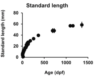
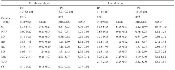
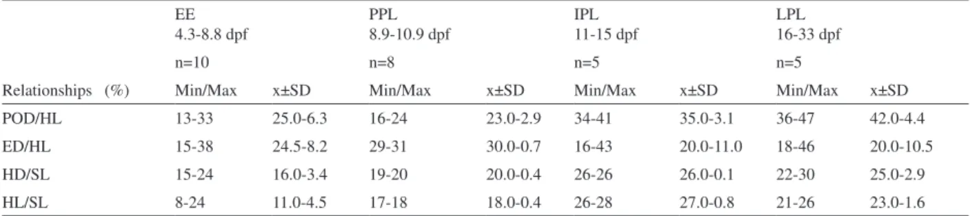
General growth, maximal standard length and morphometrics. Corydoras aff. paleatus growth was represented as standard length ± standard deviation (SL±SD) as a function of age, expressed in dpf (Fig. 1). Evolution of morphometrics variables during development is shown in Tab. I, while relationships of some of these variables with standard length (SL) and head length (HL) along development are presented in Table II. The F-test applied to our morphometrics data did not allow to unequivocally discard the hypothesis which stands that isometric growth is the best model for explaining the important developmental changes in eye diameter, preorbital distance and head length (as referred to standard length) that we found during the evolution from early eleutheroembryo to protopterygiolarvae.
Morphological phases of development. Figures 2-7 illustrate individuals representative of eleutheroembryos, protopterygiolarvae, pterygiolarvae and juveniles,
while figures 8-15 include photographs of eggs (Figs
8-9), eleutheroembryos (Figs 10-11), larvae (Figs 12-14) and young adults (Fig. 15). Figures 16-20 present histological microphotographies at different ages. The eleutheroembryonic phase (5.4-6.4 mm SL) commenced with hatching (around 5.4 days post-fertilization at 23°C), 4.1-6.5 dpf and lasted until 8.8-9.0 dpf, when transition to exogenous feeding occurs. At hatching, eleutheroembryos
exhibited a yolk sac of 0.10-0.29 mm in diameter (Figs 2,
10, 11), and its head was poorly differentiated, with very small development of upper face, eyes and peri-ophthalmic regions (Fig. 2). Pre-orbital distance was very short (Tab. I) and eye pigment absent. Until 10 dpf, only two pairs of barbels were present (Fig. 11, Tab. III). In individuals in which hatching events were directly observed, both oro-pharyngeal membrane and anal region remained
Figs 2-7. Corydoras aff. paleatus (Jenyns, 1842) morphology at defined phases and periods of development: 2, start of eleutheroembyonic phase,
5.4 days post-fertilization (dpf), 3 hours after hatching, 6.1 mm standard length (SL); 3, early protopterygiolarval phase (8.9 dpf, 6.5 mm SL); 4, early pterygiolarval phase (11 dpf, 6.8 mm SL); 5, pterygiolarval phase (17 dpf, 8.8 mm SL); 6, late pterygiolarval phase (27 dpf, 10.1 mm SL); 7, juvenile period (48 dpf, 14 mm SL). Scale bars, 2 mm.
Eleutheroembryo Larval Period
EE 4.3-8.8 dpf
PPL 8.9-10.9 dpf
IPL 11-14 dpf
LPL 15-33 dpf
Variable (mm)
n=10 n=8 n=5 n=5
Min/Max x±SD Min/Max x±SD Min/Max x±SD Min/Max x±SD
SL 5.36-6.40 5.60-0.27 6.57-6.73 6.70-0.07 6.85-6.88 6.88-0.01 8.12-10.94 10.75-1.24
POD 0.09-0.22 0.20-0.04 0.21-0.31 0.28-0.03 0.63-0.81 0.66-0.08 0.66-1.25 1.12-0.26
ED 0.11-0.24 0.21-0.04 0.36-0.38 0.38-0.01 0.30-0.85 0.38-0.24 0.34-0.69 0.50-0.12
HD 0.84-1.41 0.93-0.20 1.28-1.39 1.32-0.04 1.81-1.85 1.81-0.02 2.17-3.37 2.42-0.44
HL 0.48-1.44 0.62-0.29 1.18-1.26 1.21-0.03 1.85-1.96 1.85-0.05 1.83-2.94 2.50-0.43
BD 1.03-1.44 1.24-0.13 1.31-1.43 1.35-0.04 1.85-1.85 1.85-0.04 1.96-2.69 2.25-0.24
PAD 0.20-2.44 0.25-1.07 2.73-3.07 3.03-0.12 3.24-3.27 3.25-0.01 4.09-8.40 7.62-1.52
PDD 2.77-2.85 2.85-0.04 3.42-5.00 4.58-0.59
YS 0.10-0.29 0.15-0.01 0.03-0.08 0.07-0.02
Tab. I. Values (mm) of minimal (Min) and maximal (Max) length, mean (x) and standard deviations (SD) found for morphometrics variables (BD, body depth; ED, eye diameter; HD, head depth; HL, head length; PAD, preanal distance; PDD, predorsal distance; POD, preorbital distance; PPD, prepelvic distance; SL standard length) from groups of Corydorasaff.paleatus (Jenyns, 1842) individuals (n, number of analyzed specimens) at a given developmental phase (EE, eleutheroembryo; IPL, initial pterygiolarval phase; LPL, late pterygiolarval phase; PP, protopterygiolarval phase). Absence of data in some columns is due to incomplete development.
unperforated until 10-15 hours after hatching. Until middle pterygiolarval phase, pectoral appendages were large, oval,
skin folds devoid of mesenchymatic reinforcements (Figs
2-4, 10-12).
Protopterygiolarval phase(6.4-6.6 mm SL) occurred from 8.9 to 10.9-11 dpf until appearance of lepidotrichia within the fin fold (Fig. 4). The continuous finfold constricted near the anus, whereas at the preanal region,
a pair of pelvic fin folds could be noticed (Fig. 4). Between
8.9-9 dpf, mouth and anus were opened and larvae fed mainly from external sources (Tab. IV).
Pterygiolarval phase (6.6-12.7 mm SL, Figs 4-6,
14) went from 11-33 dpf, i.e., until the median finfold was
entirely differentiated and no longer apparent (Figs 4-6). The early pterygiolarvae (11-14 dpf) exhibited external,
Figs 8-15. Corydoras aff.paleatus (Jenyns, 1842) development: 8, eggs with embryos of 4 days post-fertilization (dpf); 9, eggs with embryos at 5
dpf; 10, eleutheroembryo at 5.4 dpf, 6.1 mm SL; 11, same specimen as in Fig. 10 at higher magnification; 12, protopterygiolarvae of 9 dpf, ventral
view; 13, barbel segment from pterygiolarvae of 14 dpf; 14, late pterygiolarvae of 27 dpf; 15, young adult (ar, barbel axial rod; b, barbel; ff, median
finfold; pf, pectoral finfold; pp, barbel pyramidal projection; rf, remaining median finfold). Scale bars, Figs 8, 9, 10, 12, 14, 15: 2 mm; Fig. 11: 400
µm; Fig.13: 200 µm.
EE 4.3-8.8 dpf
PPL 8.9-10.9 dpf
IPL 11-15 dpf
LPL 16-33 dpf
Relationships (%)
n=10 n=8 n=5 n=5
Min/Max x±SD Min/Max x±SD Min/Max x±SD Min/Max x±SD
POD/HL 13-33 25.0-6.3 16-24 23.0-2.9 34-41 35.0-3.1 36-47 42.0-4.4
ED/HL 15-38 24.5-8.2 29-31 30.0-0.7 16-43 20.0-11.0 18-46 20.0-10.5
HD/SL 15-24 16.0-3.4 19-20 20.0-0.4 26-26 26.0-0.1 22-30 25.0-2.9
HL/SL 8-24 11.0-4.5 17-18 18.0-0.4 26-28 27.0-0.8 21-26 23.0-1.6
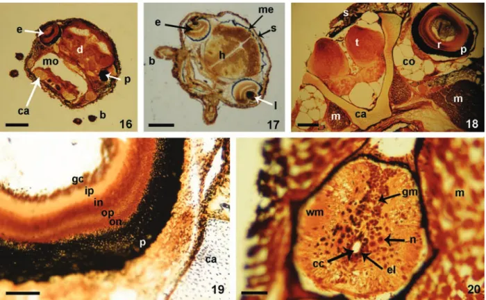
Figs 16-20. Coronal histological sections from decalcified, silver-impregnated specimens of Corydoras aff.paleatus (Jenyns, 1842): 16, protopterygiolarvae of 9.4 days post-fertilization (dpf); 17, pterygiolarvae of 12 dpf; 18, late pterygiolarval phase, 32 dpf; 19, sector of retina and orbit from a late pterygiolarvae of 32 dpf; 20, spinal cord from the same pterygiolarvae as in Fig. 19 (b, barbel; ca, cartilaginous tissue; cc, central canal; co, connective tissue; d, diencephalon; e, eye; el, ependymal lining; gc, ganglion cell layer; gm, grey matter; in, inner nuclear layer; ip, inner plexiform layer; l, lens; m, muscle; me, meninges; mo, mouth; n, neuron; on, outer nuclear layer; op, outer plexiform layer; p, pigmentary layer of
retina; s, skin; t, telencephalon; wm, white matter). Scale bars, Figs 16, 17 and 18: 300 µm; Fig. 19: 10 µm; Fig. 20: 50 µm.
Period or phase Anatomy and stereomicroscopy Histology of sense organs and central nervous system
EE 4.3-8.8 dpf 4.3-6.4 mm
Yolk sac present
Small face Two pairs of barbels Unpigmented small eyes Finfold pectoral appendages
Barbelar mechano-receptors present
Moderatedly differentiated inner ear’s hair cells No layer segregation at retina and brain areas
PP 8.9-10.9 dpf 6.5-6.7 mm
Mouth and anus open
Yolk sac disappearing
Finfold-type pectoral appendages
Eye lens forming
Highly differentiated inner ear hair cells
Rhombencephalon shows maximal differentiation among brain areas Mauthner cells receive several synapses
Barbelar pyramidal projections present
PL 11-33 dpf 6.8-10.7 mm
Highly vascularized external gills Pectoral appendages with radii Three pairs of barbels Dermal scutes developing
Cartilaginous cranial bones Differentiated muscular tissue Developed quimioreceptors
Increased cytoarchitectural complexity of cortical brain areas Highly differentiated giant Mauthner cells receive hundreds of giant synaptic terminals (32 dpf)
Cortical cytoarchitectonics similar to adults (32 dpf) Differentiated stellate spinal neurons (32 dpf) All retinal layers recognizable (32 dpf)
JP 34-60 dpf 10.8.-18.0 mm
Temporary fin folds fully replaced
by definitive fins Fully developed lateral line systemBrain cytoarchitectonics similar to adults
completely reabsorbed yolk sac (Tabs I, III). At 15-16 dpf, the first radii appear at pectoral appendages, marking the commencement of the final step of pterygiolarval phase (Figs 5-7). At 27 dpf, the finfold looked thinner (Figs 6, 14) whereas adipose and anal fin profiles were distinguishable inside it. At 33 dpf, all fins were fully detached, except
the adipose one, and histological analysis demonstrated
ossification of the axial skeleton, whereas main parts of
all cephalic bones are still constituted by cartilage (Figs 18, 19).
At juvenile period, from 34 to 60 dpf,all fins were fully differentiated and most temporary organs had been
replaced by definite ones (Fig. 7). This may occur between 43 and 50 dpf. The first bony dermal plates or scutes, typical
for Callichthyidae, started developing in connection with development of lateral line system channels.
Sensory areas and brain development. At the eleutherembryonic phase, eyes were small (Tab. I) and devoid of pigment (Figs 2, 10, 11). Future retinas and most brain regions remain thin and undifferentiated (Tab.
III). In contrast, at seven dpf, a remarkable increase
in differentiation of barbel (Fig. 13) and inner ear mechanoreceptors occurs, as in utricular and saccular maculi (Tab. III).
During the protopterygiolarval phase eyes diameter duplicate, growing from 0.21±0.04 mm in the eleutheroembryo to 0.38±24 mm in the early pterygiolarvae (see Tab I). Simultaneously, histological analysis locate eye pigments at the future pigmentary layer (Figs 2, 16) and demonstrate that diencephalic and retinal cytoarchitectonics continued exhibiting scarce development of synaptic neuropil layers (Fig. 17, Tab. III). Mauthner cells were easily recognizable and received synaptic contacts, as shown by our histological sections of silver impregnation methods.
At the pterygiolarvalphase (11-33 dpf), retina and
all cortical brain areas underwent remarkable progress
in morphogenesis, as denoted by nuclear and cortical cytoarchitectural differentiation of the central nervous system (Figs 18-20) (Tab. III). At the end of this phase, all retinal layers were recognizable in histological sections (Fig. 19). Brain morphogenesis underwent considerable progress with respect to previous phase, while rhombencephalic structures continued to lead brain differentiation (Tab. III). Giant Mauthner cells, for instance, showed structural differentiation close to that found in the adult brain and received numerous afferent “giant” terminals on their surface. At the level of the spinal cord, silver impregnation methods evidenced the relative advancement in development of the grey matter in comparison to former phases. This
included the presence of polygonal neuronal perikarya in
the spinal grey matter (Fig. 20) some of them receiving
numerous synaptic profile contacts, which where absent
at the protopterygiolarvae (Tab. III).
Development of individual behavior.The appearance of sensory and locomotor capabilities and behaviors at different times upon fertilization is schematically shown in
Table II. The behavioral repertory of the just-hatched(5-6
dpf) eleutheroembryo is extremely limited. During the first
24 h post-hatching, they were usually still, supported by
barbels and pectoral finfolds on the aquarium floor (see Tab.
II). They rested most of the time hidden under objects or leaves, without interacting with its siblings. When moving,
they did it alone. At “rest”, the long and soft, wing-like pectoral appendages, moved alternatively with flag-like
undulatory movement. Sometimes they exhibit “burst and coast swimming” behavior but, in addition, occasionally
perform quicker, jerky, short (few centimeters) horizontal swimming, which during the first 20 or 30 post-hatching h end frequently shocking obstacles (see Tab. II).
At the beginning of the pterygiolarval phase (11
dpf) larvae already demonstrate a perfect balance, quick
swimming capacity and ability to stop suddenly in front of obstacles (Tab. IV). At 12 dpf, larvae responded readily to visual, auditive and hydromechanical stimuli, they were able of rapid negative reotropism, both in antero-posterior and lateral directions to oppose water movement or vibration. They executed spontaneous - in the absence of detectable external stimuli - horizontal swimmings, as well as slanting, ascending swimming that reach the water surface. Upon some of these ascending swimming some
larvae executed their first recorded aeration behavior. Due
to space reasons, further evolution of Corydoras paleatus
individual behavior is synthesized in Table IV.
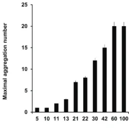
Evolution of social behavior: aggregation, shoaling and schooling. From hatching to 10 dpf, no larval aggregations were seen, i.e., “maximal aggregation number” (MAN) equals 1 (Fig. 21). First social interactions appear at 11-12 dpf, i.e., at the beginning of pterygiolarval phase; initially, larvae appear still in the open space, frequently in pairs, at 1-body-length distance (Tab. IV). One day later
Fig. 21. Maximal aggregation size (MAS) as a function of fish age of
Corydoras aff.paleatus (Jenyns, 1842). At different ages, from 5 to 100 days post-fertilization (dpf), the number of individuals present in
each detected fish aggregation was computed. The maximal number of
Phase/Period Age (dpf) Behavior Eleutherembryonic phase
4.3 to 6.4 mm SL
4-7 Mostly immobile, hidden, alone still
4-7 Photophobia
4-7 Flag-like undulation of pectoral fin folds at rest
5 Anguilliform swimming
5 Freezing upon new stimuli
6 Occasional burst and coast swimming
Protopterygiolarval phase 6.5-6.7 mm SL
8.9 External feeding begins
9 Exploratory behavior
9 Visually-triggered escape reflex
Pterygiolarval Phase 6.8-10.7 mm SL
11 Pairs, still
11-12 Perfect balance
12 Aeration behavior
13 Free swimming in horizontal direction 13 Few seconds of physical contact 13 Patrolling and foraging in pairs 18 Exploration and foraging on plants 18 Resting on elevated plant branches
19 Exploratory swimming in small shoaling groups 20-33 Switch to carangiform swimming
25 Vertical swimming to surface and aeration 26 Foraging in small shoaling groups
Juvenile period 10.8-18.0 mm SL
34 Shoaling still
34 Foraging big shoaling groups
34 Schooling
35 Exploratory swimming along the walls up to the surface
Tab. IV. Age-dependent emergence of individual of Corydoras aff.paleatus (Jenyns, 1842) and social behavior during development. Individual movements and behaviors as well as spatial patterns of aggregations are described as a function of age (dpf, days post-fertilization) and growth (SL, standard length).
(13 dpf), aggregations of three or four individuals were
seen for the first time, and next days, group size gradually
augment, as shown in Fig. 21. At 21 dpf, pterygiolarvae form aggregates exhibiting MAN of seven individuals,
and the first shoals appear. The most frequent organization
pattern is a loose still shoal (possible visual contact). From 22 to 100 dpf, they are frequently arranged as shoal still (Tab. IV) and the number of individuals integrating a shoal
grows as age increases, from the first to the third month of
life (see Fig. 21). Bi-dimensional (plane) foraging shoals of 6-10 individuals were seen after 40 dpf, and tri-dimensional foraging schools of up to 12 individuals after 60 dpf.
DISCUSSION
A first review of our Corydoras aff. paleatus growth curves shows that our samples reach mean SL sizes higher than those formerly published (Froese & Pauly, 2013)
and this may suggest that our work included the largest
specimens ever reported. This may be due to the fact that our study follows lifetimes of specimens acquired in 2011
as well as their first generation of descendants, born at our
laboratory, for almost three years. In our growth curves, SL seems to approach (asymptotically) to an apparent L
Infinity (L¥) value close to 71 mm, which would represent
the maximum length of an infinitely old fish of the given stock (Froese & Binohlan, 2000). Mean values included
in our tables yolk sac diameters as a function of SL. We have found some very remarkable developmental
changes in relationships between body distances, as is the case of growth in eleutheroembryo’s eye diameter or preorbital distance with respect to head length, which sometimes duplicate their size in a short time period, we were unable to demonstrate an allometric growth, a change which has been related to feeding adaptations that might
mark a switch in developmental stage, as suggested by
tagutiet al. (2009) for Pyrrhulina australis Eigenmann & Kennedy, 1903 (Characiformes, Lebiasinidae). Another
remarkable feature of larval period is the persistence of
some embryonic organs which are later replaced by different definitive organs of similar function (huysentruyt
et al., 2009). In Corydoras aff. paleatus, this sort of metamorphosis concentrates at the early, initial step of the pterygiolarval phase, when embryonic organs start to be not necessary anymore. This is the case of pectoral
“wing-like” appendages, which are completely different from the definitive strong, ray-armed pectoral fins. The former is not supported by fin rays and can be viewed as a large skin fold. It has been suggested for Corydoras aeneus
to help reducing the lateral movement of the head during swimming movements (thorstenet al., 2004).
Comparative aspects. The stages in which we divided Corydoras aff. paleatus post-hatching ontogeny were comparable to those found in the related species
Corydoras aeneus (Gill, 1858) by huysentruytet al. (2009), although the former exhibits a slightly more protracted development, at least at temperatures used
by us. This makes C. aeneus and C. aff. paleatus easily comparable to each other, adding heuristic value to our data. Post-hatching development of Siluriformes has deserved many studies which illustrate a wide variety of developmental adaptation strategies, from species without true larval phases (limaet al., 2012) to other exhibiting many post-hatching phases, similar to the species of our study, as shown in the cascadu, Hoplosternum littorale
(Hanckock, 1828) by ramnarineet al. (1994). Taking into account exclusively the Pimelodidae family, a great variety of developmental stages, phase names and incubation times can be found, including hatching times as short as 14-16 h as reported by Buzolloet al. (2011) and nogueiraet
al. (2012), 30 h (Pereiraet al., 2006), 50 h (honJiet al., 2012) and extremely prolonged incubations, as found in
some marine catfishes (limaet al., 2012). It is generally accepted that in most teleosts, larger eggs will produce,
as a rule, larger larvae with larger yolk sacs and therefore,
more developed organisms at hatching (sancheset al., 1999). In Characiformes, species having small oocytes present short incubation periods and consequently, very limited development at hatching time (oliveiraet al., 2012). For that reason, it is very interesting that Corydoras aff. paleatus, although produce small (not guarded) eggs,
and eleutheroembryos looking at hatching structurally and functionally very immature, quickly starts external feeding and yolk sac absorption.
Development of sensory systems. Our data show that during the first days after hatching, while eyes are undifferentiated, facial isolated mechanoreceptive neuromasts, barbelar and inner ear mechanoreceptors, plus facial and barbelar chemoreceptors are the systems developing at higher pace. Photophobia, isolation and hiding from predation are important characteristics of this phase, which impose that energy accumulated in the
yolk sac is almost not employed in locomotion, but mainly
directed to morphogenetic and differentiation processes, very intense during these early life stages (van sniket
al., 1997). Rapid morphogenesis and differentiation are
necessary for developing the sensory, motor and reflex
circuits essential for basic survival in the open aquatic
environment where larvae will seek for external nutrients, as to assure that growth will be optimized to increase fitness
(Fukuhara, 1992).
Advanced protopterygiolarvae has already well developed inner ear mechanoreceptors and a set of mechanoreceptive free neuromasts, necessary not only for balance and swimming, but also for allowing adequate
reflex defensive responses against predators. In connection
with this, larvae show well developed Mauthner cells and circuits, which are necessary for the C-start or Mauthner
reflex, fundamental for larval survival (kalueFFet al., 2013), whereas rhomboencephalon and spinal cord lead neural differentiation. The visual system accelerates its differentiation during the proptoterygiolarval phase, but is only at the pterygiolarval phase when all retinal layers become recognizable. Morphological development of their respective systems suggest that this is a step of increase
in the efficacy of respiratory, digestive and hydrodynamic
structures. At the pterygiolarvae, priorities change towards osteomuscular development.
The rhombencephalon was the brain region showing major advances in differentiation, in line to balance and swimming control.
Behavioral changes. Swimming mode is also dependent on developmental age. It switches from the anguilliform to the carangiform mode. Carangiform and vertical swimming appear to be relevant for gaining higher
swimming efficacy, necessary for the high level of social
interaction typical of this species. Ascending swimming
is necessary for aeration behavior, recorded for the first
time it was similar to the behavior described in the adult by Burgess (1989).
As our data show, social behavior, expressed as maximal aggregation number and group cohesion also increase with age. At the end of the protopterygiolarval phase, simple resting and loose foraging aggregations are seen. Resting and foraging shoals grow in complexity and number of participants during pterygiolarval phase, but particularly during juvenile period. Social behavior is a complex phenomenon whose biological mechanisms and development are not well understood in vertebrates, but there is in general agreement that shoaling and schooling behavior
are beneficial for the participating individuals (Buske &
gerlai, 2011). Fishes benefit from shoal membership
through increased hydrodynamic efficiency, and augmented
security and foraging success (Pitcher & Parrish, 1993). Some studies have devoted attention to development of behavior and sensory organs in Siluriformes (KhoDaet
al., 1995; Pruzsinszky & laDich,1998; maguitet al., 2010; mukaiet al.,2010; rahmahet al., 2011) but we
believe this is the first study demonstrating developmental
changes in aggregation size and in shoaling behavior in
Corydoras aff. paleatus.
Concluding remarks and perspectives.As a whole, our study suggests that Corydoras aff. paleatus etho-developmental evolution exhibits a strong correlation with phases of morphological development that we have described. Overall, we believe that our study may represent an useful departure point for future integrative analysis of correlative development. However, much more detailed studies on ontogeny of C. aff. paleatus neuroanatomy and behavior will be necessary before establishing a more
definitive and heuristic correlation between structure,
function and behavior of this species. Finally, since
development, a projection of the results of this study onto the whole species should be considered with caution.
Acknowledgements. The authors are grateful to PEDECIBA Postgraduate Program (Biology) for supporting M.Sc. studies of GdP and
for continued financial support to DRI’s laboratory. We are indebted to
Luis Pérez Córdova for his invaluable help with photography of living
specimens. We wish to thank Mario Lalinde and Joaquín Carrique for
help with illustrations, and Andrea Etchartea and Alicia Soza for help with specimen measurement and photography.
REFERENCES
Baigún, c.; lóPez, g.; Dománico, a.; Ferriz, r.; sverliJ, s. & DelFino schenke, r. 2002. Presencia de Corydoras paleatus (Jenyns, 1842), una nueva especie brasílica en el Norte de la Patagonia (Río Limay) y consideraciones ecológicas relacionadas con su distribución. Ecología Austral 12:41-48.
Balon, e. k. 1975. Terminology of intervals in fish development. Journal of the Fisheries Research Board of Canada 32:1663-1670. _____. 1999. Alternative ways to become a juvenile or a definitive
phenotype (and on some persisting linguistic offenses. Environmental Biology of Fishes 56:17-38.
Belanger, s. e.; Balon, e. k. & rawlings, J. m. 2010. Saltatory ontogeny
of fishes and sensitive early life stages for ecotoxicology tests.
Aquatic Toxicology 97:88-95.
Britto, m. R. 2003. Phylogeny of the subfamily Corydoradinae
Hoedeman, 1952 (Siluriformes: Callichthyidae), with a definition
of its genera. Proceedings of the Academy of Natural Sciences of Philadelphia 153:119-154.
Burgess, w. e. 1989. Family Callichthyidae (Armored Catfish). In: reD, r. ed. An Atlas of Freshwater and Marine Catfishes. A
Preliminary Survey of the Siluriformes.Neptune City, T.F.H. Publications. p.326-367.
Buske, c. & gerlai, r. 2011. Shoaling develops with age in zebrafish (Danio rerio). Progress in Neuro-Psychopharmacology & Biological Psychiatry 35:1409-1415.
Buzollo, h.; veríssimo-silveira, r.; oliveira-almeiDa, i. r.; alexanDre, J. s.; okuDa, h. t. & ninhaus-silveira, a. 2011. Structural analysis of the Pimelodus maculatus (Lacépède, 1803) embryogenesis (Siluriformes: Pimelodidae). Neotropical Ichthyology 9(3):601-616. cazenave, J.; Bistoni, m. a.; Pesce, s. F. & wunDerlin, D. A. 2006.
Differential detoxification and antioxidant response in diverse organs
of Corydoras paleatus experimentally exposed to microcystin-RR. Aquatic Toxicology 76:1-12.
De castilhos ghisi, n. & cestari, m. m. 2013. Genotoxic effects of
the herbicide Roundup(®) in the fish Corydoras paleatus (Jenyns 1842) after short-term, environmentally low concentration exposure.
Environmental Monitoring and Assessment 185(4):3201-3207. Durán, i.; marí-BeFFa, m.; santamaría, J. a.; Becerra, J. & santos -ruiz, l. 2011. Actinotrichia collagens and their role in fin formation.
Developmental Biology 354:160-172.
Ferraris Jr., C. J. 2007. Checklist of catfishes, recent and fossil (Osteichthyes: Siluriformes), and catalogue of siluriform primary types. Zootaxa 1418:1-300.
Froese, r. & Binohlan, n. 2000. Empirical relationships to estimate
asymptotic length, length at first maturity and length sat maximum yield per recruit in fishes, with a simple method to evaluate length
frequency data. Journal of Fish Biology56:758-773.
Froese, r. & Pauly, D. 2013. Corydoras paleatus (Jenyns 1842) Peppered Corydoras. FishBase. World Wide Web electronic publication.
Available at: <http://www.fishbase.org/summary/Corydoras-paleatus.
html>.Accessed on:27 November 2013.
Fukuhara, o. 1992. Study on the development of functional morphology and behaviour of the larvae of eight commercially valuable teleost
fishes. Contributions to the Fisheries Researches in Japan Sea Block 25:1-122.
Fuller, i. a. 2001. Breeding Corydoradine Catfishes. Kidderminster, Ian Fuller Enterprises. 248p.
guiloski, i. c.; rossi, s. c.; Da silva, c. a. &De assis, h. c. 2013.
Insecticides biomarker responses on a freshwater fish Corydoras paleatus (Pisces: Callichthyidae). Journal of Environmental Science and Health, Part B 48(4):272-277.
honJi, r. m.; tolussi, c. e.; mello, P. h.; canePPele, D. & moreira, r. g. 2012. Embryonic development and larval stages of Steindachneridion parahybae (Siluriformes: Pimelodidae): implications for the conservation and rearing of this endangered Neotropical species
Neotropical Ichthyology10(2):313-327.
huysentruyt, F.; moerkerke, B.; Devaere, s. & aDrieaens, D. 2009.
Early development and allometric growth in the armored catfish
Corydoras aeneus (Gill,1958). Hydrobiology 627:45-54.
kalueFF, a. v.; geBharDt, m.; stewart, a. M.; cachat, J. m.; Brimmer, m.; chawla, J. s.; craDDock, c.; kyzar, e. J.; roth, a.; lanDsman, s.; gaikwaD, s.; roBinson, k.; BaatruP, e.; tierney, k.; shamchuk, a.; norton, w.; miller, n.; nicolson, t.; BrauBach, o.; gilman, c. P.; Pittman, J.; rosemBerg, D. B.; gerlai, r.; echevarria, D.; lamB, e.; neuhauss, s. c.; weng, w.; Bally-cuiF, l. & schneiDer,
H. 2013. Towards a comprehensive catalog of zebrafish beahavior
1.0 and beyond. Zebrafish 10(1):70-86.
kaPoor, B. g. & khanna, B. 2004. Ichthyology Handbook. New Delhi, Springer. 1072p.
khoDa, m.; tanimura, m.; kikue-nakamura, m. & yamagishi, S. 1995.
Sperm drinking by female catfishes: a novel mode of insemination. Environmental Biology of Fishes 43:1-6.
kovács, v.; coPP, g. h. & Francis, m. P. 1999. Morphometry of the stone loach Barbatula barbatula: do mensural characters reflect
the species’ life history thresholds? Environmental Biology of Fishes 56:105-115.
kramer, D. l. & mcclure, m. 1980. Aerial respiration in the catfish,
Corydoras aeneus (Callychhthydae). Canadian Journal of Zoology 58:1984-1991.
lima, a. r. a.; Barletta, m. ; Dantas, D. v.; Possato, F. e.; ramos, J. a. a. & costa, m. F. 2012. Early development and allometric shifts
during the ontogeny of a marine catfish (Cathorops spixii-Ariidae). Journal of Applied Ichthyology 28(2):217-225.
maguit, Q.; olivier, D.; vanDewalle, n. & vanDewalle, P. 2010. Ontogeny of swimming movements in bronze corydoras (Corydoras aeneus). Canadian Journal of Zoology 88:378-389.
mukai, y.; tuzan, a. D.; shaleh, s. r. m. & manJaJi-matsumoto, B. m. 2010. Development of sensory organs and changes of behavior
in the larvae of the sutchi catfish, Pangasianodon hypopthalmus.
Fisheries Science 76:921-930.
nogueira, l. B.; azeveDo, P. g.; canelhas, m. r.; BeDore, a. g.; loPes, J. m. & goDinho, h. P. 2012. Induced spawning and early ontogeny
in hatchery-reared catfish Zungaro jahu (Siluriformes: Pimelodidae).
Neotropical Ichthyology 10(1):89-98.
novomeská, a.; katina, s.; coPP, g. h.; PeDicillo, g.; lorenzoni, m.; PomPei, l.; coucherousset, J. & kovács, v. 2013. Morphological
variability of black bullhead Ameiurus melas in four non-native European populations. Journal of Fish Biology 82:1103-1118 oliveira, F. g.; Bialetzi, a.; gomes, l. c.; santin, m. & taguti, t. L.
2012. Desenvolvimento larval de Brycon hilarii (Characiformes, Characidae). Iheringia, Série Zoologia 102(1):62-70.
Pereira, c. r.; Barcellos, l. J. g.; kreutz, l. c.; QueveDo, r. m.; ritter, F. & silva, l. B. 2006. Embryonic and larval development of Jundiá (Rhambdia quelen, Quoy & Gaimard, 1842, Pisces, Teleostei) a South
American catfish. Brazilian Journal of Biology 66(4):1057-1063. Pesce, s. e.; cazenave, J.; monFerran, m. v.; FreDe, s. & wunDerlin, D.
a. 2008. Integrated survey on toxic effects of lindane on neotropical
fish: Corydoras paleatus and Jenynsia multidentata. Environmental Pollution 156:775-783.
Pistone, g.; eguren, g. & roDriguez-ithurralDe, D. 2012. Inhibition, recovery and field responses of Astyanax fasciatus (Cuvier, 1819) brain cholinesterases upon exposure to azinphos-methyl. Ecotoxicology and Environmental Contamination 7(2):93-102. Pitcher, t. J. 1983. Heuristic definitions of shoaling behavior. Animal
Pitcher, t. J. & Parish, k. J. 1993. Functions of schoaling behavior in teleosts. In:Pitcher, J. J. ed. Behavior of teleost fishes. London, Chapman & Hall. p.363-439.
Pruzsinszky, i. & laDich, F. 1998. Sound production and reproductive
behavior of the armoured catfish Corydoras paleatus ( Callichthyidae).
Environmental Biology of Fishes 53:183-191.
rahmah, s.; yamamoto, s.; nakagawa, y.; kato, k.; senoo, s. & murata, o. 2011. Development of morphology, sensory organs and behavior
of larval bagrid catfish Mystus nemurus.Aquaculture Science
59(3):443-450.
ramnarine, i. w. 1994. Larval culture, development and growth of the cascade, Hoplosternum littorale (Hancock 1828; Callichthyidae).
Aquaculture 126(9):291-298.
ramóny caJal, s. &De castro, F.1972. Elementos de Técnica Micrográfica del Sistema Nervioso.Barcelona, Salvat. 283p. reis, r. e. 2003. Family Callichthyidae (armored catfishes). In: reis, r.
e.; kullanDer, s. o. & Ferraris Jr., c. J. eds. Check List of the Freshwater Fishes of South and Central America.Porto Alegre, Edipucrs. p.291-309.
roDriguez-ithurralDe, D.; maruri, a. & roDríguez, x. 1998. Motor neurone acetylcholinesterase release precedes neurotoxicity caused by systemic administration of excitatory amino acids and strychnine.
Journal of the Neurological Sciences 160(S1):S80-S86. sanches, P. v.; nakatani, k. & Bialetzki, a. 1999. Morphological
description of the developmental stages of Parauchenipterus galeatus
(Linnaeus, 1766) (Siluriformes, Auchenipteridae) on the floodplain
of the upper Paraná River. Revista Brasileira de Biologia 59(3):429-438.
sarikaya, r.; selvi, m. & erkoç, F. 2004. Investigation of acute toxicity of fenitrothion on peppered corydoras (Corydoras paleatus) (Jenyns, 1842). Chemosphere 56:697-700.
shimaBukuro-Dias, C. K.; oliveira, c.; reis, r. e. & Foresti, F. 2004.
Molecular phylogeny of the armored catfish family Callichthyidae
(Ostariophysi, Siluriformes). Molecular Phylogenetics and Evolution 32:153-163.
sokal, r. r. & rohlF, F. J. 1981. Biometry. The principles and practice of statistics in biological research. New York, W. H. Freenan and Company. 859p.
taguti, t. l.; kiPPer, D.; Bialetzki, a.; sanches, P. v.; cavicchioli- makrakis, m.; Baumgartner, g.; FernanDes, r.; FernanDes, g. & early, r. 2009. Desenvolvimento inicial de Pyrrhulina australis
Eigenmann & Kennedy, 1903 (Characiformes, Lebiasinidae). Biota
Neotropica 9(4):59-65.
tencatt, l. F. c..; Britto, m. r. & Pavanelli, c. s. 2014. A new species of Corydoras Lacépède, 1803 (Siluriformes: Callichthyidae) from the upper rio Paraná basin, Brazil. Neotropical Ichthyology 12(1):89-96. thorsten, D. h.; cassiDy, J. J. & hale, m. e. 2004. Swimming of larval
zebrafish: fin-axis coordination and implications for function and
neural control. Journal of Experimental Biology 207(24):4175-4183.
van snik, g. m. J.; vanDen Boogaart, J. g. m. & osse, J. w. M. 1997.
Larval growth patterns in two ostariophysian fish (Cyprinus carpio
and Clarias gariepinus) with attention to the finfold. Journal of Fish Biology 50:1339-1352.