Original Article
Long-term evaluation of endothelial function in Kawasaki
disease patients
Fa´tima F. Pinto,1 Se´rgio Laranjo,1 Filipa Parame´s,1 Isabel Freitas,1 Miguel Mota-Carmo2 1
Servic¸o de Cardiologia Pedia´trica; 2Servic¸o de Cardiologia, Hospital de Santa Marta, Centro Hospitalar de Lisboa Central, EPE, Lisboa, Portugal
Abstract Background: Kawasaki disease is an acute systemic vasculitis. Cardiac complications are frequent and include endothelial dysfunction in patients with coronary anomalies. So far, the presence of endothelial dysfunction in patients with no coronary lesions has not been demonstrated. Peripheral arterial tonometry (Endo-PAT) measures the microvascular function in response to local ischaemia and has been validated in adult population, but its use in children is scarce. Aim: To evaluate endothelial dysfunction in children as a long-term complication after Kawasaki disease using Endo-PAT. Methods: We evaluated two groups of subjects: (1) Kawasaki disease patients over 11 years of age, diagnosed for .5 years, with no coronary lesions, or any other risk factors for cardiovascular disease; (2) control group of individuals without cardiovascular risk factors. Patients and controls were clinically accessed. Endo-PAT was performed to determine reactive hyperaemia index and augmentation index. Results: A total of 35 individuals (21 males, age 21 6 6 years) were evaluated (group 1: 19; controls: 16). Kawasaki disease patients presented significant lower reactive hyperaemia index (1.68 6 0.49 versus 2.31 6 0.53; p 5 0.001). Augmentation index was similar in both groups (210 6 7 versus 211 6 5; p . 0.005). Most patients with Kawasaki disease disclosed endothelial dysfunction (68%) compared with only 12% in controls. Conclusions: Endo-PAT is feasible and reproducible in the child population. Endothelial dysfunction is a frequent long-term complication in patients after Kawasaki disease with normal appearing coronary arteries. However, these results need validation in a larger population.
Keywords: Kawasaki disease; endothelial function; Endo-PAT
Received: 12 December 2011; Accepted: 10 August 2012; First published online: 8 October 2012
K
AWASAKI DISEASE IS THE LEADING CAUSE OFacquired heart disease in the developed world.1,2 It is an acute, systemic vasculitis of uncertain aetiology, occurring predominantly in infants and children younger than 5 years of age.3 Coronary artery aneurysms or ectasia develop in 15–25% of Kawasaki disease patients, during its acute stage, and may lead to myocardial infarction, sudden death, or chronic coronary insufficiency.4 In addition to causing acute coronary artery aneurysm and stenosis, Kawasaki disease has also been shown to have deleterious effects on coronary artery
function years after the acute presentation. Another issue that raises concerns is vascular abnormalities other than those involving coronary arteries. Several reports indicated abnormal changes in the systemic vascular physiology in Kawasaki disease patients with persistent or regressed coronary artery lesions, includ-ing increased wall thickness and decreased distensibi-lity of the carotid arteries,5,6 and increased stiffness of central and peripheral arteries.7 Histological and pathological studies have identified aneurysms at other vascular tree sites, involving mainly small- to medium-sized vessels, including the femoral, iliac, renal, axillary, and brachial arteries.8
Although persistent coronary artery aneurysm only occurs in a minority of children with acute Kawasaki disease,9 considerable attention has been focused in the past few years on the long-term Correspondence to: Fa´tima F. Pinto, MD, Servic¸o de Cardiologia Pedia´trica,
Hospital de Santa Marta, Centro Hospitalar de Lisboa Central, EPE, Rua de Santa Marta, 1169-024 Lisboa, Portugal. Tel: 1213594332; Fax: 1213594034; E-mail: [email protected]
outcome of this subgroup, mainly because of concerns regarding increased risk for morbidity and mortality, late after resolution of acute Kawasaki disease. Arterial function has been assessed in multiple studies in which the authors evaluated myocardial and coronary flow reserve with angiography,4,10–12 invasive flow measurements,13–15 positron emission tomography,16,17 and transthoracic Doppler ultra-sound of the coronary arteries.18 Abnormalities have been observed in both endothelial-mediated and endothelial-independent coronary artery vasodilation. In spite of the main focus on Kawasaki disease patients with coronary artery involvement, a small number of studies have also shown that even in patients with a history of Kawasaki disease without coronary artery dilation or ischaemia coronary flow reserve is impaired,10,16–18 although conflicting results showed no difference from normal control subjects.4,11,12 These results, and the decreased number of new patients with Kawasaki disease with coronary artery aneurysm, revived the research interest in diagnostic assays for the assessment of endothelium dysfunction in children with Kawasaki disease with-out coronary artery aneurysm.15,19
In the healthy state, the endothelium produces a number of factors, including nitric oxide, that are essential for maintaining vascular homoeostasis. Several risk factors damage endothelial cells, lowering nitric oxide bioavailability. An impaired endothelial function has been associated with an increased risk of cardiovascular events. In spite of its relevance, endothelial function assessment has not yet been incorporated into routine risk stratification of patients with Kawasaki disease, mainly because of technical limitations.20–22 The use of novel digital tonometry device to measure endothelial function offers the possibility of an easily performed, rapid assessment of microvascular function, overcoming these limitations. The Endo-PAT device (Itamar Medical, Caesarea, Israel) is a commercially available system, consisting of a fingertip plethysmograph capable of sensing volume changes in the digit with each arterial pulsation. It has been validated in adult population to assess microvascular function,23but its use among children is scarce.24,25 Therefore, our aim was to evaluate for endothelial dysfunction in children and young adults as a long-term complication after Kawasaki disease using the Endo-PAT, comparing the results with matched normal controls.
Methods Study population
The patients, selected from our outpatient clinic, were all aged .11 years and have had a diagnosis of
acute Kawasaki disease for .5 years previously, diagnosed by standard criteria.2,26 Patients were excluded if they have had coronary artery lesions during the acute Kawasaki disease stage, or if they had potentially confounding risk factors for endo-thelial damage, such as smoking, elevated LDL cholesterol, hypertension, diabetes mellitus, or a family history of premature cardiovascular disease, or if they were taking vasoactive medication. No other selection criteria were applied. In all, 19 patients (12 males) aged 15–27 years (mean age, 21 6 6 years) were studied. Management of the acute Kawasaki disease was determined by the patient’s own physicians. In addition, 16 healthy age- and sex-matched control subjects (9 males, mean age, 21 6 6 years) were studied. These subjects were normal individuals without risk factors for endothelial damage as listed above and were recruited from family and friends of hospital staff. None were taking medications or had a history of significant illness.
Study protocol
A history was obtained from all subjects, and a physical examination was performed, including supine blood pressure. All subjects had an electrocardiogram and an echocardiogram performed. The medical records of the Kawasaki disease patients were reviewed for details of their acute illness. Endothelial function was assessed through peripheral arterial tonometry, as described below; the peripheral arterial tonometry study was repeated 1 month later, in order to assess the reproducibility of the data.
Peripheral arterial tonometry
Endothelial function tests were performed in a dedicated laboratory, in a quiet environment, with controlled temperature and humidity, during the morning period, between 9:00 and 12:00 am, at least two hours after a light breakfast without ingestion of caffeine or other xanthines. Pulse wave amplitude was assessed before and during reactive hyperaemia by peripheral arterial tonometry (Endo-PAT 2000, Itamar Medical Limited, Israel), as previously described in detail (Fig 1).21,23 In brief, baseline pulse wave amplitude data were collected using plethysmographic finger cuffs, placed on the index finger of both hands, for a period of 5 minutes. An ischaemic stimulus was induced by cuff occlusion, that is, inflation of a brachial cuff to a supra-systolic pressure of 200 mmHg for 5 minutes, and the pulse wave amplitude – reactive hyperaemia index was calculated as the ratio of the average pulse wave amplitude value, over a 1-minute epoch starting after cuff release, to the pre-occlusion baseline pulse wave
amplitude value. The augmentation index, a measure of arterial stiffness, was also computed, as the difference from the two systolic peaks, derived from the baseline period’s mean peripheral arterial tono-metry waveform ensemble. A reactive hyperaemia index cut-off value of 1.67 was used for diagnosing endothelial dysfunction, as previously defined by Bonetti et al.27
Statistical analysis
Continuous variables are expressed as mean 6 standard deviation, unless otherwise specified. Categorical
variables are given as frequencies and percentage of patients. Normality of the distributions of the continuous variables was analysed with the Kolmogorov–Smirnov test. Student’s t-test was used to assess all paired data in the same group. Comparisons between groups were made using the Mann–Whitney U-test. The chi-square test was used for categorical variables. A value of p , 0.05 was considered statistically significant. Data were analysed using SPSS software version 18 (IBM Corporation, United States of America).
Results
Table 1 summarises the patient characteristics of each group. Groups were comparable in terms of gender distribution (women: 37% versus 43%), age (21 6 6 years), and body surface mass (22.8). All subjects were well, with normal clinical and electrocardio-gram evaluation. The Kawasaki disease patients had normal left ventricular function and normal coronary arteries on echocardiography. The endothelium function studies disclosed endothelium dysfunction in the majority (68%) of Kawasaki disease patients, when compared with a significantly lower percentage of control subjects (12%). There were significant group differences in pulse wave amplitude-derived reactive hyperaemia index, with Kawasaki disease patients having a significantly lower reactive hyperaemia index when compared with the control group (1.67 6 0.49 versus 2.31 6 0.53; p 5 0.001, Fig 2). Pulse wave amplitude-derived augmentation index was similar in both groups (210 6 7 versus 211 6 5; p . 0.005). The reactive hyperaemia index values were reproducible; no statistically significant changes were found at the 1-month Endo-PAT retest (Table 2; p 5 0.7856).
Discussion
This study has shown that endothelial dysfunction is a long-term complication in patients after Kawasaki disease, even in patients without detect-able coronary involvement during the acute illness. This is the first study, to our knowledge, to show
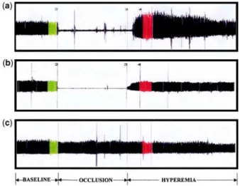
Figure 1.
(a and b) Pulse wave amplitude recordings with peripheral arterial tonometry at baseline (green) during supra-systolic cuff occlusion and during reactive hyperaemia (red) in the ischaemic arm are demonstrated. (c) Peripheral arterial tonometry recordings from the contralateral finger not undergoing reactive hyperaemia testing are shown. (a) Healthy individual with no cardiovascular risk factors showing a steady-state peripheral arterial tonometry signal (baseline), complete disappearance of the signal during cuff inflation (occlusion), followed by an increased peripheral arterial tonometry signal during recovery (hyperaemia). (b) Individual with coronary artery disease showing a blunted finger peripheral arterial tonometry response during reactive hyperaemia. (c) Peripheral arterial tonometry recording from the contralateral finger in the same patient (reproduced with permission from Kuvin et al. Assessment of peripheral vascular endothelial function with finger arterial pulse wave amplitude, Am Heart J, 2003; 146: 168–174).
Table 1. Demographic characteristics of the subjects.
Kawasaki disease patients Control group p-value
Number of patients (n) 19 16
Male gender (n) 12 9
Age (years) 21 6 6 21 6 6 0.81
Weight (Kg) 61 6 15 62 6 14 0.94
Height (cm) 166 6 9 168 6 10 0.4
microvascular endothelial dysfunction in Kawasaki disease patients, as all the previous studies assessed the arterial and endothelial function of large- and medium-sized vessels. We have also demonstrated that peripheral arterial tonometry is a feasible, safe, and reproducible technique, in the child population, to study endothelial function.
Kawasaki disease occurs worldwide in both endemic and community-based epidemic forms, in
children of all races. It is a medium-vessel vascu-litis with predominant involvement of the coronary arteries. The search for an aetiologic agent has been wide ranging. Some investigators believe that an infectious agent triggers a typical immune response, in phenotypically susceptible individuals, resulting in the clinical manifestations of the disease. Regard-less of the initiating event, Kawasaki disease is accompanied by significant derangements in the immunoregulatory system28–30that lead to coronary inflammation and coronary artery abnormalities, dilatation, aneurysm formation, and giant aneurysms in some patients. Endothelial cell damage appears to occur as a result of this increased immune activity.31 In addition to causing coronary artery aneurysm and stenoses, Kawasaki disease has also been shown to have deleterious effects on coronary artery function years after the acute presentation. In patients with persistent coronary artery aneurysm, coronary artery function has repeatedly been shown to be impaired compared with control subjects.4,11,12,32,33 Coronary flow reserve is also abnormal in patients with a history of transiently dilated coronary arteries.4,34 Even in patients with a history of Kawasaki disease without antecedent coronary artery dilation or ischaemia, impaired coronary flow reserve has been observed.10,13,16,34 The inflammatory insult asso-ciated with acute Kawasaki disease has the potential to affect not only the coronary arteries, but also all components of the cardiovascular system. Our finding of late functional abnormalities, of the endothelial-dependent microvascular relaxation, in the majority of the Kawasaki disease group in our study may be a consequence of this systemic inflammation. These findings raise concerns that the risk of late cardiovascular complications may not be confined to those patients with detectable early coronary artery aneurysm.
Although the first study of this kind was conducted more than a decade ago, only recently the interest in this area was revived, and a number of similar studies have appeared in the literature. Most of these studies were conducted in children with Kawasaki disease having coronary artery aneurysm. Our results are inline with the one from Dhillon et al,35which demonstrated abnormal flow-mediated dilation of the brachial artery in 20 male subjects, who were studied 5–17 years after the onset of Kawasaki disease, irrespective of whether they had developed aneurysms during the acute phase. Deng et al36 and Dalla Pozza et al37 have reported endothelial dysfunction in a small number of children with Kawasaki disease, but without coronary artery aneurysms. Our study adds evidence to these findings and suggests that the occurrence of Kawasaki disease may have lifelong consequences
Figure 2.
Pulse wave amplitude-derived reactive hyperaemia index in Kawasaki disease and control groups. The reactive hyperaemia index values were significantly lower in the Kawasaki disease patients than those in the male control subjects.
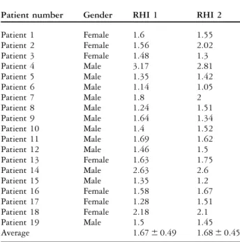
Table 2. RHI values for the Kawasaki disease group.
Patient number Gender RHI 1 RHI 2
Patient 1 Female 1.6 1.55 Patient 2 Female 1.56 2.02 Patient 3 Female 1.48 1.3 Patient 4 Male 3.17 2.81 Patient 5 Male 1.35 1.42 Patient 6 Male 1.14 1.05 Patient 7 Male 1.8 2 Patient 8 Male 1.24 1.51 Patient 9 Male 1.64 1.34 Patient 10 Male 1.4 1.52 Patient 11 Male 1.69 1.62 Patient 12 Male 1.46 1.5 Patient 13 Female 1.63 1.75 Patient 14 Male 2.63 2.6 Patient 15 Male 1.35 1.2 Patient 16 Female 1.58 1.67 Patient 17 Female 1.28 1.51 Patient 18 Female 2.18 2.1 Patient 19 Male 1.5 1.45 Average 1.67 6 0.49 1.68 6 0.45
RHI 5 reactive hyperaemia index
Values shown are mean 6 SD. RHI 1 – basal reactive hyperaemia index value; RHI 2 –reactive hyperaemia index value, 1 month after the first test.
even when there is no discernible coronary artery involvement. The issue, however, is rather conten-tious. In studies of Japanese subjects, of comparable characteristics, only the ones who developed coronary artery aneurysms during the acute stage of Kawasaki disease had abnormal flow-mediated dilation.38,39 The authors of a recent study40 of 52 Canadian Kawasaki disease patients concluded that there was no evidence of long-term endothelial cell dysfunction late after Kawasaki disease. It is unclear whether these conflicting results are best explained by methodological issues or genetic differences of the populations studied.
Children with Kawasaki disease, with or without coronary artery lesions, may have a more adverse cardiovascular risk profile. Whether this is a cause or a consequence of an abnormal endothelial function has not yet been assessed.40,41What is noteworthy is the fact that, in our sample, almost 70% of our Kawasaki disease group had an abnormal endothelial function, when compared with only 12% of the control group subjects.
Our findings may have surveillance and thera-peutic implications for the Kawasaki disease patients who had no coronary artery involvement during the acute disease. In the current management practices, long-term follow-up of these patients is seldom undertaken. Our results suggest that children with Kawasaki disease may have long-term sequelae, even when there is no overt coronary artery involvement in the acute stage of the disease. Affected children should therefore be kept on long-term follow-up, and instructed to avoid known cardiovascular risk factors, such as sedentary lifestyle, overweight, high-lipid and -caloric meals, alcohol or tobacco use. Prospective work is required, in a larger population, to assess the potential benefit of therapeutic approaches to these patients. The availability of newer, simple, and reproducible non-invasive techniques will allow a more rigorous follow-up of the endothelial function in these patients, allowing a close monitoring of the therapeutical effectiveness.
Acknowledgement
The authors thank Technician Lurdes Medroa for her contribution performing the endopat studies and Eduardo da Cruz, MD, PHD, for his scientific support.
References
1. Taubert KA, Rowley AH, Shulman ST. Nationwide survey of Kawasaki disease and acute reactive hyperaemia rheumatic fever. J Pediatr 1991; 119: 279–282.
2. Newburger JW, Takahashi M, Gerber MA, et al. Diagnosis, treatment, and long-term management of Kawasaki disease: a statement for health professionals from the Committee on
Rheumatic Fever, Endocarditis and Kawasaki Disease, Council on Cardiovascular Disease in the Young, American Heart Association. Circulation 2004; 110: 2747–2771.
3. Kawasaki T, Kosaki F, Okawa S, Shigematsu I, Yanagawa H. A new infantile acute febrile mucocutaneous lymph node syndrome (MLNS) prevailing in Japan. Pediatrics 1974; 54: 271–276. 4. Iemura M, Ishii M, Sugimura T, Akagi T, Kato H. Long term
consequences of regressed coronary aneurysms after Kawasaki disease: vascular wall morphology and function. Heart 2000; 83: 307–311.
5. Noto N, Okada T, Yamasuge M, et al. Noninvasive assessment of the early progression of atherosclerosis in adolescents with Kawasaki disease and coronary artery lesions. Pediatrics 2001; 107: 1095–1099.
6. Cheung YF, Wong SJ, Ho MH. Relationship between carotid intima-media thickness and arterial stiffness in children after Kawasaki disease. Arch Dis Child 2007; 92: 43–47.
7. Cheung YF, Yung TC, Tam SC, Ho MH, Chau AK. Novel and traditional cardiovascular risk factors in children after Kawasaki disease: implications for premature atherosclerosis. J Am Coll Cardiol 2004; 43: 120–124.
8. Amano S, Hazama F, Hamashima Y. Pathology of Kawasaki disease: II. Distribution and incidence of the vascular lesions. Jpn Circ J 1979; 43: 741–748.
9. Singh S, Bansal A, Gupta A, Kumar RM, Mittal BR. Kawasaki disease: a decade of experience from North India. Int Heart J 2005; 46: 679–689.
10. Suzuki A, Yamagishi M, Kimura K, et al. Functional behaviour and morphology of the coronary artery wall in patients with Kawasaki disease assessed by intravascular ultrasound. J Am Coll Cardiol 1996; 27: 291–296.
11. Sugimura T, Kato H, Inoue O, Takagi J, Fukuda T, Sato N. Vasodilatory response of the coronary arteries after Kawasaki disease: evaluation by intracoronary injection of isosorbide dinitrate. J Pediatr 1992; 121: 684–688.
12. Yamakawa R, Ishii M, Sugimura T, et al. Coronary endothelial dysfunction after Kawasaki disease: evaluation by intracoronary injection of acetylcholine. J Am Coll Cardiol 1998; 31: 1074–1080.
13. Hamaoka K, Onouchi Z, Ohmochi Y. Coronary flow reserve in children with Kawasaki disease without angiographic evidence of coronary stenosis. Am J Cardiol 1992; 69: 691–692.
14. Hamaoka K, Onouchi Z, Kamiya Y, Sakata K. Evaluation of coronary flow velocity dynamics and flow reserve in patients with Kawasaki disease by means of a Doppler guide wire. J Am Coll Cardiol 1998; 31: 833–840.
15. Mitani Y, Okuda Y, Shimpo H, et al. Impaired endothelial function in epicardial coronary arteries after Kawasaki disease. Circulation 1997; 96: 454–461.
16. Hauser M, Bengel F, Kuehn A, et al. Myocardial blood flow and coronary flow reserve in children with ‘‘normal’’ epicardial coronary arteries after the onset of Kawasaki disease assessed by positron emission tomography. Pediatr Cardiol 2004; 25: 108–112.
17. Muzik O, Paridon SM, Singh TP, Morrow WR, Dayanikli F, Di Carli MF. Quantification of myocardial blood flow and flow reserve in children with a history of Kawasaki disease and normal coronary arteries using positron emission tomography. J Am Coll Cardiol 1996; 28: 757–762.
18. Cicala S, Galderisi M, Grieco M, et al. Transthoracic echo-Doppler assessment of coronary microvascular function late after Kawasaki disease. Pediatr Cardiol 2008; 29: 321–327. 19. Ghelani SJ, Singh S, Manojkumar R. Endothelial dysfunction in a
cohort of North Indian children with Kawasaki disease without overt coronary artery involvement. J Cardiol 2009; 53: 226–231. 20. Patvardhan EA, Heffernan KS, Ruan JM, Soffler MI, Karas RHI, Kuvin JT. Assessment of vascular endothelial function with
peripheral arterial tonometry: information at your fingertips? Cardiol Rev 2010; 18: 20–28.
21. Nohria A, Gerhard-Herman M, Creager MA, Hurley S, Mitra D, Ganz P. Role of nitric oxide in the regulation of digital pulse volume amplitude in humans. J Appl Physiol 2006; 101: 545–548.
22. Kuvin JT, Karas RH. Clinical utility of endothelial function testing: ready for prime time? Circulation 2003; 107: 3243–3247. 23. Kuvin JT, Patel AR, Sliney KA, et al. Assessment of peripheral vascular endothelial function with finger arterial pulse wave amplitude. Am Heart J 2003; 146: 168–174.
24. Selamet Tierney ES, Newburger JW, Gauvreau K, et al. Endothelial pulse amplitude testing: feasibility and reproduci-bility in adolescents. J Pediatr 2009; 154: 901–905.
25. Haller MJ, Stein J, Shuster J, et al. Peripheral artery tonometry demonstrates altered endothelial function in children with type 1 diabetes. Pediatr Diabetes 2007; 8: 193–198.
26. Ayusawa M, Sonobe T, Uemura S, et al. Revision of diagnostic guidelines for Kawasaki disease (the 5th revised edition). Pediatr Int 2005; 47: 232–234.
27. Bonetti PO, Pumper GM, Higano ST, et al. Noninvasive identifica-tion of patients with early coronary atherosclerosis by assessment of digital reactive hyperemia. JACC 2004; 44: 2137–2141.
28. Duong TT, Silverman ED, Bissessar MV, Yeung RS. Super-antigenic activity is responsible for induction of coronary arteritis in mice: an animal model of Kawasaki disease. Int Immunol 2003; 15: 79–89.
29. Rowley AH. The etiology of Kawasaki disease: superantigen or conventional antigen? Pediatr Infect Dis J 1999; 18: 69–70. 30. Wang CL, Wu YT, Liu CA, Kuo HC, Yang KD. Kawasaki
disease: infection, immunity and genetics. Pediatr Infect Dis J 2005; 24: 998–1004.
31. Leung DY. Immunologic aspects of Kawasaki syndrome. J Rheumatol Suppl 1990; 24: 15–18.
32. Burns JC, Shike H, Gordon JB, Malhotra A, Schoenwetter M, Kawasaki T. Sequelae of Kawasaki disease in adolescents and young adults. J Am Coll Cardiol 1996; 28: 253–257.
33. Kato H, Sugimura T, Akagi T, et al. Long-term consequences of Kawasaki disease. A 10- to 21-year follow-up study of 594 patients. Circulation 1996; 94: 1379–1385.
34. Furuyama H, Odagawa Y, Katoh C, et al. Altered myocardial flow reserve and endothelial function late after Kawasaki disease. J Pediatr 2003; 142: 149–154.
35. Dhillon R, Clarkson P, Donald AE, et al. Endothelial dysfunction late after Kawasaki disease. Circulation 1996; 94: 2103–2106.
36. Deng YB, Xiang HJ, Chang Q, Li CL. Evaluation by high-resolution ultrasonography of endothelial function in brachial artery after Kawasaki disease and the effects of intravenous administration of vitamin C. Circ J 2002; 66: 908–912. 37. Dalla Pozza R, Bechtold S, Urschel S, Kozlik-Feldmann R, Netz
H. Subclinical atherosclerosis, but normal autonomic function after Kawasaki disease. J Pediatr 2007; 151: 239–243. 38. Kadono T, Sugiyama H, Hoshiai M, et al. Endothelial function
evaluated by flow-mediated dilatation in pediatric vascular disease. Pediatr Cardiol 2005; 26: 385–390.
39. Ikemoto Y, Ogino H, Teraguchi M, Kobayashi Y. Evaluation of preclinical atherosclerosis by flow-mediated dilatation of the brachial artery and carotid artery analysis in patients with a history of Kawasaki disease. Pediatr Cardiol 2005; 26: 782–786.
40. McCrindle BW, McIntyre S, Kim C, Lin T, Adeli K. Are patients after Kawasaki disease at increased risk for accelerated athero-sclerosis? J Pediatr 2007; 151: 244–248.
41. Noto N, Okada T, Karasawa K, et al. Age-related acceleration of endothelial dysfunction and subclinical atherosclerosis in subjects with coronary artery lesions after Kawasaki disease. Pediatr Cardiol 2006; 30: 262–268.
content may not be copied or emailed to multiple sites or posted to a listserv without the copyright holder's express written permission. However, users may print, download, or email articles for individual use.