Revista Portuguesa de
Cardiologia
Portuguese Journal of Cardiology
www.revportcardiol.org
CASE REPORT
The proarrhythmic effect of cardiac resynchronization
therapy: An issue that should be borne in mind
夽
Nuno Cabanelas
a,∗, Mário Oliveira
b, Manuel Nogueira da Silva
b, Pedro Cunha
b,
Bruno Valente
b, Ana Lousinha
b, Sofia Santos
b, Luísa Branco
b, Rui Ferreira
baServic¸o de Cardiologia, Hospital de Santarém, Santarém, Portugal
bServic¸o de Cardiologia, Hospital Santa Marta, Centro Hospitalar de Lisboa Central, Lisboa, Portugal
Received 14 December 2012; accepted 20 January 2014 Available online 18 June 2014
KEYWORDS Cardiac resynchronization; Epicardial pacing; Repolarization heterogeneity
Abstract The demonstrated benefits of cardiac resynchronization therapy (CRT) in reducing
mortality and hospitalizations for heart failure, improving NYHA functional class and inducing reverse remodeling have led to its increasing use in clinical practice. However, its potential contribution to complex ventricular arrhythmias is controversial.
We present the case of a female patient with valvular heart failure and severe systolic dys-function, in NYHA class III and under optimal medical therapy, without previous documented ventricular arrhythmias. After implantation of a CRT defibrillator, she suffered an arrhythmic storm with multiple episodes of monomorphic ventricular tachycardia (VT), requiring 12 shocks. Subsequently, a pattern of ventricular bigeminy was observed, as well as reproducible VT runs induced by biventricular pacing.
Since no other vein of the coronary sinus system was accessible, it was decided to implant an epicardial lead to stimulate the left ventricle, positioned in the left ventricular mid-lateral wall. No arrhythmias were detected in the following six months.
This case highlights the possible proarrhythmic effect of biventricular pacing with a left ventricular lead positioned in the coronary sinus venous system.
© 2012 Sociedade Portuguesa de Cardiologia. Published by Elsevier España, S.L. All rights reserved.
PALAVRAS-CHAVE
Ressincronizac¸ão cardíaca;
Pacing epicárdico;
Terapêutica de ressincronizac¸ão cardíaca e efeito pró-arrítmico: um problema que deve ser lembrado
Resumo Os benefícios demonstrados com a terapêutica de ressincronizac¸ão cardíaca (TRC)
na reduc¸ão da mortalidade e hospitalizac¸ão por ICC, melhoria da classe funcional e obtenc¸ão
夽 Please cite this article as: Cabanelas N, Oliveira M, Nogueira da Silva M, et al. Terapêutica de ressincronizac¸ão cardíaca e efeito pró-arrítmico: um problema que deve ser lembrado. Rev Port Cardiol. 2014;33:309.e1---309.e7.
∗Corresponding author.
E-mail address:[email protected](N. Cabanelas).
Heterogeneidade da repolarizac¸ão
de remodelagem inversa em doentes selecionados com insuficiência cardíaca (ICC), têm con-tribuído para a crescente utilizac¸ão destes dispositivos na prática clínica.
No entanto, permanece controverso o impacto da TRC como fator causador de arritmias ventriculares complexas. Apresentamos o caso duma doente com cardiopatia valvular oper-ada, disfunc¸ão sistólica grave e ICC classe III da NYHA, com terapêutica médica otimizada, sem documentac¸ão prévia de arritmias ventriculares significativas. Após implantac¸ão do sis-tema de TRC com cardioversor-desfibrilhador, desenvolveu quadro de tempestade arrítmica com múltiplos episódios de taquicardia ventricular monomórfica (TV) e necessidade de 12 choques, mantendo padrão de bigeminismo ventricular reprodutível e induc¸ão de salvas de TV pelo pac-ing biventricular. Dada a inacessibilidade a outra veia tributária do seio coronário foi decidido implantar elétrodo epicárdico em localizac¸ão diferente (de veia póstero-lateral para posic¸ão lateral-mediana), sem registo de recorrência de arritmias num follow-up de seis meses. Este caso sugere que a TRC pode contribuir para um efeito pró-arrítmico com consequências clínicas potencialmente graves.
© 2012 Sociedade Portuguesa de Cardiologia. Publicado por Elsevier España, S.L. Todos os direitos reservados.
Introduction
The benefits of cardiac resynchronization therapy (CRT) in reducing mortality and hospitalizations for heart failure (HF), improving NYHA functional class and inducing reverse remodeling have been amply demonstrated in various mul-ticenter trials in the last 10 years, leading to a considerable expansion of indications for biventricular (BiV) pacing.1---8
CRT can have adverse effects, most of which are related to procedural complications, infection and system malfunc-tion. In recent years there has also been debate concerning the possible contribution of BiV pacing to the occurrence of complex ventricular arrhythmias.
Case report
A 58-year-old female patient with controlled mild hyperten-sion, type 2 diabetes and dyslipidemia was being followed in the cardiology outpatient clinic for valvular HF and perma-nent atrial fibrillation (AF). She had previously undergone mitral valve replacement with a mechanical valve due to severe mitral stenosis.
During follow-up, progressive clinical deterioration was seen to NYHA class III under optimal medical therapy (OMT). The ECG showed QRS interval of 150 ms and com-plete left bundle branch block. She had no history of ventricular arrhythmias during follow-up. Serial echocardio-grams showed steadily worsening global systolic function, ejection fraction (EF) falling from 24% to 13%. Six years after valve replacement surgery, she had severely impaired global systolic function, with left ventricular (LV) end-diastolic diameter of 82 mm, EF estimated at 13% by the modified Simpson’s rule, and echocardiographic criteria of intraventricular dyssynchrony, with tissue synchronization imaging showing septal-lateral delay of 100 ms, two-dimensional strain imaging showing radial strain of 448 ms with inferior-anteroseptal delay but no ventricular dyssyn-chrony (pulmonary and aortic pre-ejection times of 78 ms and 105 ms, respectively). Right ventricular (RV) function
Figure 1 Angiogram of the coronary sinus, showing sparse venous system.
was also impaired, with tricuspid annular plane systolic excursion of 5 mm.
A VVIR mode CRT defibrillator (CRT-D) was implanted with the LV lead positioned in a posterolateral vein (Figure 1) with a different ostium from that of the coronary sinus, the venous system of which was sparse, consisting of small and markedly angulated vessels (Figure 2). In our center, the posterolateral vein is often used when the branches of the coronary sinus are technically difficult to access, although it is generally difficult to characterize. However, this vein is only used as an alternative, since the distance between it and the RV apex gives insufficient time for myocardial activation. Furthermore, the fact that both leads activating the ventricular mass are relatively close could trigger new dyssynchrony by the late activation of more distant areas of the myocardium.
Figure 2 Angiogram of the posterior vein.
One month after implantation, the patient returned to our department after suffering 12 shocks in 24 hours. Device interrogation revealed these to have been appropri-ate shocks in response to an arrhythmic storm of multiple episodes of rapid ventricular tachycardia (VT) with a mean cycle length of 250 ms (Figure 3). At the same time repro-ducible ventricular bigeminy and VT runs induced by BiV pacing were seen. Various programming modes were tested with different pulse polarities, amplitudes and widths, as well as left ventricular pacing alone, none of which elimi-nated the ventricular extrasystoles. In view of the failure of these attempts, pacing via the left ventricular lead
was switched off and the patient regained natural rhythm without ventricular extrasystoles, with RV pacing set to a minimum of 40 bpm. The CRT-D was programmed with three zones: VT-1, from 171 bpm with two bursts of antitachycar-dia pacing; VT-2, from 182 bpm, with two ramps followed if necessary by a 40-J shock; and VF, from 200 bpm, with up to six 40-J shocks.
Holter 24-hour monitoring performed two months later showed natural rhythm (AF) in 99% of the record, with only 195 isolated dimorphic ventricular extrasystoles, one pair and one 4-complex run. The patient was under medication with amiodarone 200 mg/day and carvedilol 6.25 mg twice daily.
Echocardiography five months after switching off LV pac-ing showed continupac-ing very low EF (13%) with evidence of intra- and interventricular dyssynchrony; the patient remained in NYHA class III.
It was decided to make another attempt to implement CRT. Since no other branch of the coronary sinus could be catheterized, an epicardial lead was implanted in the lateral LV wall (Figure 4). The alternative of repositioning the RV lead to a septal position was not chosen because, before CRT implantation, the segments with the latest activation were those of the lateral wall, the distance between the leads would not be significantly increased by this change, and in order to begin pacing via the LV lead it would have to be implanted in a less arrhythmogenic position.
An epicardial lead was implanted between the second and third obtuse marginals by submammary thoracotomy.
One month later, the patient was still in NYHA class III, with poor global systolic function. The LV pacing percentage was 89% and the RV pacing percentage was 13%. The low percentage of BiV pacing is explained by the rapid intrinsic rates resulting from the patient’s AF. However, no ventricu-lar arrhythmias were recorded by the device.
LV mV RV A A RV LV
Predetection Time (seconds)
Detection VF Burst VF 258 VF 258 VF 258 VF 258 VF 250 VF 234 VF 250 VF 242 VF 250 VF 250 VF 289 Vs 531 Vs 531 Vp Vp LV p LV p LV p LV p LV s 367 Onset LV s 266 LV s 250 LV s 250 LV s 250 LV s 250 LV s 250 LV s 258 LV s 258 LV s 258 LV s 242 LV s 266 LV s 273 VT1 329 VF 273 1 -2 -3 -4 -5 -6 - 0
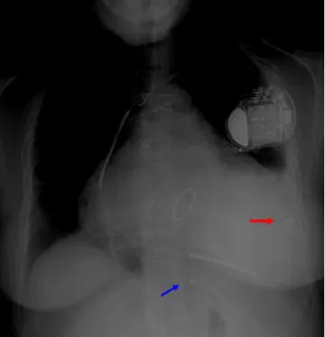
Figure 4 Chest X-ray, anteroposterior view, showing pacing lead in the posterior vein (blue arrow) and epicardial lead in lateral position (red arrow).
Digoxin was added to the patient’s therapeutic regime to control her intrinsic ventricular rate, the minimum pac-ing rate was raised to 80 bpm, and she was enrolled in a cardiac rehabilitation program. We opted for a conservative approach to maximize BiV pacing rate through optimizing medical therapy and reprogramming the device, given the theoretically greater risk of loss of capture by the epicardial lead than if the lead had been positioned in the coronary sinus venous system. If this strategy failed, the next step would be ablation of the atrioventricular node. If the LV lead had been in the coronary venous system, then nodal ablation would have been the first option.
At six months after implantation of the epicardial LV lead and after an increase in the percentage of BiV pacing, the patient was still in NYHA class III, but reported slight symp-tomatic improvement in her day-to-day activities, and her EF had risen to 19%. No new episodes of decompensated HF occurred and no ventricular arrhythmias were detected by remote monitoring during this period.
Discussion
Initial reports of the impact of CRT on the incidence of ventricular arrhythmias show an antiarrhythmic effect,9,10
which could be due to reduction of wall stress caused by the inverse remodeling induced by BiV pacing, decreased dispersion of ventricular repolarization resulting from dual depolarization wave fronts,11,12 and reduced sympathetic
nervous system activation.13
Other studies suggested that this antiarrhythmic effect is due to reductions in the number of ventricular extrasystoles,14 the incidence of tachyarrhythmic events15
and the inducibility of sustained VT.16More recent research
has indicated that this effect is mainly seen in patients who respond to CRT, as a consequence of the hemodynamic improvement induced by BiV pacing,17,18 but not in
non-responders.
Indications for CRT have widened in the last 10 years and research into its consequences has intensified, but its elec-trophysiological effects on the incidence of sudden death and ventricular arrhythmias are still poorly understood.
Cases have been reported of increased arrhythmic events in some patients when treated with BiV pacing.19---21 In
2005, Shukla et al.19 described a series of 145 consecutive
patients, five of whom developed arrhythmic storm after implantation of a CRT-D, which was permanently resolved by discontinuing LV pacing. Similarly, Medina-Ravell et al.20
assessed 29 patients who received a CRT-D, four of whom developed ventricular extrasystoles with BiV pacing, elimi-nated when LV pacing was ended. The same phenomenon is seen in the case presented here.
Further evidence is found in the two largest randomized clinical trials to assess the effects of CRT in patients with and without an implantable defibrillator, which showed a reduc-tion in all-cause mortality compared to OMT (COMPANION4
and CARE-HF5), the percentage of sudden death as a cause
of death was slightly higher in those without a defibrillator.22
In the COMPANION trial, there was a statistically sig-nificant reduction of 36% in all-cause death (p=0.003) in patients under OMT and CRT compared to OMT only. How-ever, the incidence of sudden death was higher in those under CRT with pacing only plus OMT than in those with OMT only (7.8% vs. 5.8%), while in those with CRT-D and OMT it was only 2.9%. Analysis of causes of death in the three groups shows that sudden death was responsible in 36.6% of patients with CRT with pacing only plus OMT, 23.4% of those with OMT only, and only 16.2% of those with CRT-D and OMT.22
In the CARE-HF trial, although mortality was lower in patients undergoing CRT than in those receiving OMT only, the percentage of the former suffering sudden death was higher (35.4% vs. 31.7%).
However, evidence to the contrary recently came from the REVERSE study, which assessed the incidence of VT/ventricular fibrillation and sustained VT in patients with CRT-D devices, one group with BiV pacing on and the other with pacing off. After two years of follow-up, the incidence of arrhythmic events was similar in the two groups (18.7% vs. 21.9%, p=0.84).23
In the normal sequence of myocardial activation, the endocardium is depolarized before the more epicardial lay-ers, while repolarization travels in the opposite direction (from epicardium to endocardium). BiV pacing is normally effected via an endocardial lead placed in the RV and a lead placed inside the coronary sinus or one of its branches, and so the LV myocardium is stimulated via the epicardium. Epicardial pacing involves a non-physiological activation sequence in which the vector of the transmural propagation is reversed, resulting in delayed endocardial depolarization and earlier epicardial depolarization.24 Experimental
stud-ies have shown that epicardial-endocardial conduction time is significantly longer than endocardial-epicardial conduc-tion time, due to a zone of myocardial wall between the deep subendocardium and mid-myocardial layers.25,26 The
and JT intervals and the interval between the peak and the end of the T wave.25,27
Furthermore, a small number of predisposed patients may be more likely to suffer reentrant phenomena. The presence of cardiomyopathy, use of QT-prolonging drugs, and autonomic dysregulation can also promote these phenomena.28,29
Identification of factors that predispose to ventricular arrhythmias caused by BiV pacing is thus of considerable clinical importance in the assessment of candidates for this therapy and in the decision whether to implant a CRT-D system.
The predictors of arrhythmias in these patients have not been fully identified. In a study of 75 patients undergoing CRT, variation in QT dispersion before and after implantation was an independent predictor of major arrhythmic events; in a follow-up of 807 days, in the group with increased QT dispersion the incidence of events was 29%, while in those in whom it decreased, the incidence was 3% (p=0.0017).30
The same study30assessed the interval between the peak
and the end of the T wave (Tpeak-end), another marker of dis-persion of repolarization. Patients who suffered arrhythmic events during follow-up had a significantly smaller reduction in Tpeak-end than those who were event-free (−1.5±12.8 ms vs.−20.0±5.4 ms, p=0.047).
The mechanisms involved in this differing QT dispersion response to CRT have not been investigated. It is thought that factors such as severe systolic dysfunction,29perfusion
disturbances31 and stimulation of LV myocardium in areas
close to fibrotic tissue32 could in theory be related to
changes in QT dispersion.
Dilated cardiomyopathy is associated with ventricu-lar fibrosis, changes in muscle tissue architecture and abnormalities of cellular ultrastructure, particularly in cell membranes.33,34The electrophysiological properties of
car-diomyopathic myocardium are also altered by lines of conduction block resulting from fibrosis and areas of abnor-mal conductibility and refractoriness.35 The zones with
altered electrophysiological properties are not homoge-nous throughout the dysfunctional myocardium,36 and the
patterns of their distribution appear to differ between ischemic and non-ischemic dilated cardiomyopathy,36 with
more diffuse involvement, mainly in the basal segments, in the latter compared to the former, in which these zones tend to be restricted to particular arterial territories and typically affect the endocardium more extensively.37,38
In the case presented, the patient had severe valvu-lar disease and had undergone mitral valve replacement, which may have altered the tissue architecture and hence the distribution of zones with altered electrophysiological properties.
When a pro-arrhythmic effect of BiV pacing is suspected, an alternative site for LV stimulation can reduce arrhythmo-genicity. In our patient, the lack of options led us to adopt a surgical approach, and during thoracotomy threshold tests were performed in different parts of the epicardium and the induction of ventricular extrasystoles by BiV pacing was assessed.
Various strategies have been tried to overcome the potential arrhythmic risk of LV stimulation via branches of the coronary sinus, including endocardial LV pacing with the lead positioned via transseptal puncture. Initial results of
this procedure, still in the early stages of evaluation, are promising.39,40
Assessment of heterogeneity of repolarization during implantation is considered of little value, since no cor-relation has been established between changes in these parameters and adverse events, and any pro-arrhythmic effect may only be manifested hours or days after implantation.41
Conclusion
CRT is not without adverse effects. Although its benefits clearly outweigh the risks in patients with indication for this therapy, in rare cases BiV pacing may induce arrhyth-mias. Reversal of the physiological depolarization sequence increases dispersion of repolarization, promoting reentry phenomena and increasing the incidence of ventricular arrhythmias in some patients.
Although the paradoxical increase in arrhythmogenicity with CRT is uncommon, it can have serious clinical conse-quences that reprogramming alone cannot prevent. In the case presented, only removal of the pacing lead from the coronary sinus eliminated the ventricular arrhythmia. Since ventricular resynchronization was necessary, an alternative site for the LV pacing lead had to be found, and an epicardial approach was the one chosen.
Ethical disclosures
Protection of human and animal subjects. The authors
declare that no experiments were performed on humans or animals for this study.
Confidentiality of data. The authors declare that they have
followed the protocols of their work center on the publica-tion of patient data.
Right to privacy and informed consent. The authors have
obtained the written informed consent of the patients or subjects mentioned in the article. The corresponding author is in possession of this document.
Conflicts of interest
The authors have no conflicts of interest to declare.
References
1. Linde C, Leclercq C, Rex S, et al. Long-term benefits of biven-tricular pacing in congestive heart failure: results from the MUltisite STimulation in cardiomyopathy (MUSTIC) study. J Am Coll Cardiol. 2002;40:111---8.
2. Abraham W, Fisher W, Smith A, et al., The MIRACLE Study Group. Cardiac resynchronization in chronic heart failure, Mul-ticenter Insync Randomized Clinical Evaluation. N Engl J Med. 2002;346:1845---53.
3. Abraham W, Young J, Leon A, et al., on behalf of the Multicenter InSync ICD II (MIRACLE ICD II) Study Group. Effects of cardiac resynchronization on disease progression in patients with left ventricular systolic dysfunction, an indica-tion for an implantable cardioverter-defibrillator, and mildly
symptomatic chronic heart failure. Circulation. 2004;110: 2864---8.
4. Bristow M, Saxon L, Boehmer J, et al., Comparison of Medical Therapy. Pacing and Defibrillation in Heart Failure (COMPAN-ION) Investigators. Cardiac resynchronization therapy with or without an implantable defibrillator in advanced chronic heart failure. N Engl J Med. 2004;350:2140---50.
5. John G, Cleland M, Daubert J, et al., for the Cardiac Resyn-chronization - Heart Failure (CARE-HF) Study Investigators. The effect of cardiac resynchronization on morbidity and mortality in heart failure. N Engl J Med. 2005;352:1539---49.
6. Linde C, Abraham W, Gold M, et al. Randomized trial of cardiac resynchronization in mildly symptomatic heart failure patients and in asymptomatic patients with left ventricular dysfunc-tion and previous heart failure symptoms. J Am Coll Cardiol. 2008;52:1834---43.
7. Solomon S, Foster E, Bourgoun M, et al., (MADIT-CRT Investiga-tors). Effect of cardiac resynchronization therapy on reverse remodeling and relation to outcome. Multicenter Automatic Defibrillator Implantation Trial-Cardiac Resynchronization Ther-apy. Circulation. 2010;122:985---92.
8. Tang A, Wells G, Talajic M, et al., Resynchronization-Defibrillation for Ambulatory Heart Failure Trial Investigators. Cardiac resynchronization therapy for mild-to-moderate heart failure. N Engl J Med. 2010;363:2385---95.
9. Walker S, Levy T, Rex S, et al. Usefulness of suppression of ven-tricular arrhythmia by bivenven-tricular pacing in severe congestive cardiac failure. Am J Cardiol. 2000;86:231---3.
10. Zagrodzky J, Ramaswamy K, Page R, et al. Biventricular pac-ing decreases the inducibility of ventricular tachycardia in patients with ischemic cardiomyopathy. Am J Cardiol. 2001;87: 1208---10.
11. Kies P, Bax J, Molhoek S, et al. Effect of left ventricular remod-eling after cardiac resynchronization therapy on frequency of ventricular arrhythmias. Am J Cardiol. 2004;94:130---2. 12. Martinelli Filho M, Pedrosa A, Costa R, et al. Biventricular pacing
improves clinical behavior and reduces prevalence of ventricu-lar arrhythmia in patients with heart failure. Arq Bras Cardiol. 2002;78:110---3.
13. Hamdan M, Zagrodzky J, Joglar J, et al. Biventricular pacing decreases sympathetic activity compared with right ventricular pacing in patients with depressed ejection fraction. Circulation. 2000;102:1027---32.
14. Martinelli Filho M, Pedrosa A, Costa R, et al. Biventricular pacing improves clinical behaviour and reduces prevalence of ventricu-lar arrhythmia in patients with heart failure. Arq Bras Cardiol. 2002;78:110---3.
15. Yu C, Bleeker C, Fung J, et al. Left ventricular reverse remodeling, but not clinical improvement predicts long-term survival after cardiac resynchronization therapy. Circulation. 2005;112:1580---6.
16. Zagrodsky J, Ramaswamy K, Page R, et al. Biventricular pac-ing decreases the inducibility of ventricular tachycardia in patients with ischemic cardiomyopathy. Am J Cardiol. 2001;87: 1208---10.
17. Shahrzad S, Soleiman N, Taban S, et al. The effect of left ventricular (LV) remodelling on ventricular arrhythmia in cardiac resynchronization therapy (CRT-D) patients (antiar-rhythmic effect of CRT). Pacing Clin Electrophysiol. 2012;35: 592---7.
18. Barsheshet A, Wang P, Moss A, et al. Reverse remodeling and the risk of ventricular tachyarrhythmias in the MADIT-CRT (Multicenter Automatic Defibrillator Implantation Trial --- Cardiac Resynchronization Therapy). J Am Coll Cardiol. 2011;57:2416---23.
19. Shukla G, Chaudhry G, Orlov M, et al. Potential proarrhyth-mic effect of biventricular pacing: fact or myth? Heart Rhythm. 2005;2:951---6.
20. Medina-Ravell V, Lankipalli R, Yan G, et al. Effect of epicar-dial or biventricular pacing to prolong QT interval and increase transmural dispersion of repolarization: does resynchronization therapy pose a risk for patients predisposed to long QT or tor-sade de pointes? Circulation. 2003;107:740---6.
21. Rivero-Ayerza M, Vanderheyden M, Verstreken S, et al. Images in cardiovascular medicine. Polymorphic ventricular tachycardia induced by left ventricular pacing. Circulation. 2004;109:2924---5.
22. Carson P, Anand I, O’Connor C, et al. Mode of death in advanced heart failure: the Comparison of Medical, Pacing and Defibril-lation Therapies in Heart Failure (COMPANION) trial. J Am Coll Cardiol. 2005;46:2329---34.
23. Gold M, Linde C, Abraham W. The impact of cardiac resynchro-nization therapy on the incidence of ventricular arrhythmias in mild heart failure. Heart Rhythm. 2011;8:679---84.
24. Di Diego J, Belardinelli L, Antzelevitch C. Cisapride-induced transmural dispersion of repolarization and torsade de pointes in the canine left ventricular wedge preparation during epicar-dial stimulation. Circulation. 2003;108:1027---33.
25. Fish J, Di Diego J, Nesterenko V, et al. Epicardial activation of left ventricular wall prolongs QT interval and transmural dis-persion of repolarization: implications for biventricular pacing. Circulation. 2004;109:2136---42.
26. Poelzing S, Dikshteyn M, Rosenbaum D. Transmural conduction is not a two-way street. J Cardiovasc Electrophysiol. 2005; 16:455.
27. Bai R, Yang X, Song Y, et al. Impact of left ventricular epi-cardial and biventricular pacing on ventricular repolarization in normal-heart individuals and patients with congestive heart failure. Europace. 2006;8:1002---10.
28. Akar F, Rosenbaum D. Transmural electrophysiological hetero-geneities underlying arrhythmogenesis in heart failure. Circ Res. 2003;93:638---45.
29. Ray I, Fendelander L, Singh J. Cardiac resynchronization therapy and its potential proarrhythmic effect. Clin Cardiol. 2007;30:498---502.
30. Chalil S, Yousef Z, Muyhaldeen S, et al. Pacing-induced increase in QT dispersion predicts sudden cardiac death following cardiac resynchronization therapy. J Am Coll Cardiol. 2006;47:2486---92. 31. Bonnemeier M, Wiegand U, Bode F, et al. Impact of infarct-related artery flow on QT dynamicity in patients undergoing direct percutaneous coronary intervention for acute myocardial infarction. Circulation. 2003;108:2979---86.
32. Leyva F, Paul F. Is cardiac resynchronisation therapy proarrhyth-mic? Indian Pacing Electrophysiol J. 2008;4:268---80.
33. Unverferth D, Baker P, Swift S, et al. Extent of myocardial fibro-sis and cellular hypertrophy in dilated cardiomyopathy. Am J Cardiol. 1986;57:816---20.
34. Sugrue D, Holmes D, Gersh B, et al. Cardiac histologic find-ings in patients with life-threatening ventricular arrhythmias of unknown origin. J Am Coll Cardiol. 1984;4:952---7.
35. de Bakker J, van Capelle F, Janse M, et al. Fractionated elec-trograms in dilated cardiomyopathy: origin and relation to abnormal conduction. J Am Coll Cardiol. 1996;27:1071---8. 36. Nakahara S, Tung R, Ramirez R, et al. Characterization of
the arrhythmogenic substrate in ischemic and nonischemic cardiomyopathy: implications for catheter ablation of hemody-namically unstable ventricular tachycardia. J Am Coll Cardiol. 2010;55:2355---65.
37. Hsia H, Callans D, Marchlinski F, et al. Characterization of endocardial electrophysiological substrate in patients with nonischemic cardiomyopathy and monomorphic ventricular tachycardia. Circulation. 2003;108:704---10.
38. Bogun F, Desjardins B, Good E, et al. Delayed-enhanced mag-netic resonance imaging in nonischemic cardiomyopathy: utility for identifying the ventricular arrhythmia substrate. J Am Coll Cardiol. 2009;53:1138---45.
39. Whinnett Z, Bordachar P. The risks and benefits of transseptal endocardial pacing. Curr Opin Cardiol. 2012;27:19---23. 40. Scott P, Yue A, Watts E, et al. Transseptal left ventricular
endo-cardial pacing reduces dispersion of ventricular repolarization. Pacing Clin Electrophysiol. 2011;34:1258---66.
41. Cazeau S, Leclercq C, Lavegne T, et al. The Multisite Stim-ulation in Cardiomyopathies (MUSTIC) Study I. Effects of multisite biventricular pacing in patients with heart failure and intraventricular conduction delay. N Engl J Med. 2001;344: 873---80.