Impact of intravesical hyaluronic acid treatment on bladder
inflammation in interstitial cystitis rat model
_______________________________________________
Ilker Fatih Sahiner 1, Hakan Soylu 2, Erhan Ates 3, Nuray Acar 2, Ismail Ustunel 2, Ahmet Danısman 1
1 Department of Urology, Akdeniz University School of Medicine, Antalya, Turkey; 2 Department of
Histology and Embryology, Akdeniz University School of Medicine, Antalya, Turkey; 3 Department of Urology, Adnan Menderes University School of Medicine, Aydin, Turkey
ABSTRACT
ARTICLE
INFO
______________________________________________________________ ______________________
Objective: To evaluate the effect of intravesical hyaluronic acid (HA) treatment on inflammatory cells and the severity of inflammation in an interstitial cystitis rat model created with hydrogen chloride (HCL) via immunohistochemical studies and myeloperoxidase activity for the first time in the literature.
Materials and Methods: A total of 30 adult female white Rattus Norvegicus rats were divided into 3 groups as the HCL group, hyaluronic acid treatment (HCL-HA) group and control group. Chemical cystitis was created by administering HCL(400 microL,10 mM) except control group. A single dose of intravesical HA(0.5 mL,0.8 mg/mL) was administered to the treatment group. The bladder tissues of all subjects were immuno-histochemically stained. The cell surface markers were used to evaluate inflammatory cell infiltration. Mast cell activation and IL-6 was evaluated to assess the inflammation and severity of inflammation, respectively. Myeloperoxidase activity was measured as it shows neutrophil density. Statistical significance was accepted as P<0.05.
Results: It was observed that there was rich monocyte, T lymphocyte, B lymphocyte, and Natural Killer cells infiltration and high IL-6 levels in the bladder tissue after the intravesical hydrogen chloride instillation, especially in the stroma layer(p<0.005). In the HCL-HA group, severity of inflammation had statistically significantly regressed to the levels of the control group(p<0.005). An increase was observed in the bladder myeloperoxidase activity of the HCL group compared to the other two groups(p<0.05).
Conclusions: Single dose intravesical hyluronic acid instillation reduces inflammatory cell infiltration and the severity of bladder inflammation in the rat model of bladder pain syndrome/interstitial cystitis.
INTRODUCTION
The European Society for the Study of Interstitial Cystitis (ESSIC) and the European As-sociation of Urology (EAU) define Bladder Pain Syndrome (BPS)/Interstitial Cystitis (IC) as the complaint of suprapubic pain associated with filling of the bladder accompanied by other symp-toms such as increased frequency of daytime and
nighttime urination, in the absence of a urinary infection or other pathology (1, 2).
The etiology of BPS/IC is still not fully understood. Causes of urothelial dysfunction such as glycosaminoglycan (GAG) layer disorders and inhibition of urothelial cell proliferation have been reported as etiological factor (3, 4). Intra-vesical hyaluronic acid (HA), a treatment option recommended by guidelines, strengthens the
uri-Keywords:
Interstitial cystitis; Inflammation; Hyaluronic Acid
Int Braz J Urol. 2018; 44: 1014-22
_____________________
Submitted for publication: December 29, 2017
_____________________
Accepted after revision: March 26, 2018
_____________________
ne-tissue barrier by integrating with the GAG layer and creates an anti-inflammatory effect by inhibiting the leukocyte migration, adhesion of immune complexes, and bonding with spe-cific receptors (I-CAM 1) that occur during the inflammatory process (5, 6).
Lymphocytes and other cells express a large number of different marker molecules that can be used to identify cells and characterize the cell type on their surfaces. These marker molecules were named CD (Cluster of Differentiation) by the International Human Leukocyte Differentiation Antigens (HLDA) Study Group (7). IL-6 is a pro--inflammatory cytokine produced by a variety of cell types including endothelial cells, macropha-ges, fibroblasts, and mast cells and indicates the severity of inflammation (8). Studies revealed that mast cells have a critical role on many inflamma-tory diseases. Increases in mucosal damage and activated mast cells in biopsies of patients with IC have been shown (9). Also, mast cells have been shown to be active in bladder injury models ge-nerated by intravesical instillation of protamine sulfate (10).
Myeloperoxidase is an enzyme found in neutrophil granules. A direct correlation has been shown to exist between the measurement of MPO activity in tissue samples and neutro-phil counts (11).
Histopathologically, it has been shown that there is primarily an inflammatory cellular infiltration limited to the lamina propria which consists of lymphocytes and plasma cells in IC cases (12). Studies on animal models of IC have revealed neutrophil infiltration and activation of some inflammatory cytokines in the bladder (13).
In our study, we tried to show the effects of treatment on the rat model of IC treated with intravesical single dose HA with myeloperoxidase activity and by making an immunohistochemical assessment with polyclonal antibodies.
MATERIALS AND METHODS
The Animal Model
Following approval from the animal ethics committee of the Akdeniz University (2013.10.02), 30 adult female white Rattus norvegicus rats
lower abdominal midline incision. Some of the bla-dder samples obtained were embedded in paraffin and placed in 4% formalin. The others were then stored in liquid nitrogen to perform a myeloperoxi-dase assay.
HISTOPATHOLOGICAL ASSESSMENT
Immunohistochemical analysis
We used CD (Cluster of Differentiation) system to detect cell surface markers in the area of inflammation in our study. CD3 was used as a T lymphocyte surface marker, CD14 as a monocyte surface marker, CD19 as a B lymphocyte surface marker, and CD56 as a natural killer surface marker. We used IL-6 to assess the severity of inflammation.
The samples which were set in 4% formal-dehyde for about 12 hours were then washed in tap water for 2 hours and dehydrated in ascending al-cohol series. They were then made transparent in xylol and embedded in paraffin. In order to make routine light microscopic observations and to per-form an immunohistochemical technique, sections with a thickness of 5 µm were placed on polylysine--coated slides. After paraffin removal, the sections were boiled for 7 min (4 min + 3 min) in citrate buffer (pH: 6.0) for antigen retrieval and allowed to cool at room temperature for 20 min. The sections were then left to wait for 20 minutes in 3% hydro-gen peroxide to block endohydro-genous peroxidase acti-vity. After this procedure, Ultra V block (Lab-Vision, Fremont, CA, USA) was applied to the sections for 7 min in a humidified chamber at room tempera-ture. After removal of excess serum, sections were incubated overnight in a humidified chamber at 4ºC with primary antibodies: CD3 rabbit polyclonal an-tibody (Bioss Biotechnology; bS-0765R) in a 1:500 dilution; CD14 rabbit polyclonal antibody (Bioss Biotechnology; bs-1192R) in a 1:500 dilution; CD19 rabbit polyclonal antibody (Bioss Biotechnology; bs-4755R) in a 1:400 dilution; CD 56 rabbit polyclo-nal antibody (Bioss Biotechnology; bS-0805R) in a 1:500 dilution; and IL-6 rabbit polyclonal antibody (Bioss Biotechnology; bs-4540R) in a 1:500 dilu-tion. The following day, the sections were washed 3 times with PBS solution for 5 minutes and then incubated for 30 minutes at room temperature in a humidified chamber with a biotin marked secondary
antibody; anti-rabbit IgG (BA-1000) in a 1:500 dilu-tion. The slides were washed 3 times for 5 min with PBS and the emissions obtained were developed with diaminobenzidine (K3466; Dako). The sections were counterstained with hematoxylin, dehydrated in ascending alcohol series, and examined under a light microscope (Zeiss, Oberkochen, Germany) after closing them with Kaiser’s glycerin gelatine (Merck; OB514196, NJ, USA).
Semi-quantitative evaluation
The immunoreactive cells positively stai-ned with the markers studied in all groups and their immunostaining densities were semi-quantitatively assessed by two observers with the method mentio-ned below.
Detection and count of mast cell
Toluidine blue powder (0.5 g) (Toluidine Blue O, Merck, 115930) was dissolved in 100 mL dis-tilled water. Formalin fixed paraffin embedded sam-ples were cut into 5 µm sections and placed on sli-des. After deparaffinization, sections were taken to water and were stained with this solution for 1 min. They were rinsed in water for 3-5 min, differentia-ted respectively in 95% alcohol, absolute alcohol, and were cleared. Finally, the sections were taken through graded alcohols to xylene and mounted in entellan. Sections were examined by light micros-copy (Zeiss, Oberkochen, Germany). Photographs of PBS, HCL and HCL+HA groups were taken with an Axioplan® microscope (Zeiss, Oberkochen,
Ger-No staining = Grade 0
Cellular staining between 0-25%
= Grade 1
Cellular staining between 25%-50%
= Grade 2
Cellular staining between 50%-75%
= Grade 3
Cellular staining between 75%-100%
many) and mast cells were counted through the use of the Image J (http://imagej.nih.gov/ij/) program.
Measuring of Myeloperoxidase activity
Because the measurement of myeloperoxi-dase in the environment indirectly indicates neutro-phil concentration, MPO was used as a neutroneutro-phil marker in our study.
The other half of the collected tissues was stored in a nitrogen tank at -196ºC. The Myelope-roxidase Activity Assay Kit (Abcam, ab111749) was used to detect neutrophil accumulation and activity in tissues. The tissues were removed from the nitro-gen tank and placed in 400 µL of assay buffer to be homogenized with a sonicator and were then centri-fuged for 10 minutes at 14000 g. The supernatants were taken into Eppendorfs and 50 µL of each su-pernatant was added to the 96-well microwell plate. 50 µL of the positive control was added to one mi-crowell and fluorescein standard was added so as to be 0, 10, 20, 30, 40, and 50 pmoL/microwell in it in the given order in 6 microwells so as to form a stan-dard curve. Five minutes after the stanstan-dard micro-wells were stirred, they were scanned at 485/525 nm by an ELISA Reader. Finally, 46 µL of assay buffer, 2 µL of myeloperoxidase substrate solution, and 2 µL of myeloperoxidase probe were added to the other microwells except for the standards to start the re-action and these were scanned at 485/525 nm. After this scanning, the plate was incubated in a dark pla-ce for 30 minutes and re-scanned at 485/525 nm to determine the MPO concentrations of the samples.
Statistical analysis
The immunohistochemistry datas obtained from Image J analysis were compared with Stu-dent’s t-test. Comparisons were made among PBS, hydrochloric acid and hydrochloric acid+hyaluronic acid structures. Probability values of less than 0.05 were considered significant. All statistical analyses were performed using Sigma Stat 3.5 (Statcon, Wit-zenhausen, Germany).
RESULTS
Immunohistochemical and Semi-quantitative Evaluation Results
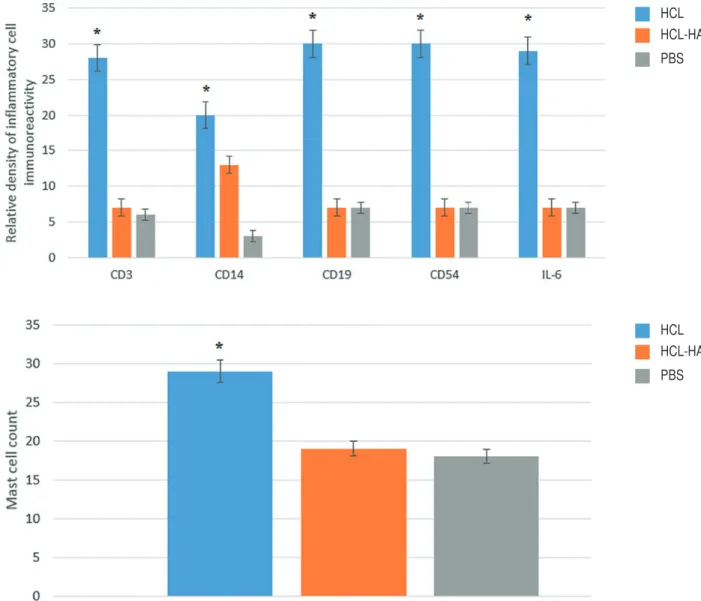
The immunohistochemical assessment sec-tions were semi-quantitatively evaluated by two di-fferent observers. After immunohistochemical stai-ning, it was observed that there was infiltration rich in monocyte cells and very rich in T lymphocyte, B lymphocyte, and natural killer cells in the blad-der tissue of the HCl group, especially in the stroma layer. Levels of IL-6 correlated with the severity of inflammation were elevated to peak levels in the HCl group, especially in the stroma and smooth muscle layer. In the HA-treated group which was treated with single dose intravesical HA, it was observed that the rich infiltration formed in the stroma layer of the bladder by inflammation cells and the se-verity of inflammation had statistically significance regressed to the levels of the PBS group (p<0.005) (Figure-1). Likewise, the increase in the number of activated mast cells in the HCL group significantly declined to level of the PBS group after intravesi-cal HA treatment (p<0.005) (Figure-2). In light of these findings, it has been determined that 0.5 mL, 0.8 mg/mL of intravesical HA instillation adminis-tered to the subjects in the treatment of chemical cystitis statistically significantly suppresses the inflammatory response (p<0.005) (Figure-3).
Myeloperoxidase Assay Results
An increase was observed in bladder MPO activity in the HCl group compared to the HA-tre-ated and PBS group (p<0.05). MPO activity levels in the HA-treated and PBS groups were found to be close to each other (Figure-4). It was observed that a result close to the pre-disease condition was achieved with HA treatment.
DISCUSSION
Figure 1 - Images representing immunohistochemical staining of CD3, CD14, CD19, CD56 and IL-6 in HCl group (A) and HCl-HA group (B). Black arrows indicate stained infl ammatory cells. It is observed that infl ammatory cell infi ltration is more common in stroma and smooth muscle.
Figure 3 - Relative density of CD3, CD14, CD19, CD56 and IL-6 immunoreactivity. Infl ammatory response was found to regress to the control group (PBS) levels signifi cantly compared with HCl group after intravesical HA treatment. The number of activated mast cells is signifi cantly reduced after intravesical HA treatment.
*P <0.005.
reported functions of the surface GAG layer are the prevention of surface entrapment of substan-ces and the regulation of transepithelial molecule movements. These polysaccharides containing ne-gatively charged sulfate form a thin layer in the form of a physical barrier between the cell surfa-ce and urine because they are highly hydrophilic (15). Parsons has shown that the GAG layer is sig-nifi cantly reduced in BPS/IC patients and that the normal permeability barrier is thereby impaired
(16). Subsequent animal experiments have shown that destruction of the GAG layer of urine can lead to bladder infl ammation and hyperactivity (17).
The fact that urologists are prone to drug administration into the bladder, the inability to achieve the desired level of response from oral tre-atments, and the belief that this disease is based on impairment of the bladder mucosa have brou-ght up the topic of intravesical treatments. The advantages of intravesical therapy are that it can
HCL
HCL HCL-HA
HCL-HA PBS
provide more intensive use of therapeutic agents in the bladder and limit systemic side effects while being invasive and carrying the risk of infection are its disadvantages. Many agents have been in-vestigated in BPS/IC treatment with the purpose of intravesical use. When heparin, one of the in-travesical agents that support the GAG layer, was intravesically administered at 25000 IU in 5 cc sterile water, improvements were observed in 72% of patients (18). In a non-randomized study with chondroitin sulfate, another agent that supports the GAG layer, approximately 70% improvement in symptom scores was observed (19). In the stu-dy by Kallestrup et al. (20) on HA, another GAG supporting agent which is accepted for its long--term efficacy, improvement in the symptoms was observed in 65% percent of female patients who underwent instillation treatment once weekly du-ring the first month and once monthly afterwards, and full cure was achieved after 3 years in 50% of patients. Another study involving 48 patients is also noteworthy due to demonstrating that HA treatment is also urodynamically effective and having a long follow-up period of 5 years (21). The formula for increasing the amount of HA by administering it from the outside can restore the integrity of the GAG layer by strengthening the
barrier function. However, there is no objective as-sessment tool other than some surveys to assess the effectiveness of GAG replacement therapy (22). To the best of our knowledge, there are no other stu-dies in the literature which demonstrate the results of HA treatment by assessing inflammatory cells in the tissue via immunohistochemical studies and MPO activity. In this respect, our study carries the distinction of being the first to do so.
Until recently, the only clinical material could be used in IC research and animal models has been created with the advent of experimen-tal studies. It has been shown that when chemi-cal cystitis models which have been created were compared in terms of both leukocyte counts and mast cell counts, they were found to be similar to IC and that therefore an animal model of IC can be created and studies can be done in this way. The IC model can be created with protamine sulfate (PS), cyclophosphamide, HCl, acetic acid, lipopolysac-charide, and uroplakin (23). In our study, we used HCl to create IC and pathological similarity with IC was found to be consistent with the literature.
Studies on animal models of IC have poin-ted at neutrophil infiltration, activation of cer-tain inflammatory cytokines in the bladder, and increased expression of inflammatory genes as
Figure 4 - Image representing MPO activity increase HCl group
HCL
HCL-HA
PBS
the source of symptoms (13, 24). Lv et al. (25) assessed results after intravesical HA in a PS--induced rat model of chemical cystitis with the immunohistochemical method and looked at IL-6 levels for the severity of inflammation. IL-6 le-vels which correlate with the severity of inflam-mation reached peak levels in the chemical cys-titis induced group, while a significant drop was observed in the IL-6 levels of the group which was treated with single dose HA after chemical cystitis. We also found a significant decrease in mast cell count and IL-6 levels in our study with single dose intravesical HA treatment compared to the disease group and obtained a result consis-tent with the literature. In addition, the MPO ac-tivity level and the other immunohistochemical markers, namely CD3, CD14, CD19, and CD56, were found to have reached the level of control group rats in the group receiving HA treatment after the procedure and near normal mucosa, in-tact epithelium, and basal membrane layer pre-sence were observed.
The limitation of our study included small number of experiment animals, evaluation of effi-cacy of single dose treatment and lack of histopa-thological results of long-term treatment. Long--term symptomatic remission with intravesical hyaluronic acid treatment has been shown in IC (26). Future works should investigate the long--term effects of intravesical hyaluronic acid treat-ment on bladder inflammation in patients with IC.
CONCLUSIONS
Our study has shown that single dose in-travesical HA instillation in the rat model of IC is effective in treatment by reducing inflammatory cell infiltration and the severity of inflammation, preserves the mucosal integrity of the bladder, and thus achieves histopathological improvement. The low cost and the lack of side effects may enable HA to be among the first drugs to be preferred for the treatment of IC. However, extensive research is needed to determine its functional effects on blad-der capacity and compliance, and to determine to which extent its combined use with other methods enabling GAG layer repair will affect the topical effects of HA that we have identified.
ACKNOWLEDGEMENTS
This study was supported by the Akdeniz University Scientific Research Projects (project number: 2014.04.0103.002).
CONFLICT OF INTEREST
None declared.
REFERENCES
1. van de Merwe JP, Nordling J, Bouchelouche P, Bouchelouche K, Cervigni M, Daha LK, et al. Diagnostic criteria, classification, and nomenclature for painful bladder syndrome/interstitial cystitis: an ESSIC proposal. Eur Urol. 2008;53:60-7. 2. Fall M, Baranowski AP, Elneil S, Engeler D, Hughes J,
Messelink EJ, et al. EAU guidelines on chronic pelvic pain. Eur Urol. 2010;57:35-48.
3. Grover S, Srivastava A, Lee R, Tewari AK, Te AE. Role of inflammation in bladder function and interstitial cystitis. Ther Adv Urol. 2011;3:19-33.
4. Davis NF, Brady CM, Creagh T. Interstitial cystitis/painful bladder syndrome: epidemiology, pathophysiology and evidence-based treatment options. Eur J Obstet Gynecol Reprod Biol. 2014;175:30-7.
5. Hanno PM, Burks DA, Clemens JQ, Dmochowski RR, Erickson D, Fitzgerald MP, et al. AUA guideline for the diagnosis and treatment of interstitial cystitis/bladder pain syndrome. J Urol. 2011;185:2162-70.
6. Leppilahti M, Hellström P, Tammela TL. Effect of diagnostic hydrodistension and four intravesical hyaluronic acid instillations on bladder ICAM-1 intensity and association of ICAM-1 intensity with clinical response in patients with interstitial cystitis. Urology. 2002;60:46-51.
7. Bernard A, Boumsell, L, Dausset J, Milstein C, Schlossman SF. Leucocyte Typing. Berlin, Germany: Springer-Verlag; 1984.
8. Kishimoto T. The biology of interleukin-6. Blood. 1989;74:1-10.
9. Theoharides TC, Sant GR, el-Mansoury M, Letourneau R, Ucci AA Jr, Meares EM Jr. Activation of bladder mast cells in interstitial cystitis: a light and electron microscopic study. J Urol. 1995;153(3 Pt 1):629-36.
11. Bradley PP, Priebat DA, Christensen RD, Rothstein G. Measurement of cutaneous inflammation: estimation of neutrophil content with an enzyme marker. J Invest Dermatol. 1982;78:206-9.
12. Lynes WL, Flynn SD, Shortliffe LD, Stamey TA. The histology of interstitial cystitis. Am J Surg Pathol. 1990;14:969-76. 13. Smaldone MC, Vodovotz Y, Tyagi V, Barclay D, Philips BJ,
Yoshimura N, et al. Multiplex analysis of urinary cytokine levels in rat model of cyclophosphamide-induced cystitis. Urology. 2009;73:421-6.
14. Gülpınar O, Kayış A, Süer E, Gökçe MI, Güçlü AG, Arıkan N. Clinical comparision of intravesical hyaluronic acid and hyaluronic acid-chondroitin sulphate therapy for patients with bladder pain syndrome/interstitital cystitis. Can Urol Assoc J. 2014;8:E610-4.
15. Lilly JD, Parsons CL. Bladder surface glycosaminoglycans is a human epitelial permeability barrier. Surg Gynecol Obstet. 1990;171:493-6.
16. Parsons CL. The role of a leaky epithelium and potassium in the generation of bladder symptoms in interstitial cystitis/overactive bladder, urethral syndrome, prostatitis and gynaecological chronic pelvic pain. BJU Int. 2011;107:370-5.
17. Chuang YC, Chancellor MB, Seki S, Yoshimura N, Tyagi P, Huang L, et al. Intravesical protamine sulfate and potassium chloride as a model for bladder hyperactivity. Urology. 2003;61:664-70. 18. Kuo HC. Urodynamic results of intravesical heparin therapy
for women with frequency urgency syndrome and interstitial cystitis. J Formos Med Assoc. 2001;100:309-14.
19. Steinhoff G, Ittah B, Rowan S. The efficacy of chondroitin sulfate 0.2% in treating interstitial cystitis. Can J Urol. 2002;9:1454-8.
20. Kallestrup EB, Jorgensen SS, Nordling J, Hald T. Treatment of interstitial cystitis with Cystistat: a hyaluronic acid product. Scand J Urol Nephrol. 2005;39:143-7.
21. Daha LK, Riedl CR, Lazar D, Hohlbrugger G, Pflüger H. Do cystometric findings predict the results of intravesical hyaluronic acid in women with interstitial cystitis? Eur Urol. 2005;47:393-7; discussion 397.
22. Porru D, Leva F, Parmigiani A, Barletta D, Choussos D, Gardella B, et al. Impact of intravesical hyaluronic acid and chondroitin sulfate on bladder pain syndrome/interstitial cystitis. Int Urogynecol J. 2012;23:1193-9.
23. Song PH, Chun SY, Chung JW, Kim YY, Lee HJ, Lee JN, et al. Comparison of 5 Different Rat Models to Establish a Standard Animal Model for Research Into Interstitial Cystitis. Int Neurourol J. 2017;21:163-70.
24. Chung MK, Butrick CW, Chung CW. The overlap of interstitial cystitis/painful bladder syndrome and overactive bladder. JSLS. 2010;14:83-90.
25. Lv YS, Yao YS, Lin ME, Rong L, Deng BH, Huang J, et al. Interleukin-6 levels in female rats with protamine sulfate-induced chronic cystitis treated with hyaluronic acid. Int J Urol. 2013;20:1017-22.
26. Engelhardt PF, Morakis N, Daha LK, Esterbauer B, Riedl CR. Long-term results of intravesical hyaluronan therapy in bladder pain syndrome/interstitial cystitis. Int Urogynecol J. 2011;22:401-5.
_______________________ Correspondence address: Erhan Ates, MD Department of Urology Adnan Menderes University School of Medicine, 09010, Aydin, Turkey