Eiji KobayashiI
New trends in translational microsurgery
1Abstract
Technological advances such as optical instruments and surgical tools have enabled the considerable contributions of microsurgery to surgical therapies. Accordingly, surgical therapeutics has provided the latest information across a wide range of medical specialties, including immunology and pharmacology, despite specialization according to organs and organ systems. The International Society for Experimental Microsurgery, an academic organization, has utilized experimental microsurgery technology in the identification of curative concepts for diseases that remain difficult to treat. For this publication to mark the 32nd anniversary of the Brazilian Surgical Society, I introduced the following types of technology related to the further development of microsurgical technological innovations in the future: high-resolution three-dimensional (3D) video and touch-sensitive microsurgery robots.
Key words: Microsurgery. Video-Assisted Surgery. Robotics.
IMD, PhD, Department of Organ Fabrication, Keio University School of Medicine, Tokyo, Japan. Conception and design of
the study, manuscript writing, critical revision.
lives by transplanting new organs into patients who had experienced lethal organ failure2.
Brazil was a leading clinical center worldwide during the early stages of transplantation therapy; here, pioneers in the field were performing organ transplantation in cases that were otherwise difficult to treat3. However, it
was difficult to achieve good clinical results with transplantation, and better immunosuppressive and organ preservation methods were urgently
■
Introduction
Table 1 - The year and place in which each conference was held, as well as the names of the chairman
of each conference.
to promote the creation of new treatments5.
Currently, revolutionary progress in the field of medical optical instruments is ongoing, and surgical techniques are beginning to incorporate robotic technology. This year, as the Brazilian Journal of Surgery celebrates its 32nd birthday, we would like to introduce two new types of technology that we hope will have great impacts on the world of microsurgery.
■
Changes in the International
Society for Experimental Microsurgery
(ISEM)
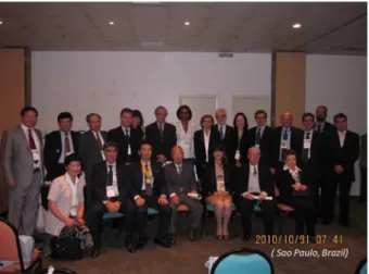
Table 1 shows the locations of all previous international conferences, as well as the respective chairmen. In 2010, 26 years after the founding of this organization, the 10th ISEM international conference was held in Sao Paulo, Brazil. At that conference, Professor Saul Goldenberg served as the honorary organizer, Professor Edna Montero and Professor Murched O. Taha served as co-hosts, and I served as the 10th ISEM chairman (Figure 1). models that were similar to humans. Dr. Sun
Lee, the founder of the International Society for Experimental Microsurgery (ISEM), was the first person to develop a rat organ transplant model and disseminate that technology worldwide4.
However, it remained necessary to overcome the difficulties encountered when integrating medical specialties based on organs and organ systems and thus use an interdisciplinary approach that combined immunology, pharmacology and physiology. In 1992, Dr. Lee started the ISEM as an organization that aimed to take a more interdisciplinary approach and address all steps in the research process, from foundational research to clinical application. In 2010, ISEM held its 10th
Figure 1 - Group photo of the ISEM board members
in Sao Paulo, Brazil in 2010.
These ISEM academic conferences are held every 2 years, and topics such as foundational research and cutting edge clinical achievements, as well as microsurgery-related education, are routinely discussed. In the next section, I will discuss two types of microsurgery technology on which I have recently worked.
■
Application of high-resolution 3D
video microscopes to microsurgery
Microsurgery with a conventional optical microscope provides a limited field of view and imposes serious strain on surgeons during long operations. Accordingly, an ophthalmological surgical technique known as “heads-up surgery” was recently developed; here, the field of view is displayed on a screen via a high-resolution camera6.This method
is now being applied to various types of
advanced microsurgery.
In our specialty, arterial reconstruction for living-donor pediatric liver transplantation, microsurgery has the following characteristics5,7:
1. Narrow field of view;
2. Deep operating field (1 and 2 also apply to brain surgery);
3. Respiratory movement (variable focal depth);
4. Requirement for the use of a microscope (microscopy setup: ~15–30 minutes). To overcome these issues, the following improvements can be made using the heads-up method with a 3D-4K video system.
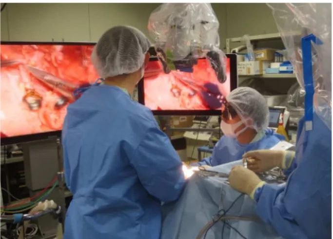
Figure 3 - Testing of the latest three-dimensional (3D)-4K videoscope on a porcine model.
The photo depicts surgeons performing a microsurgery operation (seated) and their assistants (standing). Both groups can look directly at the 3D image (Photo: Kyoto University School of Medicine, Animal Experimentation Laboratory).
3. A 3D view of the operating field is available. Accordingly, a dual-eye (two HD sensors) system will provide greater evolutional views; 4. Given the viewing distance, the
focal depth becomes pan-focal and is not greatly disturbed by pulsing blood vessels or movement (e.g., respiratory);
5. The video system can be used in conjunction with fluorescence imaging, such as indocyanine green, to measure blood flow and identify malignancies.
We hope that this new 3D-4K video system will reduce surgical complications and increase patient safety. This system should simultaneously relieve the stress and neck pain experienced by operators.
■
Development of robot-assisted
microsurgical forceps with haptic
feedback
Today, many surgical procedures
are performed arthroscopically. Surgical techniques are often practiced using dummies, cadavers and live pigs (Figure 4).
Figure 4 - Arthroscopic surgery training using a
porcine model. This type of surgical practice has been common since the early 2000s. In recent years, it has also been used for robotic surgery training (Photo: Jichi Medical University CDAMTec).
technology8.
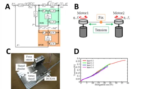
Bilateral control, an engineering methodology used to transmit haptic sensation, has been developed via senseless technology (Figure 5)9. With this method, haptics can be
controlled mutually between the “Master,” which is operated by a human, and the “Slave,” which directly manages surgical sites. We have further extended our research to the topic of
“scaling bilateral control,” wherein the haptic force is magnified between the “Master” and “Slave”. A novel device equipped with scaling bilateral control system has been partially developed9. An attempt was made to prove
the ability of a magnifying tensile force to identify the extremely thin threads used in microsurgery. We successfully demonstrated the ability to pull a thread of spider silk (Figure 6).
Figure 5 - The haptic system developed by the Faculty of Science and Technology at Keio University. A. Overview
diagram; B. Simple micro-threader; C. Actual micro-threader; D. Micro-threading results (from reference 9).
8441345.
3. Murray JE. The Nobel Lectures in Immunology. The Nobel Prize for Physiology or Medicine, 1990. The first successful organ transplants in man. Scand J Immunol. 1994;39(1):1-11. PMID: 8290887.
4. Okumura M, Mester M. The coming of age of small bowel transplantation: a historical perspective. Transplant Proc. 1992;24(3):1241-2. PMID: 1604601.
5. Nemeth N. Tribute to the “Father” of experimental microsurgery, Professor Sun Lee (1920-2015). Microsurgery. 2016;36(2):97-8. doi: 10.1002/micr.30015. 6. Kobayashi E, Haga J. Translational
microsurgery. A new platform for transplantation research. Acta Cir Bras. 2016;31(3):212-7. doi: 10.1590/S0102-865020160030000010.
7. Eckardt C, Paulo EB. Heads-up surgery for vitreoretinal procedures: an experimental and clinical study. Retina. 2016;36(1):137-47. doi: 10.1097/IAE.0000000000000689. 8. Sanada Y, Wakiya T, Hishikawa S, Hirata
Y, Yamada N, Okada N, Ihara Y, Urahashi T, Mizuta K, Kobayashi E. Risk factors and treatments for hepatic arterial complications in pediatric living donor liver transplantation. J Hepatobiliary Pancreat Sci. 2014;21(7):463-72. doi: 10.1002/jhbp. 9. Deshpande N, Chauhan M, Pacchierotti
C, Prattichizzo D, Caldwell DG, Mattos LS. Robot-assisted microsurgical forceps with haptic feedback for transoral laser
microsurgery. Conf Proc IEEE Eng Med Biol Soc. 2016;2016:5156-9. doi: 10.1109/ EMBC.2016.7591888.
10. Oda K, Ohnishi K, Kobayashi E. Verification of a developed testing machine for suture threads through measurement of tension and elongation. 14th International Workshop on Advanced Motion Control, AMC 2016. Institute of Electrical and Electronics Engineers Inc; 2016. p.186-91. doi: 10.1109/AMC.2016.7496348.
■
Acknowledgements
The 3D-4K project has received support from the Japanese government (AMED) for a 3-year period (2017–2019), in collaboration with Mitaka Kohki and Panasonic Ltd. in the industrial sector and the Faculty of Medicine at Kyoto University (Dr. Shintaro Yagi and Professor Shinji Uemoto) and the Keio University School of Medicine in the academic sector.
This project (principal investigator: Kawana DDS, Keio University School of Medicine) is also supported by the Japanese government (AMED) for a 3-year period (2017– 2019), in collaboration with the Department of System Design Engineering, Keio University (Professors Takahiro Nozaki and. Kouhei Ohnishi).
Correspondence:
Eiji Kobayashi
Department of Organ Fabrication, Keio University School of Medicine
35 Shinanomachi, Shinjuku-ku, Tokyo 160-8582 Japan
Phone: 81-3-5315-4090 organfabri@keio.jp
Received: May 08, 2018 Review: July 05, 2018 Accepted: Aug 03, 2018
Conflict of interest: none Financial source: none
1Research performed at Department of Organ