UNIVERSIDADE DA BEIRA INTERIOR
Faculdade de Ciências
Detection of biogenic amines in urine and plasma
by liquid chromatography coupled to
electrochemical detection (HPLC-ED) using
microextraction in packed syringe (MEPS)
David Jerónimo Oppolzer
Dissertação para obtenção do Grau de Mestre em
Bioquímica
(2º ciclo de estudos)
Orientador: Prof
a. Doutora Maria Eugénia Gallardo Alba
Orientador: Prof. Doutor Luis António Paulino Passarinha
iii
Resumo alargado
As aminas biogénicas são neurotransmissores com importantes funções ao nível do sistema endócrino e ao nível do sistema nervoso central. Alterações e distúrbios na sua função normal estão frequentemente associadas a várias doenças e desordens psicológicas, nestas situações ocorre uma alteração, por excesso ou défice, dos níveis normais circulantes destes compostos. Estes níveis podem também estar alterados em pacientes sujeitos a terapias com fármacos psicoterapêuticos (por exemplo antidepressivos e antipsicóticos) e em casos de consumo de algumas drogas de abuso. Ainda, elevados níveis de aminas biogénicas são importantes marcadores para certo tipo de tumores que secretam estes compostos pelas suas propriedades hormonais. Deste modo, é de interesse clínico e neuroquímico a existência de métodos de deteção e quantificação destes compostos em amostras biológicas.
O objectivo deste trabalho foi o desenvolvimento e validação de um método analítico para a deteção e quantificação das aminas biogénicas, serotonina (5-HT), dopamina (DA) e norepinefrina (NE) em amostras de urina e plasma, usando a microextração em seringa empacotada (MEPS) e cromatografia líquida acoplada à deteção eletroquímica de coulometria (HPLC-ED). A 3,4-dihidroxybenzilamina (DHBA) foi utilizada como padrão interno. A optimização do protocolo de MEPS foi efectuada com recurso ao desenho experimental (DOE) e os compostos foram detectados num sistema de cromatografia líquida de ultra performance acoplada a um detector de fluorescencia (UPLC-FLD). O uso de um desenho factorial fraccionado (2k-1) demonstrou ser uma ferramenta útil para a optimização do método de extração, reduzindo o número de experiências, minimizando o tempo de processamento da amostra, reagentes e trabalho laboratorial. O protocolo de MEPS optimizado inclui 8 aspirações da amostra, sem passo de lavagem e duas eluições de 100 µL de metanol.
O método foi completamente validado de acordo com critérios internacionalmente aceites, da Food and Drug Administration (FDA). O método demonstrou ser linear na gama de 50 a 1000 ng/mL para a 5-HT, e de 5 a 1000 ng/mL para a DA e NE, com coeficientes de determinação (R2) superiores a 0.99 para todos os compostos. Os limites de deteção e quantificação foram, respectivamente, de 20 e 50 ng/mL para a 5-HT, e de 2 e 5 ng/mL para a DA e NE. Para todos os compostos, a precisão intra- e interdia variou entre 1 a 10 % e a exactidão esteve no intervalo de ±15%.
A eficácia do método foi avaliada, por aplicação da metodologia validada a amostras reais de urina e plasma. Foi possível detetar e quantificar os 3 compostos em amostras de urina e a 5-HT em plasma. Por tudo anteriormente citado, podemos afirmar que o método desenvolvido é apropriado para a determinação destes compostos em análises laboratoriais de rotina nestas matrizes biológicas.
iv Este é o primeiro estudo que utiliza uma coluna de MEPS comercializada para a determinação simultânea de 5-HT, DA e NE em amostras de urina, sendo também o primeiro a aplicar MEPS para a determinação de 5-HT em amostras de plasma.
Como método de extração, MEPS demonstrou ser uma técnica de preparação da amostra simples e rápida, com baixo consumo de solventes, poupando tempo, dinheiro e trabalho aos laboratórios.
Palavras chave
vi
Abstract
Biogenic amines are neurotransmitters involved in several physiological processes. Changes and disturbs in their function are associated to several neurological disorders or diseases, as well as consumption of psychotherapeutic or psychotropic drugs. The detection of these compounds in biological fluids is therefore of clinical and neurochemical interest.
The goal of this work was to develop and validate and analytical method for the detection and quantification of the biogenic amines serotonin (5-HT), dopamine (DA) and norepinephrine (NE), using microextraction in packed syringe (MEPS) and liquid chromatography coupled to electrochemical detection (HPLC-ED) in both urine and plasma samples. The internal standard used was 3,4-dihydroxybenzylamine (DHBA). The MEPS extraction procedure was optimized using the design of experiments (DOE) tool, and the final conditions of 8 strokes, no washing, and two elutions of 100 µL methanol were chosen.
The method was fully validated according to internationally accepted guidelines from the Food and Drug Administration. Linearity was established between 50-1000 ng/mL for 5-HT and between 5-1000 ng/mL for DA and NE, with determination coefficients (R2) higher than 0.99 for all compounds. The limits of detection and quantification were respectively 20 and 50 ng/mL for 5-HT, and 2 and 5 ng/mL for DA and NE. Intra- and interday precision ranged from 1 to 10 %, while accuracy was within a ±15% interval for all compounds.
Authentic urine and plasma samples were analysed by the validated method, and the three compounds were detectable and quantifiable in urine, while only 5-HT was detected and quantified in plasma.
This is the first time that a commercially available MEPS column was used for the simultaneous detection of biogenic amines in urine, and also the first time that 5-HT was detected and quantified in plasma using MEPS. MEPS proved to be fast to perform with the use of less solvent volumes, saving time and money and being less laborious. The validated method is useful for the determination of biogenic amines in laboratorial routine.
Keywords
viii
Index
Resumo alargado ... Abstract ... Index of figures ... Index of tables ... List of abbreviations ...Justifications and objectives ... I. Revision of literature ... 1. Biogenic amines and amine transmission ...
1.1. Physiological properties of biogenic amines ... 1.1.1 Serotonin ... 1.1.2 Catecholamines ... 1.2. Biogenic amine secretion, metabolism and re-uptake ... 1.3. Disturbs in normal biogenic amine transmission ... 1.3.1. Diseases and psychiatric disorders and biogenic amine transmission ... 1.3.2. Medications and drugs of abuse interfering with biogenic amines pathways ...
2. Biogenic amine detection ...
2.1. Biological fluids ... 2.2. Extraction techniques ... 2.2.1. Microextraction in packed syringe ... 2.3. Separation and detection techniques ... 2.3.1 HPLC coupled to electrochemical detection ...
II. Experimental part ... 1. Materials and methods ...
1.1. Reagents and standards ... 1.2. Instrumentation ... 1.3. Internal standard ... 1.4. Stock and working solutions ... 1.5. Chromatographic and detection systems ... 1.6. Chromatographic and detection conditions... 1.7. Biological samples ... 1.8. Optimized extraction procedure ...
2. Results and discussion ...
2.1. Optimization of the mobile phase ... 2.1.1. Amperometric system ... 2.1.2. Fluorescence system ... 2.1.3. Coulometric system ... iii vi xi xiii xv xix 1 1 1 1 2 3 4 6 7 8 8 9 10 12 13 17 17 17 17 18 18 18 19 20 20 21 21 21 23 23
ix 2.2. Optimization of the extraction procedure ... 2.2.1. Design of experiments (DOE) ... 2.3. Method validation ... 2.3.1. Selectivity ... 2.3.2. Linearity ... 2.3.3. Limits of detection (LOD) and quantification (LOQ) ... 2.3.4. Precision and accuracy ... 2.3.5. Recovery ... 2.4. Method application to urine and plasma samples ...
III. Conclusions ... IV. References ... 25 26 31 32 32 33 34 37 38 41 43
xi
Index of figures
Figure 1: Biosynthesis process and structures of serotonin (a) and the catecholamines
(dopamine, norepinephrine, epinephrine) (b)... 3
Figure 2: The serotonergic (A) and noradrenergic (B) synapses... 5
Figure 3: A MEPS syringe (250 µL) and BIN from SGE, and a scheme showing the composition of the extraction column... 10
Figure 4: Schematic representation of the different steps in a MEPS extraction procedure... 11
Figure 5: Chromatogram of 5-HT, DA, NE and E at 1 µg/ml using a mobile phase with 12.5 % methanol... 22
Figure 6: Chromatogram of 5-HT, DA, NE, E and internal standard (DHBA) at 1 µg/mL using a mobile phase with 10 % methanol... 22
Figure 7: Chromatogram obtained by fluorescent detection of 5-HT, DA, NE and internal standard (DHBA) at 50 ng/mL using the final optimized conditions... 23
Figure 8: Chromatogram showing the baseline signal of an injection of mobile phase (a) and the peak of NE at 50 ng/mL (b)...24
Figure 9: Chromatogram of 5-HT, DA, NE (50 ng/mL) and internal standard (DHBA, 250 ng/mL)... 25
Figure 10: Pareto charts of the effects for 5-HT, DA and NE... 29
Figure 11: Main effect plots for 5-HT, DA and NE... 30
Figure 12: Interaction plot of the experimental factors for 5-HT, DA and NE... 30
Figure 13: Chromatogram of an extracted urine sample (Urine 1)... 38
xiii
Index of tables
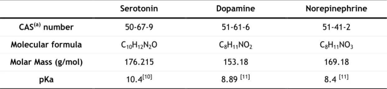
Table 1: Chemical properties of the monoamines serotonin, dopamine and norepinephrine... 3
Table 2: Normal range levels of NE, DA and 5-HT in urine and plasma... 12
Table 3: An overview of recent published techniques for biogenic amine determination in urine and plasma... 14
Table 4: Retention time variation of 5-HT, DA and NE with different methanol contents in the mobile phase... 21
Table 5: Factor levels in the design for MEPS optimization... 27
Table 6: Experimental design matrix showing the experimental factors, and response to each factor... 28
Table 7: Response surface matrix of the experimental factors and the respective response... 31
Table 8: Linearity data for 5-HT, DA and NE (n=5)... 33
Table 9: Within-run precision and accuracy (n=6) for all compounds... 35
Table 10: Between-run precision and accuracy (n=5) for all compounds... 36
Table 11: Combined within- and between-run precision and accuracy (n=15) for all compounds... 37
Table 12: Recovery at three different concentrations (n=3) for all compounds... 37
xv
List of abbreviations
5-HIAA 5-hydroxyindoleacetic acid 5-HT 5-hydroxytryptamine (serotonin) AD Alzheimer’s disease
BIN Barrel insert and Needle assembly C2 Ethylsilane
C8 Octylsilane C18 Octadecylsilane CAT Catecholamines
CAS Chemical Abstracts Service CB1 Cannabinoid receptor type 1 CE Capillary electrophoresis COMT Catechol-O-methyl transferase CV Coefficient of variation DA Dopamine
DAT Dopamine transporter DC Direct current
DHBA 3,4-dihydroxybenzylamine DOE Design of experiments DPBA Diphenyl boronic acid DS Down’s syndrome E Epinephrine
ED Electrochemical detection EDTA Ethylenediaminetetraacetic acid FDA Food and drug administration FLD Fluorescence detection GC Gas chromatography HClO4 Perchloric acid
xvi HPLC High Performance Liquid Chromatography
HVA Homovanillic acid
ICH International conference on harmonization IS Internal standard
KH2PO4 Potassium dihydrogen phosphate
LC Liquid chromatography LLE Liquid-Liquid extraction LOD Limit of detection
(L)LOQ (Lowest) limit of quantification
M1 Mixed mode with C8 and cation exchanger
MAO Monoamine oxidase MCX Mixed cation exchanger ME Metanephrine
MEPS Microextraction in packed syringe
MDMA 3,4-methylenedioxy-N-methylamphetamine MIP Molecular imprinted polymer
MS Mass spectrometry NE Norepinephrine
NET Norepinephrine transporter NME Normetanephrine
OSA Sodium octyl sulphate PD Parkinson’s disease R2 Determination coefficient RAM Restricted-access media RSM Response surface methodology SCX Strong cation exchanger SERT Serotonin transporter SPE Solid phase extraction SPME Solid phase microextraction
xvii THC ∆9-tetrahydrocannabinol
TOF Time-of-flight UV Ultraviolet
VMA Vanillylmandelic acid
xix
Justifications and Objectives
Neurotransmitters, including biogenic amines are since long known to play several important roles in the endocrine and nervous systems of the human organism.
Several psychiatric disorders and diseases (among others, depression, schizophrenia, Parkinson’s disease and tumours), are related to some extend with disturbs or changes in biogenic amine transmission. Biogenic amines are also the main target of several psychiatric medications and psychotropic drugs. All this situations affect the neurotransmitters either at their production, storage, release, re-uptake and metabolism or their receptors; as a result the normal concentrations of circulating biogenic amines are altered.
For these reasons, the development of a method for detection of biogenic amines in biological fluids is thus a very useful tool for the diagnosis of psychiatric disorders and diseases, and for the monitoring of patients undergoing psychiatric medication or cases of psychotropic drug consumption.
Therefore, the objective of this work was to develop an analytical method for the clinical determination of three biogenic amines, serotonin (5-HT), dopamine (DA) and norepinephrine (NE) in urine and plasma, using microextraction in packed syringe (MEPS) and high performance liquid chromatography coupled to electrochemical detection (HPLC-ED). The three transmitters were chosen based on their importance in the situations mentioned above and compatibility with the electrochemical detection.
Detection of biogenic amines in urine and plasma by HPLC-ED using MEPS
1
I. Revision of literature
1. Biogenic amines and amine transmission
1.1. Physiological properties of Biogenic amines
Biogenic amines are neurotransmitters and neuromodulator molecules with various
important functions at the central and peripheral nervous system. They are involved in several psychological and hormonal signalling processes, ranging from homeostatic functions to emotional and motor behaviours. Defects in their function are often associated with psychiatric disorders and most drugs of abuse interfere with amine transmission [1-7]. This has made the pharmacology of biogenic amine synapses the target of several medications in modern pharmacology [1].
Biogenic amines are of basic nature, with pKa values ranging from 8 to 11 (table 1), and are therefore protonated at physiological conditions. Structurally, they are composed of an amino group connected to an aromatic ring and so they are also called as monoamines (figure 1). The catecholamines (CATs), dopamine (DA), norepinephrine or noradrenalin (NE), epinephrine or adrenalin (E) and the indolamines serotonin (5-hydroxytryptamine, 5-HT) and histamine are the major endogenous monoamines in most animal species including humans [1-4,6].
1.1.1. Serotonin
Serotonin was first identified in 1948 as a serum tonic factor, released by platelets
during the clotting of blood, only latter was its function associated to a neurotransmitter molecule. The chemical structure of serotonin and related compounds contains a substituted indole moiety, consisting of a benzene ring and a nitrogen containing five-member ring, bonded to an amino group. The synthesis occurs through enzymatic hydroxylation and descarboxylation of the dietary amino acid L-tryptophan (figure 1a) [1-4,8]. Serotonergic neurons in the brain and enterochromaffin cells in mucosa of the gastrointestinal tract have the necessary enzymes for the biosynthesis process. Synthesis can also occur at the pineal gland, but as a precursor of melatonin. Most of the serotonin in the organism is stored at the serotonergic neurons, enterochromaffin cells and blood platelets (by uptake of circulating serotonin rather than secretion) [2,3].
Detection of biogenic amines in urine and plasma by HPLC-ED using MEPS
2 In the nervous system transmission serotonin’s action is mediated when it binds to one of its receptors on the postsynaptic neuron. A large number of receptors have been identified, and the synaptic transmission response is determined by the receptor to which serotonin binds and activates. Several behaviours have been associated to serotonin, as emotions, mood, sexuality, circadian rhythms, motor behaviours and mental state arousal. Other functions are also associated, such as the smooth muscle contraction, blood pressure regulation, gastrointestinal physiology, mediation of satiety and decreased food consumption [1-4,8,9].
1.1.2. Catecholamines
Catecholamines are structurally different from indolamines, mainly due to the fact
that instead of a central indole ring, as in serotonin, they have a central catechol ring with an amine side chain. The major endogenous catecholamines are DA, NE and E, and their synthesis occurs at the adrenal medulla, sympathic nervous system and brain. Synthesis of the three CATs occurs by a biochemical pathway starting from the amino acid tyrosine. In mammals both phenylalanine and tyrosine are precursors of CATs, since an enzyme, phenylalanine hydroxylase, located in the liver allows the conversion of dietary phenylalanine into tyrosine. The process occurs by a series of hydroxylation and decarboxylation reactions, originating sequentially dopamine, norepinephrine and epinephrine (figure 1b) [1-4]. Neurons only contain the necessary enzymes for their own production of neurotransmitters, thus the biosynthesis process ends either on DA, NE, or E in DA-, NE-, E-releasing neurons, respectively [2,4].
The catecholamines play important roles as hormones and as excitatory neurotransmitters, reflected by their synthesis and action at hormonal and nervous tissues. The CATs influence and control several physiological processes of the nervous system and systemic circulation as the stress system, heartbeat rate and blood pressure regulation. Dopamine has an essential role in the coordination of body movements, and is also involved in motivation, the reward system and reinforcement. NE and E functions have been implicated in control of arousal, attention, learning, memory and stress response influencing sleep and wakefulness, and also feeding behaviours [1,2,4,7].
Detection of biogenic amines in urine and plasma by HPLC-ED using MEPS
3
Table 1: Chemical properties of the monoamines serotonin, dopamine and norepinephrine. Serotonin Dopamine Norepinephrine
CAS(a) number 50-67-9 51-61-6 51-41-2
Molecular formula C10H12N2O C8H11NO2 C8H11NO3
Molar Mass (g/mol) 176.215 153.18 169.18
pKa 10.4[10] 8.89 [11] 8.4 [11]
aCAS – Chemical Abstract Service.
1.2. Biogenic amine secretion, metabolism and re-uptake
After synthesis, serotonin and CATs are stored in vesicles, by the vesicular monoamine transporter (VMAT) through active transport from the cytoplasm. The neurotransmission occurs through the release of the monoamines from the vesicles into the synaptic cleft and binding to different several different receptors (5-HT, dopamine and adrenergic receptors) on the postsynaptic neuron [1-5]. The interaction with the relevant receptors initiates a biochemical cascade, resulting in several physiological responses [12].
(a) (b)
Figure 1: Biosynthesis process and structures of serotonin (a) and the catecholamines (dopamine,
norepinephrine, epinephrine) (b). The enzymes involved in the several steps are shown as well. (Adapted from Purves D., et al., 2004) [1]
Detection of biogenic amines in urine and plasma by HPLC-ED using MEPS
4 The excess of neurotransmitter that remains in the synaptic cleft is removed mainly by a mechanism of re-uptake into the nerve terminal, through binding to specific membrane-associated monoamine transporter proteins, namely the serotonin (SERT), dopamine (DAT) and norepinephrine transporters (NET) [1-4].
The breakdown of monoamines is mediated mostly by the enzymes monoamine oxidase (MAO) and catechol-O-methyl transferase (COMT) [1-4,8,13-17], which are located in the cytoplasm in all mammalian tissues [2,13-16]. This is the main reason why monoamines have a short half life in the free form, and have to be stored into synaptic vesicles after synthesis [1-4]. Serotonin is metabolized by MAO to 5-hydroxyindoleacetic acid (5-HIAA) while CATs are metabolized both by MAO and COMT. The main metabolite of DA is homovanillic acid (HVA), NE and E originate normetanephrine (NME) and metanephrine (ME) respectively, and vanillylmandelic acid (VMA) as a final metabolite [2,4,14-17].
1.3. Disturbs in normal biogenic amine transmission
The normal action of biogenic amines in the organism may be compromised in a variety of situations that affect their synthesis, storage, re-uptake or catabolism (figure 2). Since biogenic amines are involved in such a variety of endocrine and neuronal functions, defects in their normal function often occurs in several neuropsychiatric and pathological situations [1-5]. This has made the pharmacology of amine synapses a crucial target for psychiatric therapy, with the development of drugs affecting the synthesis, binding to receptor and catabolism of these neurotransmitters [3]. On the other hand, amine synapse transmission is a target for most drugs of abuse, mainly psychoactive drugs (figure 2) [1,2,4,7].
Detection of biogenic amines in urine and plasma by HPLC-ED using MEPS
5
Figure 2: The serotonergic (A) and noradrenergic (B) synapses. Sites where most neuropsychiatric
disorders, psychotherapeutic medicines and drugs of abuse have effect are shown as well. (Adapted from Kandel R.E. et. al., 2000) [5]
Detection of biogenic amines in urine and plasma by HPLC-ED using MEPS
6
1.3.1. Diseases and psychiatric disorders and biogenic amine transmission
Biogenic amines are implicated in several affective behaviours as euphoria, depression and anxiety. These responses provide important biological function and can become disordered in such a manner to constitute an illness, generally termed as mood disorders [5]. A decrease in the serotonergic and noradrenergic tone in the brain is commonly associated with depression, resulting in lack of the behavioural effects euphoria and pleasure. In patients with this disorder, low circulating levels of 5-HT and CATs are identified [3-5,8,9]. High levels of monoamine transmission, specially 5-HT and DA, are characteristic to psychosis, which can also occur as a long term side effect of several stimulatory psychotropic drugs [1,4]. Anxiety disorders and schizophrenia are strongly associated with altered levels of biogenic amines, often reflected by dysfunction of the catabolic enzymes MAO and COMT [1,3,14].
The underlying biochemistry of the major neuropsychiatric disorders is often not completely understood, the therapeutic effects of antidepressants and antipsychotic have mostly provided valuable information of the neurochemical functions of monoamines in these disorders. In fact, serotonin alone is administered as an antidepressant in some cases of depression [4].
Neurodegenerative diseases also result in either an excess or deficit in monoamine availability, most notable are Parkinson’s disease (PD) and Alzheimer’s disease (AD). In PD a destruction of the dopaminergic neurons of the substantia nigra occurs, with subsequent loss of the capacity to coordinate movements. Locus coeruleus, the principle site of the brain of NE synthesis is lost in AD, but also in PD and Down’s syndrome (DS); compromising NE availability [2,3,14].
Some diseases of tumourigenic nature also affect normal biogenic amines function, and/or secretion. The hormonal effects of these neurotransmitters are also used as advantage for several tumours and blastomas. Notably serotonin is secreted in large amounts by carcinoid tumours, facilitating blood vessel irrigation of the tumour, resulting in a condition called carcinoid syndrome [9,15]. Neuroblastomas and other tumours affecting the brain will result in elevated monoamine levels. In fact, catecholamines are currently considered very important markers for neuroblastomas and pheochromocytomas diagnosis. 5-HT also plays a relevant role in several blood disorders, and both 5-HT and the CATs are involved and altered in cardiovascular diseases [9,14-19].
Detection of biogenic amines in urine and plasma by HPLC-ED using MEPS
7
1.3.2 Medications and drugs of abuse interfering with biogenic amines
pathways
As shown on figure 2, drugs can interfere in several steps of either serotonin or catecholamines pathway. Generally, drugs interfering with these pathways can be classified as depressant drugs and antidepressant drugs, either stimulating or inhibiting the monoamines transmission path [1,4,5,7,17].
Most drugs of abuse interact at some level with amine neurotransmission, including opiates, tranquilizers, hypnotics, designer drugs, stimulant drugs, hallucinogens, tryptamines and piperazines. Stimulant psychotropic drugs like cocaine and amphetamines stimulate monoamine release from the nerve terminal and block the monoamine transporters DAT and NET. The 3,4-methylenedioxy-N-methylamphetamine (MDMA) or “ecstasy” blocks both membrane SERT and vesicular VMAT transporters, preventing the re-uptake process, resulting longer period of the monoamines 5-HT, DA and NE availability in the synaptic cleft [1,2,4,7]. The Δ9-tetrahydrocannabinol (THC), a hallucinogenic drug, interacts with a presynaptic
receptor, CB1, where it reduces the release of neurotransmitters, exhibiting a neuromodulator function [2,4,5].
Psychotherapeutic drugs also have their major effect on aminergic transmission pathways. Based on their effects, they can be divided into antipsychotics, antidepressants, stimulants and antianxiety drugs [1].
Antipsychotics reduce the amount of monoamines available for nervous transmission. Reserpine, a traditional folk medicine used for hypertension and psychoses, inhibits the VMAT transporter, thereby keeping NE and DA in the cytoplasm where they undergo catabolism by MAO. Other commonly used antipsychotic drugs reduce monoamine levels by blocking specific receptors on the postsynaptic nerve [3,8].
Antidepressant drugs, as the tricyclic compounds, raise the circulating monoamine levels by inhibiting the re-uptake of both serotonin and norepinephrine. Another class of antidepressants selectively inhibit the re-uptake of serotonin or dopamine, by blocking their re-uptake receptors, SERT and DAT. These are called as selective serotonin re-uptake inhibitors [1,5]. Monoamine oxidase inhibitors block the activity of the enzyme MAO, thus preventing breakdown of biogenic amines. The consequence of this inhibition is longer persistence (longer half life) of monoamines in the organism, and is one of the mechanisms used for the treatment of anxiety disorders and depression [1,5].
Another class of therapeutic agents, not directly focused on the treatment of neuropsychiatry disorders, can significantly affect in a definite manner brain structures specific for biogenic amine transmission. Two methods used for the treatment of brain tumors and leukemias, radiotherapy and chemotherapy, are the most notable. Survivors of this type of treatment may develop neurocognitive deficits, primarily in the area of attention, resulting in loss of biogenic amine availability, especially DA [20,21].
Detection of biogenic amines in urine and plasma by HPLC-ED using MEPS
8
2. Biogenic amine detection
The quantification of monoamines in biological samples is of great interest for clinical areas and neurochemistry sciences. The evaluation of circulating concentrations of neurotransmitters in the organism is a very useful tool for the diagnosis of neuropsychiatric diseases and malignant tumours. Even if not to directly diagnose the disease, the detection of circulating monoamines can give an important idea of the neuronal signalling pathway affected and evaluate the physiological extent of the disease. With the same principle, biogenic amine detection can also be a very handful tool to accompany patients undergoing neuropychiatric and cardiovascular medications [16-18,22-24]. At last, knowledge of the circulating biogenic amine concentration could also be used as a secondary tool to monitor cases related to the consumption of drugs of abuse.
2.1 Biological fluids
After secretion monoamines are distributed through various tissues in the organism. For a successful quantification of the circulating levels in the body, it’s important the use of a sample or biological fluid of easy acquisition and usable for routine clinical tests. On the other hand, the sample matrix should have a good correlation between its monoamine concentration levels and the concentration in the brain and other functional organs.
Clinical analysis of biogenic amines in humans is mostly done in urine and plasma. Urine is obtained non-invasively and high volumes are often obtained, oppositely to plasma. Urinary monoamine concentrations reflect the activity of the sympatho-adrenal system and are useful markers for the diagnosis of several types of tumors. Plasma monoamines also provide very useful information about normal and pathological sympatho-adrenal activity. This sample is especially used to detect biogenic amines for the prognosis of several cardiovascular diseases and neuroendocrine tumors [9,12,14-19,22-25]. It has been found that levels of biogenic amines and metabolites in urine and plasma are increased in patients with cancer diseases, and decreasing levels directly reflect the level of applied therapy [14]. Both samples are not easy to adulterate, but several factors can influence the monoamine concentration, like food consumption and states of anxiety [15]. In plasma, sample collection has to take into account the ease of oxidation of the amines by MAO, and it is necessary to add antioxidants [17].
Detection of biogenic amines in urine and plasma by HPLC-ED using MEPS
9
2.2 Extraction techniques
When handling with complex samples like urine and plasma, a preparation step is crucial for a successful analysis [27,28]. The purpose of this step can be resumed to isolation of the compounds of interest from the matrix and concentration, since they may be present only in vestigial quantities. Removal of interfering compounds is an important step since they can have similar properties to the analytes of interest thus interfering with detection. Urine and plasma samples also contain macromolecules like proteins, which are incompatible with some chromatographic systems. A good extraction procedure must therefore be reproducible, selective, easy to automate and have a high recovery rate [26,29].
Samples can be submitted to a previous step before extraction, depending on the compound of interest, hydrolysis of conjugates and protein precipitation are often used before extraction protocols. In urine 5-HT, DA, E and NE are present in the free form, as sulphoconjugates and as glucoronides; in some studies determining total monoamine concentration, dissociation of this complexes is required [30,31].
In humans, urine and plasma monoamines are classically extracted by absorption on alumina or on boric acid gel and liquid-liquid extraction (LLE) [12,19,22,23]. But some limitations may be assisted with these methods, like lack of selectivity, low extraction yields, labor intensive and time consuming procedures, large volume of samples, and the use of toxic organic solvents in LLE. Recent works have focused mostly on solid phase extraction (SPE) for its easier automatization, high recovery rates, relatively fast procedures and use of lower organic solvent volumes [12,19,24,25,32-34].
SPE is presently the most commonly used sample preparation technique for several compounds, including biogenic amines. In SPE, the extraction, cleanup and concentration of compounds from the sample matrix occurs on a solid support [35]. Several papers have been published employing SPE in urinary and plasmatic biogenic amine sample preparation (Table 3). The recovery rate is often above 90 %, varying with the kind of cartridge used. Most used columns employed for monoamine SPE sample preparation are silica-based sorbents, containing octadecylsilane (C18) or octylsilane (C8) groups, and strong cation exchanger (SCX)
groups mixed with C8 (mixed cation exchanger, MCX) [12,19,24,25,32-34,36]. A specific one
step LLE procedure employing diphenyl boronic acid (DPBA) ethanolamine ester followed by SPE with a C18 sorbent is also reported [33,37].
For clinical monoamine detection, LLE and SPE are the two most frequently used sample preparation technique [22]. Table 3 resumes the most recent published techniques used for the determination of these compounds.
Detection of biogenic amines in urine and plasma by HPLC-ED using MEPS
10
2.2.1 Microextraction in packed syringe
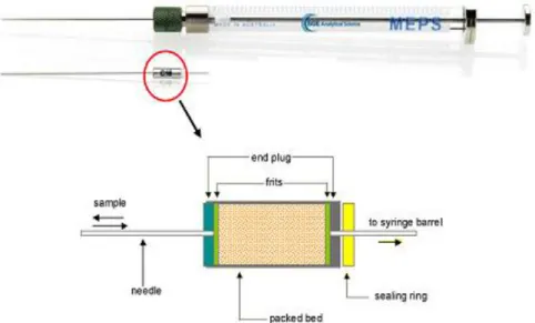
Microextraction in packed syringe (MEPS) is a recent microextraction technique developed in 2004 for AstraZeneca, Sweden. MEPS is considered as a miniaturized version of SPE, employing the basic principles of SPE but in form of small packed columns inserted into a syringe. This method is relatively fast to perform, simple and robust, and compared to SPE, MEPS has some considerable advantages. The use of organic solvents is reduced, the sample amount can be as low as 10 µL and the column can be re-used up to 100 extractions for plasma and 400 for water samples, oppositely to SPE columns which are discarded after usage. Another great achievement of MEPS is the possibility of directly injecting the eluates (typically 20-50 µL) into liquid chromatography (LC), gas chromatography (GC) and capillary electrophoresis (CE) systems without any modifications to the device, facilitating on-line coupling to any of these systems [27,28,35,38,39].
Physically MEPS consists of a syringe similar to the ones used for LC injection (100-250 µL), and a barrel insert and needle assembly (BIN) that contains the SPE column. In the BIN approximately 1 mg of solid sorbent is packed as a plug, protected by two inert frits on both sides to prevent its motion (figure 3). The sorbent chemistry of both SPE and MEPS are similar, commercially available BINs for MEPS include silica-based absorption materials [C18,
C2, C8, and M1 (mixed mode with C8 and cation exchanger)], restricted-access media (RAM)
and molecular imprinted polymers (MIPs) [27,28,38,39].
The main objective of MEPS is to simultaneously remove interfering compounds present in the matrix and to selectively isolate and concentrate the analytes of interest. The
Figure 3: A MEPS syringe (250 µL) and BIN from SGE, and a scheme showing the composition of the
Detection of biogenic amines in urine and plasma by HPLC-ED using MEPS
11 sample flows through the solid sorbent bed (extractant) by withdrawing the sample with the syringe. The analytes present in the liquid sample are isolated through retention on the solid phase. The column is then washed to remove interferences and the retained compounds are eluted with an organic solvent (figure 4) [38,39].
MEPS performance is affected by many factors, such as sample dilution, pH, composition and volumes of the washing and elution solutions, sorbent amounts and most importantly the sorbent material. The main step in the development of a MEPS extraction procedure requires optimization of these variables [35,38,39].
Initially the sorbent material is activated with an organic solvent like methanol, increasing the exposure of the functional groups for maximal interaction with the analytes in the sample. This is the conditioning step. For a successful passing of the sample through the sorbent, samples require dilution to reduce viscosity (1:5 for
urine and plasma, up to 1:25 for blood) and removal of any unnecessary macro particles by centrifugation and eventually deproteinization. The adjustment of the pH of the sample is a crucial step for reverse-phase sampling. The aspiration speed of the sample should not be too high (normally ranging from 10 to 20 µL/s) to allow a strong interaction between analytes and sorbent, as well as not to physically force and destroy the column sorbent. Repeated circles of draw-ejection from the sample can be used to increase analyte retention, thus obtaining higher recoveries [38,39].
To remove any interfering species and proteins that may have interacted with the sorbent, a washing step is usually performed with water, buffers or aqueous solutions containing a small proportion of organic solvents. The volume and constitution of this solution is very important, it should be able to remove any unwanted compounds from the stationary phase and at the same time interact to the least extent possible with the analytes of interest [38,39].
Finally an elution step is done using an organic solvent for which the analyte has affinity to disrupt the interaction between analyte an stationary phase. This solvent should be able to elute all analytes in small volumes and be miscible with the mobile phase of the chromatographic system, normally methanol, isopropanol or acetonitrile are used [38,39]. In some cases the volatile eluent can be dried under a gentle nitrogen stream and re-dissolved in the mobile phase or another solution before injection. This allows concentration of the eluted analyte.
In one recent work, El-Beqqali A. et. al. (2007) developed a protocol, for the determination of 5-HT and DA in urine, using MEPS online with LC-MS-MS detection. In this method a polystyrene polymer sorbent (silica-C8) was manually injected inside a MEPS
Figure 4: Schematic
representation of the
different steps in a MEPS extraction procedure.
Detection of biogenic amines in urine and plasma by HPLC-ED using MEPS
12 syringe, validation was done in urine and a low sample volume (30 µL) was used. The limits of quantification (LOQ) and detection (LOD) for both compounds were 50 ng/mL and 1 ng/mL respectively, and the extraction recovery was about 50 % [40]. This is the only published work that uses this extraction technique for the determination of biogenic amine neurotransmitters.
2.3 Separation and detection techniques
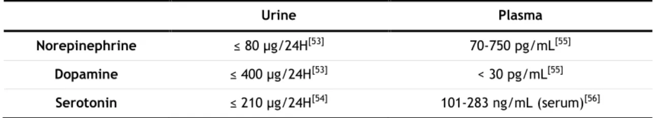
The range levels of neurotransmitters in plasma and urine vary mostly at very low concentrations, very often in the order of ng/mL and pg/mL (table 2). At this level it is required highly sensitive and selective detection and separation techniques, especially considering the chemical instability of these compounds, the complex matrix from which they are extracted and the metabolites present [12].
Historically, analysis of monoamines has been done by immunoassays [41], voltammetry [42], and gas chromatography coupled to mass spectrometry (GC-MS) [43]. But these methods have proven to be either time consuming, expensive, may require derivatization reactions or in some cases are not sensitive enough to detect the physiological concentration ranges (table 2). Due to these limitations, monoamine analysis in biological samples has focused to more rapid and sensible methodologies. High performance liquid chromatography (HPLC) is one of the most popular separation techniques used [44]. Several studies have reported methodologies using HPLC coupled to electrochemical (ED) [12,14,15,21-23,25,30,31,33,34,45,46], ultraviolet (UV) [47,48], fluorescence (FLD) [46,49] and mass spectrometry (MS) [50,51,52] detectors. Several studies also employ capillary electrophoresis (CE) coupled with UV [19,24,34], MS [34] and time of flight/mass spectrometry (TOF/MS) [24,32]. Table 3 shows an overview of recent published detection techniques for biogenic amines.
Table 2: Normal range levels of NE, DA and 5-HT in urine and plasma.
Urine Plasma
Norepinephrine ≤ 80 µg/24H[53] 70-750 pg/mL[55]
Dopamine ≤ 400 µg/24H[53] < 30 pg/mL[55]
Detection of biogenic amines in urine and plasma by HPLC-ED using MEPS
13
2.3.1 HPLC coupled to electrochemical detection
Electrochemical detectors (ECD or ED) are amongst the most sensible methods used
nowadays for monoamine detection. These detectors rely on an electrochemical reaction, wherein a certain constant potential (in DC mode) is applied by an electrochemical cell (electrode) to the flowing mobile phase. If the potential is high enough it will oxidize or reduce the elements in the mobile phase, which produces a current signal measured in function of time, originating a result given in a current vs. time chromatogram [57].
Compounds must be of electroatractive nature to be suitable for this type of detection. By selecting a specific potential under which the compound of interest undergoes reduction or oxidation, a signal is produced proportional to the quantities present in the sample. The applied potential is characteristic to each compound, and is dependent on various factors as the nature of the electrochemical cell surface, composition of the mobile phase and pH. A cyclic voltammogram consists of a peak area/potential curve, and can be obtained by injecting separately the compound of interest at a range of voltages. The resulting curve gives the information of the maximum oxidation potential of the compound, and can be used to choose a potential at which no interferences are oxidized optimizing the signal to noise ratio [57].
There are two types of electrochemical detectors, amperometric and coulometric. In an amperometric detector, the electrochemical cell has a design as which allows the mobile phase to flow over its surface, being only partly exposed to the electrode’s surface hence only part of the electroatractive species present in the flow undergo an electrochemical reaction. The corresponding signal of this fraction (typically 5-15 % of the total amount) is then converted into an estimation of the total of species present in the sample [57].
Although it’s relative high sensitivity, amperometric detectors usually do not allow reaching very low concentrations of electroatractive compounds. Enhanced sensitivity can be obtained with the use of a coulometric detector cell; in this design the flow does not pass over the surface but rather through a porous graphite electrode. Resulting in a higher exposure area of the solvent to the electrode, consequently most of the electroatractive species are oxidized or reduced, giving a higher signal without a corresponding increase of background noise, thus allowing detection of lower quantities of compound of interest. In this detection the signal typically corresponds directly to almost 100 % of the analytes present in the sample [57].
Several studies reported detection methods for serotonin and catecholamines, using HPLC coupled to amperometric or coulometric detection (table 3). Coulometric detection has shown to obtain lower detection limits, almost twice as low when compared to amperometric detection [25].
Detection of biogenic amines in urine and plasma by HPLC-ED using MEPS
14
Table 3: An overview of recent published techniques for biogenic amine determination in urine and plasma.
Compound (s) Sample Sample Preparation Detection mode
LOD (ng/mL) LOQ (ng/mL) Recovery (%) Reference
DA, NE Plasma SPE HPLC-ED
(Coulometric) 12 40
DA – 98
NE – 95 [12]
DA Urine SPE CE-UV 107 127 103.5 [19]
DA, NE Urine SPE (Amperometric) HPLC-ED NE – 8.8 DA – 7 N/A NE – 82.5 DA – 93 [22]
DA, NE Plasma Alumina Adsorption (Coulometric) HPLC-ED N/A N/A NE – 87.5 DA – 60 [23]
DA, NE Urine SPE
CE-UV NE – 20.3 DA – 17 N/A DA – 85.95 NE – 83.7
[24]
CE-TOFMS NE – 50.75 DA – 46 N/A DA – 85.95 NE – 83.7
DA, NE Plasma SPE
HPLC-ED (Amperometric) DA - 0.15 NE – 0.10 DA – 0.20 NE – 0.20 DA – 75.3 NE – 91.7 [25] HPLC-ED (Coulometric) DA - 0.06 NE – 0.05 DA – 0.12 NE – 0.10 DA – 75.7 NE - 92
5-HT Plasma SPE CE-TOFMS 26.5 N/A 71.6 [32]
DA, NE Urine LLE and SPE
HPLC-ED (Coulometric) DA – 4 NE – 2.4 DA – 9.8 NE – 5.4 DA – 92.25 NE – 87.5 [33]
Detection of biogenic amines in urine and plasma by HPLC-ED using MEPS
15
Compound (s) Sample Sample Preparation Detection mode LOD (ng/mL) LOQ (ng/mL) Recovery (%) Reference DA Urine SPE
HPLC-ED N/A N/A
44.5 [34]
CE-UV 107 N/A
CE-MS 183.8 N/A
DA, NE Plasma SPE HPLC-ED
(Coulometric)
DA – 0.01
NE – 0.02 N/A
DA – 64.6
NE – 74.6 [37]
DA, NE Urine MEPS LC-MS/MS 1 50 50 [40]
5-HT, DA … SPME HPLC-ED
(Amperometric) 0.1 N/A N/A [45]
5-HT, DA, NE Urine SPE
HPLC-FLD 8 N/A DA – 90.2 NE – 91.3 [46] HPLC-ED DA – 0.1 NE – 0.2 N/A
DA, 5-HT Urine Deproteinization with HClO4 HPLC-FLD
DA – 10 5-HT – 8 DA – 30 5-HT – 24 DA – 96.36 5-HT – 87.5 [49]
DA, NE Urine LLE LC-MS/MS N/A DA – 2.5
NE – 10 71 [50]
5-HT Urine and plasma On-line SPE LC-MS/MS N/A Urine – 5.3
Plasma – 158.6
Urine - 84
Plasma – 57.5 [51]
DA, NE Urine On-line SPE LC-MS/MS N/A DA – 1.8
NE – 2.7 N/A [52]
Detection of biogenic amines in urine and plasma by HPLC-ED using MEPS
Detection of biogenic amines in urine and plasma by HPLC-ED using MEPS
17
II. Experimental part
1. Materials and methods
1. 1. Reagents and standards
Serotonin hydrochloride, dopamine hydrochloride, (-) norepinephrine (≥98 %, crystalline) and 3,4-dihydroxybenzylamine hydrobromide (DHBA)were acquired from Sigma-Aldrich (Sintra, Portugal).
Methanol and acetonitrile (HPLC-grade) were acquired from VWR Internacional (Carnaxide, Portugal). EDTA, citric acid, OSA, dibutylamine and phosphoric acid were acquired from Sigma-Aldrich (Sintra, Portugal). Sodium acetate, triethylamine and acetic acid (HPLC-grade) were acquired from Sigma-Aldrich (Sintra, Portugal). Potassium dihydrogen phosphate and phosphoric acid were acquired from Panreac Química (Cascais, Portugal)
1.2 Instrumentation
Nylon membrane filter from Pall Corporation (VWR Internacional, Carnaxide, Portugal);
Mili-Q Advantage A10 water purifying system from Interface (Amadora, Portugal);
Analytical balance, model CP225, from Sartorius S.A. (Lisboa, Portugal);
Vortex Mixer 230V model from Labnet International (VWR Internacional, Carnaxide, Portugal);
Magnetic agitation plate, model ASINCRO, from J.P. Selecta (ILC, Oporto, Portugal);
Refrigerating chamber (at 4 °C) from Dagard Ibérica (Odivelas, Portugal);
pH meter 744 model from Metrohm (Soquímica, Lisboa, Portugal) ;
Automatic micropippetes from Gilson (VWR Internacional, Carnaxide, Portugal)., maximum volumes of 20, 200 and 1000 µL;
Ultrasound system, modelTransonic 460/H from Elma (VWR Internacional, Carnaxide, Portugal);
Heraeus Multifuge IS-R- Thermo Electron Corporation centrifuge (Osterode, Germany);
Waters XTerra MS C18 ODS reverse phase analytical column (5 µm, 250 x 4.6 mm i.d.)
Detection of biogenic amines in urine and plasma by HPLC-ED using MEPS
18
Agilent Zorbax 300SB C18 reverse phase analytical columns, (5 µm, 150 x 4.6 mm i.d.
and 5 µm, 250 x 4.6 mm i.d.) were obtained from Soquímica (Lisbon, Portugal);
MEPS syringe (100-250 µL) from SGE – Analytical Science (Australia), acquired from ILC (Oporto, Portugal);
MEPS columns (M1 and C18) from SGE – Analytical Science were acquired from ILC
(Oporto, Portugal);
1.3. Internal standard
When analysing biological samples it’s useful to add a compound of known concentration (internal standard) to the sample before extraction. This is done to correct the effect of analyte loss during sample extraction. To be effective, the internal standard (IS) should have similar physical and chemical properties to the compounds of interest. Yet the internal standard should not normally be present in the biological sample and should not have overlaying signals with any of the target analytes [61].
For biogenic amines detection in biological fluids, 3,4-dihydroxybenzylamine (DHBA) is the most common used IS. Identification and separation of this compound was successful in both electrochemical and fluorescence systems under the already optimized conditions.
1.4. Stock and working solutions
Individual stock solutions of the neurotransmitters and internal standard were prepared in methanol at a final concentration of 1 mg/mL. For this, 5 mg of each compound were weighted and dissolved in methanol, then added to a 5 mL volumetric flask, and filled up with methanol. For NE, the weighted solid was first dissolved in 200 µL of acetic acid and then filled up with methanol to 5 mL. Working solutions of NE, DA and 5-HT were obtained by direct dilution of the standards to final concentrations of 0.1, 1 and 10 µg/mL. The internal standard was diluted to a 10 µg/mL working solution. All standards and working solutions were kept in amber glass vials and stored at 4°C in the absence of light.
1. 5. Chromatographic and detection systems
For the compound detection and extraction protocol optimization, an electrochemical amperometric and a fluorescence detection system were used. Validation was done in an electrochemical coulometric system.
Detection of biogenic amines in urine and plasma by HPLC-ED using MEPS
19 For amperometric detection, a Waters HPLC system including a quaternary pump with controller (600), an in-line degasser (AF), a manual injector (Rheodyne 7725) and an electrochemical amperometric detector (ECD-2465) was used. All instrument parts were controlled by Empower software supplied by Waters (Milford, MA, USA). The separation was achieved using a XTerra MS C18 ODS reverse phase analytical column (5 µm, 250 x 4.6 mm i.d.)
and a precolumn (5 µm, 10 x 4.6 mm i.d.) from Waters. Electrochemical oxidation of the compounds was achieved with a flow-single cell equipped with a 3 mm diameter glassy carbon working electrode over and ISAAC reference electrode.
For fluorescence detection, an ultra high-performance liquid chromatography (UPLC) system from Agilent Technologies (1290) with auto sampler and binary pump, coupled to a fluorescence detector (1260) was used. The separation was achieved using a Zorbax 300SB C18
reverse phase analytical column (5 µm, 150 x 4.6 mm i.d.).
For coulometric detection, a HPLC system (1260) from Agilent, with auto sampler and quaternary pump was used. The system was coupled to a Coulochem III from ESA (Dias de Sousa S.A., Lisbon, Portugal) Compounds separation was done using a Zorbax 300SB C18
reverse phase analytical column (5 µm, 250 x 4.6 mm i.d.). Electrochemical oxidation of the compounds was achieved using a high sensitivity dual electrode analytical cell (5011A) from ESA.
Both fluorescence and coulometric chromatography systems were controlled by Chemstation software supplied by Agilent Technologies (Waldbronn, Germany).
1.6. Chromatographic and detection conditions
For the amperometric system the mobile phase was constituted by 0.145 mM EDTA, 0.1 M sodium acetate, 0.1 mM citric acid, 0.5 mM sodium octyl sulphate (OSA), 1 mM dibutylamine and 10 % methanol (v/v), adjusted to pH 3.5 with perchloric acid 70 % (v/v). An isocratic flow of 1 mL/min was used with a running time of 30 min. The applied potential of the working electrode was set at +750 mV.
For the fluorescence system a mobile phase containing 50 mM sodium dihydrogen phosphate (KH2PO4) and 1 % acetonitrile (v/v) was used. The temperature of the column oven
was 20°C. An isocratic flow of 0.5 mL/min was used with a running time of 25 min. For detection a timetable was used, with constant excitation wavelength (280 nm) but varying emission wavelength (0-10 min: 310 nm, 10-20 min: 340 nm).
Detection of biogenic amines in urine and plasma by HPLC-ED using MEPS
20 For the coulometric system, separation of the compounds was achieved with a mobile phase consisting of 75 mM KH2PO4, 1.7 mM OSA, 0.01 % Triethylamine (v/v), 25 µM EDTA and
10 % acetonitrile (v/v) adjusted to pH 3.5 with phosphoric acid. The temperature of the column oven was 23°C. An isocratic flow of 1 mL/min was used with a running time of 25 min. The potential of the analytical cell was set at +300 mV in the channel 1 (oxidation channel) and -150 mV in the channel 2 (reduction channel).
The auto sampler temperatures of both fluorescence and coulometric system were kept at 4°C.
All mobile phases were prepared with MilliQ water to a final volume of 1 or 2 L, filtered trough a 0.2 µm pore nylon membrane and degassed for a minimum of 15 min before use. Storage of the mobile phases was done under 4°C, and before use, they were allowed to reach room temperature.
1.7. Biological samples
Urine samples were obtained from laboratory staff and kept at 4°C until use. Plasma samples were obtained from the excess supplies of the Instituto Português do Sangue (outdated transfusions). These samples were stored at −20°C until analysis.
1.8. Optimized extraction procedure
Final conditions for the extractions were as following: 500 µL of sample were diluted in 500 µL of 0.1M KH2PO4 (1:2 dilution), each sample was spiked with 25 µL of IS (at 10
µg/mL). The mixture was slightly vortex-mixed for 30 s. The MEPS sorbent was activated 3 times with 250 µL methanol and 3 times with 250 µL of water. The sample was manually drawn through the sorbent and ejected in the same vial 8 times (strokes). The analytes were eluted from the stationary phase twice with 100 µL methanol. After each extraction, the sorbent was washed sequentially with 3×250 μL methanol and 3×250 μL water. This step aimed at decreasing memory effects (carry-over), conditioning simultaneously the sorbent for the next extraction.
Detection of biogenic amines in urine and plasma by HPLC-ED using MEPS
21
2. Results and discussion
2.1. Optimization of the mobile phase
In order to obtain a good chromatographic separation of the compounds, the composition of the mobile phase was optimized. Since different chromatographic systems were used, it was necessary to repeat the optimization procedure for each chromatographic system. Electrochemical methods require certain components (e.g. counter ions) which are not necessary for the fluorescence method. Also, the dimension of the columns in the three methods is not the same. Therefore different compositions for each system were tested in isocratic flow, based in relevant information found on literature.
2.1.1. Amperometric system
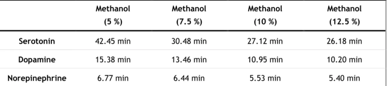
Initially a mobile phase composed of 0.145 mM EDTA, 0.1 M sodium acetate, 0.1 mM citric acid, 0.5 mM sodium octyl sulphate (OSA), 1 mM dibutylamine and 5 % methanol (v/v), at pH 3.5, was tested in isocratic mode with a flow of 1 mL/min and a potential of +750 mV as reported [58]. These conditions allowed a good separation of the compounds, but serotonin’s retention time was 42.45 minutes which would require a total running time of almost 45 minutes. In order to decrease the retention time, the content of organic solvent (methanol) was raised, as shown in table 4. Methanol percentages raging from 5-12.5 % were tested.
Table 4: Retention time variation of 5-HT, DA and NE with different methanol contents in the mobile
phase. Methanol (5 %) Methanol (7.5 %) Methanol (10 %) Methanol (12.5 %) Serotonin 42.45 min 30.48 min 27.12 min 26.18 min
Dopamine 15.38 min 13.46 min 10.95 min 10.20 min
Norepinephrine 6.77 min 6.44 min 5.53 min 5.40 min
Initially epinephrine was used as well in the method, and with methanol percentages higher than 10 %, overlay of the peaks from NE and E was observed (Figure 5).
Detection of biogenic amines in urine and plasma by HPLC-ED using MEPS
22 Since serotonin’s retention time didn’t lower significantly over 10 % of methanol and in order to keep a shorter running time, this percentage was chosen by compromise for the mobile phase. Figure 6 shows a typical chromatogram of the mobile phase with 10 % methanol.
Figure 5: Chromatogram of 5-HT, DA, NE and E at 1 µg/mL using a mobile phase with 12.5 %
methanol; an overlay of the peaks from NE and E is observable.
Figure 6: Chromatogram of 5-HT, DA, NE, E and internal standard (DHBA) at 1 µg/mL using a mobile
Detection of biogenic amines in urine and plasma by HPLC-ED using MEPS
23
2.1.2. Fluorescence system
A mobile phase composed of 50 mM KH2PO4 was used as reported [59] with 1 % of
acetonitrile added, and the temperature of the column oven set to 20°C. The compounds were detected with an excitation wavelength of 280 nm, and emission of 310 nm (catecholamines) and 340 nm (serotonin) with a flow of 0.5 mL/min. The conditions showed a good separation of 5-HT, DA, NE and the internal standard (DHBA) (Figure 7). For better intensity of the peaks, a timetable was created where the emission wavelength changed from 310 nm to 340 nm at 10 min of run.
2.1.3. Coulometric system
Initially, a mobile phase reported by Hubbard et al. [21] was used with a flow of 1 mL/min, which allowed a good separation of the compounds. But the peak of NE showed a slight overlay with an unidentified peak from the mobile phase or from the system (Figure 8). Changes in flow or column temperature did not separate those peaks, but better resolution appeared at 23°C.
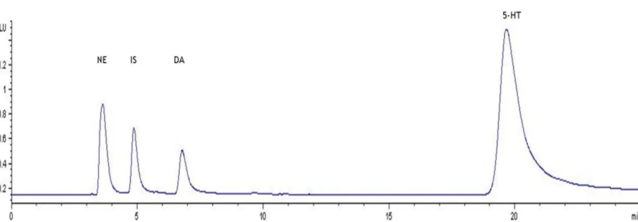
Figure 7: Chromatogram obtained by fluorescent detection of 5-HT, DA, NE and internal standard
(DHBA) at 50 ng/mL using the final optimized conditions. The retention times are 19.66 min for 5-HT, 6.77 min for DA, 3.63 min for NE and 4.85 min for DHBA.
Detection of biogenic amines in urine and plasma by HPLC-ED using MEPS
24 Afterwards, a different mobile phase designed for determination of biogenic amines was used [60]. The established composition was 75 mM sodium dihydrogen phosphate monohydrate, 1.7 mM OSA, 0.01 % triethylamine (v/v), 25 µM EDTA and 10 % acetonitrile (v/v) adjusted to pH 3.5 with phosphoric acid. The unidentified peak was still present; however, at 23°C a good separation with NE was observed (Figure 9).
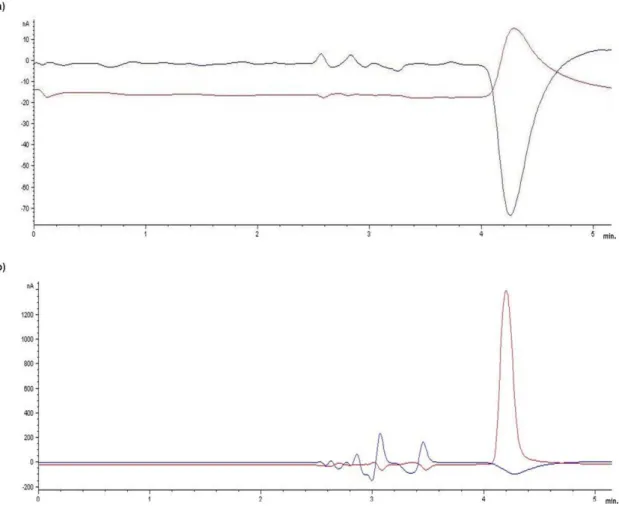
Figure 8: Chromatogram showing the baseline signal of an injection of mobile phase (a) and the peak
of NE at 50 ng/mL (b). The retention time of NE (4.20 min) overlays with an unknown peak seen in the mobile phase injection. The upper line shows the signal of channel 1 (+300 mV), and the under line the signal of channel 2 (-150 mV).
Detection of biogenic amines in urine and plasma by HPLC-ED using MEPS
25 These conditions were used for subsequent analysis, and the final retention times of the compounds were: 21.46 min for 5-HT, 8.98 min for DA, 4.92 min for NE and 6.72 min for the internal standard. As reported by Hubbard et al. [21], electrochemical oxidation of the catecholamines and serotonin was most effective with an oxidation potential of +300 mV in channel 1, -150 mV were set for the second channel. For the intended concentrations used in the calibration curve (5-1000 ng/mL), a sensitivity under 100 nA resulted in the saturation of the signal with the higher concentrations. A sensitivity of 100 nA was considered sufficient for detection in these ranges.
2.2. Optimization of the extraction procedure
The efficiency and performance of a MEPS extraction protocol depends on several variable factors, most importantly, the employed column (type of sorbent), sample dilution, number of sample drawings, washing and elution.
Based on literature [39], the conditioning step of the extraction protocol was set to 3 drawings of 250 µL of methanol and water. Washing was done only with water, since adding an amount of organic solvent to the washing solution resulted in a drastic loss of analytes. Different elution solutions were also tested (methanol with 0.1 % formic acid, isopropanol and acetonitrile), but solutions containing methanol showed better results. The presence of formic acid did not seem to affect the extraction yield, and therefore only pure methanol was used. Two types of columns were tested, a reverse phase C18 and a mixed mode (C8 and SCX,
M1). During initial extraction experiments, the area of the compounds extracted with the two
Figure 9: Chromatogram of 5-HT, DA, NE (50 ng/mL) and internal standard (DHBA, 250 ng/mL). The
upper line shows the signal of channel 1 (+300 mV), and the under line the signal of channel 2 (-150 mV).
Detection of biogenic amines in urine and plasma by HPLC-ED using MEPS
26 types of sorbent were compared. The areas resulting from extraction with M1 columns were
much lower than those observed with the C18 columns, and therefore all subsequent
experiments were done using C18 columns.
In order to obtain better yields of extraction, an experimental design or design of experiments (DOE) tool was used.
2.2.1. Design of experiments (DOE)
When developing a method, including extraction protocols, the several variable factors that have to be considered make this a very time-consuming task. Most variable factors are chosen based on existing literature, or otherwise an attempt to identify the factors is done through a trial-and-error approach or varying one factor at the time, while maintaining the other constant. Such is of course very time-consuming, exhaustive and expensive, while it can also provide misleading information since some interactions between factors may not have been taken into account [62].
DOE is a useful statistical tool for the planning of the entire procedure, assessing several intervening factors and minimizing the effects of uncontrolled factors. This spares a lot of time, work, resources and money to the laboratories, while better results are obtained [62].
With this in mind, a 2k factorial design was applied using the statistical program
MINITAB® version 15 for optimization of the extraction protocol.
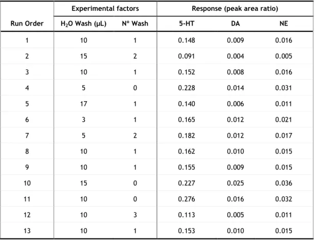
The peak area of each analyte after extraction by MEPS was divided by the peak area of the IS (added after extraction) in order to compensate injection differences (n=3); this peak area ratio was used as the measured response (dependent variable). An initial screening design was done to identify significant factors influencing the extraction response. The following factors were assessed for statistical significance: number of sample drawings (2 or 8 strokes), number of washing steps (1 or 2), washing solution volume (10 or 50 µL), number of elutions (1 or 2) and volume of elution solvent (50 or 100 µL). Each factor was studied at two levels, low (-1) and high (+1) (Table 5).
Detection of biogenic amines in urine and plasma by HPLC-ED using MEPS
27
Table 5: Factor levels in the design for MEPS optimization.
Factor Low (-1) High (+1)
Strokes 2 8
Washing volume (µL) 10 50
Number of washing steps 1 2
Methanol volume (µL) 50 100
Number of elutions 1 2
Considering these five factors, a two level full factorial design (25) would require 32
experiments and a high number of these experiments (n=16) would assess only fifth-order interactions. In order to reduce the number of experiments, the interactions of fifth order were considered negligible and a fractional factorial design (25-1) was used. This design
reduces the experiments to half (n=16) by running only part of the full factorial design, while still evaluating the main interactions, sparing time, reagents and labour.
These experiments were carried out in a random order, to avoid the influence of noise factors, minimizing systematic errors.
The experimental design matrix as well as the response in the different extraction conditions is shown in table 6.
Detection of biogenic amines in urine and plasma by HPLC-ED using MEPS
28
Table 6: Experimental design matrix showing the experimental factors, and response to each factor.
Run Order
Experimental factors Response (peak area ratio)
Strokes Washing volume (µL) Nº of Washing steps Volume of methanol eluent (µL) Nº of elutions 5-HT DA NE 1 8 10 2 50 2 0.156 0.009 0.014 2 2 10 1 100 1 0.145 0.008 0.014 3 8 50 1 100 1 0.109 0.005 0.006 4 8 50 2 50 1 0.052 0.005 0.004 5 8 10 1 50 1 0.147 0.010 0.016 6 8 10 1 100 2 0.141 0.011 0.011 7 2 10 1 50 2 0.122 0.011 0.011 8 2 50 1 100 2 0.101 0.006 0.009 9 8 50 2 100 2 0.076 0.008 0.006 10 2 50 2 50 2 0.065 0.006 0.003 11 8 10 2 100 1 0.116 0.009 0.010 12 2 10 2 100 2 0.122 0.010 0.010 13 2 10 2 50 1 0.098 0.004 0.007 14 2 50 1 50 1 0.076 0.005 0.006 15 2 50 2 100 1 0.038 0.005 0.004 16 8 50 1 50 2 0.087 0.005 0.006
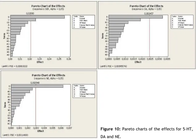
Based on these responses, the following Pareto charts were obtained showing the experimental factors in decreasing order of the effect magnitude. If a factor has a magnitude high enough to cross the red line, it’s considered statistically significant and is assumed as to influence the response.
Detection of biogenic amines in urine and plasma by HPLC-ED using MEPS
29 For all compounds the volume of water used in the washing step was found to be statistically significant, and in the case of serotonin the number of washing steps.
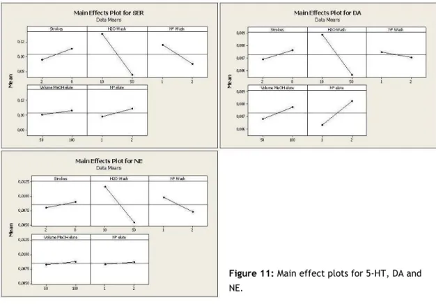
Regarding the main effects plot (figure 11), we observed that a washing volume of 10 µL originates significantly better responses for the three compounds. Generally, using one single washing step and two elutions (each with 100 µL methanol) showed better responses.
As shown in the interaction plots (figure 12), an interaction of the number of washing steps with the number of elutions is observable for 5-HT and NE. For DA, a slight interaction of volume of washing solution and number of washing steps is seen as well. However, none of those interactions had a significant effect as regards the response.
Figure 10: Pareto charts of the effects for 5-HT,
Detection of biogenic amines in urine and plasma by HPLC-ED using MEPS
30
Figure 11: Main effect plots for 5-HT, DA and
NE.
Figure 12: Interaction plot of the experimental